* To whom all correspondence should be addressed. Mobile: +8801746700196; Fax:+031682505; E-mail: [email protected]
Prevalence and In-vitro Evaluation of Probiotic Properties of
Lactobacillus delbrueckii
and
Lactobacillus plantarum
Isolated from Yoghurt in Chittagong Division, Bangladesh
Abu Sayeed Mohammad Mohamud1*, Mobarak Chowdhury2, Rezaul Karim4, Saiful Islam1, Md. Saidur Rahman1, Milton Halder1, Khorshed Alam3,
Habibur Rahman Bhuiyan1 and Faridul Islam1
1Bangladesh Council of Scientific and Industrial Research Laboratories, Chittagong, Bangladesh. 2Shahjalal University of Science and Technology, Sylhet, Bangladesh.
3Bangladesh Standards and Testing Institution.
4Institute of Food Science and Technology, Bangladesh Council of Scientific and Industrial Research.
(Received: 14 April 2014; accepted: 28 May 2014)
A total of five different Lactic acid bacterial species were identified from yoghurt samples collected from Chittagong region of Bangladesh. Among the isolates, the prevalence of Lactobacillus delbrueckii and Lactobacillus plantarum were 111 and 114cfu/ml respectively. After identification and isolation, we have optimized the suitable growth of isolates against pH, temperature and various concentration of NaCl. The results showed that better growth of organisms was observed in the presence of 3-6% NaCl. These species can tolerate up to 3% bile salt but the best tolerance found at 1% bile salt. To evaluate the antimicrobial properties of identified species, growth inhibition test has been done against some selected pathogens. It showed that Lactobacilli inhibited the growth of all pathogenic bacterial species. Inhibition by isolates and further treated with papain indicate that our isolates inhibited the growth of pathogenic microorganisms by producing Bacteriocins like substances.The selected species were subjected to Antibiotic Susceptibility testing to observe their features of carrying Antibiotic resistance genes. Among the 21 Antibiotics 17 antibiotics found resistant to Lactobacillus delbrueckii, on the other hand,
Lactobacillus plantarum were found resistant to 4 antibiotics.
Key words: Lactic Acid Bacteria,Probiotic, Lactobacillus, Prevalence, Yoghurt.
Yoghurt is a widely enjoyed dairy product that is essentially an altered form of milk containing waste products from fermentation. The lactic acid that is produced from the fermentation of lactose contributes to the sour taste of yogurt by decreasing pH and allows for the characteristic texture by acting on the milk proteins18. The acids
created by the friendly bacteria fermenting the milk
help to curb the growth of unfriendly bacteria in the yogurt. Yogurt has been continually studied for its health benefits, particularly from the addition of probiotics. Current research has been investigating how to improve yogurt both in terms of its potential as a healthy food and as an appetizing product that appeals to the general population. The probiotic efficacy of lactic acid bacteria isolated from traditional Ethiopian fermented foods6. The prevalence of lactic acid
activity of lactic acid bacteria and their effects was studied10. Potential activities of Lactobacillus
exopolysaccharides was studied on immune-modulatory and antioxidant1. The probiotic
properties of Lactobacillus isolates originating from porcine intestine and feces was also observed12.
The cancer preventing attributes of probiotics was investigated11. In Bangladesh, yoghurt is perhaps
the oldest fermented milk product known and consumed by large sectors of the population as a part of their daily diet. In most of the areas of Bangladesh, different types’ of traditional yogurts are found, but their probiotic role was not studied. Incorporation of probiotic microorganisms (isolated from indigenous yoghurts) in market yogurts can positively enhance health status of larger segment of communities of Bangladesh.
MATERIALS AND METHOD
Sample and Sampling site
The samples for the study were collected from different places of Chittagong and Jessore region in Bangladesh. The experiment was conducted at the Industrial Microbiology Research Division, Bangladesh Council of Scientific and Industrial Research (BCSIR), Chittagong during the period August, 2012 to March, 2013. All the samples were collected in sterile vials. Subsequently, transferred in to sterile screw capped test tubes containing MRS broth media. At each time of collection, precaution was taken to prevent or avoid cross-contamination of samples. After collection of the samples, they were transported to the Laboratory as soon as possible in an insulated foam box with ice to maintain a temperature ranging from 40C to 60C.
Microbiological examinations were done immediately as they arrived at the laboratory.
Isolation and Identification of Bacteria
The collected samples were inoculated into MRS broth in conical flask and incubated at 37oC for overnight5. After overnight incubation at
37oC, opaque white colored cultures were chosen
for the growth of Lactobacillus and Lactococcus sp. Culture from MRS broth (Oxoid) was then inoculated on MRS agar plates. After overnight incubation at 37oC, pure white colored colonies
were tentatively chosen to be Lactobacillus and
Lactococcus by observing their colony
morphology, physiological, sugar fermentation and as well as some biochemical characteristics. All the five selected strains were tested for their morphological, cultural and biochemical characters: the characters were compared with the standard description of Bergey,s Manual of Systematic
Bacteriology8.
Enumeration of Bacterial Load
After incubation, the deManRogosa and Sharp Agar plates (Oxoid) were placed on a colony counter (Stuart scientific, UK) and the colonies were counted. The number of colonies or viable aerobic bacterial count per ml was calculated by multiplying the average number of colonies per plate by the reciprocal of the dilution (3). The calculated results were expressed as colony forming units (cfu) per ml of sample. McIntosh and Filde’s anaerobic jar is an instrument was used in the production of an anaerobic environment.
The Fermentation Test (SAB 1957)
To determine the products of sugar fermentation, a carbohydrate fermentation broth was prepared at pH 7.4. This nutrient broth contains 0.5%-1.0% of the Carbohydrates ingredients to be tested (e.g. Lactose, Glucose, Sucrose, Mannose, Arabinose, Galactose, Starch and Mannitol), and the pH indicator phenol red. The nutrient broth, which is a light red color, supports the growth of most organisms whether they are able to ferment the sugar or not. The test organism is inoculated into a broth containing the test sugar and incubated at 37 ± 2°C for 48 to 96 hours. A bright yellow color indicates the production of enough acid products from fermentation of the sugar to drop the pH to 6.9 or less. Production of gas is determined with a Durham tube, a small inverted vial filled with the carbohydrate fermentation broth.
Growth Response at Different Concentration of NaCl
Nutrient broth containing different concentration of sodium chloride (0%, 3%, 4%, and 6%) was inoculated and incubated at37±1°C for 48 to 72 hours. The growth of Lactobacillus species at different concentration of NaCl was then compared with the control.
Preparation of Pathogenic Bacterial Suspension
bacterial culture was then mixed with the water thoroughly. A total of 11 pathogenic microorganisms were used in this method namely
Bacillus subtilis, Staphylococcus aureus. Streptococcus Group-B, Bacillus aureus, Escherichia coli, Salmonella paratyphi, Pseudomonas aeruginosa, Serratia sp., Klebsiella
Pneumonia, andNeiseria meninzitidis
Activity against pathogenic microorganisms by agar well diffusion method
In perform growth inhibition by agar well diffusion method, Mueller Hinton plates were heavily seeded (2.7×10³ cells per ml) uniformly with
the test organisms. Then a hole was made in media by gel cutter in sterile condition. Then one drop of melted agar was poured into hole and allowed to solidify to make a base layer. After that specific amount of culture filtrate (0.1 ml) was poured into the hole. Then plates were kept at low temperature (4°C) for 2-4 hours to allow maximum diffusion2.
During this time the test materials were dissolved and diffused out of the media. The diffusion occurs according to the physical law that controls the diffusion of molecules through agar gel. As a result there is a gradual change of test materials concentration in the media surrounding the discs. The plates were then incubated at 37°C for 24 hours to allow maximumgrowth of the organisms at inverted position. If the test materials have any antibacterial activity, it will inhibit the growth of microorganisms giving a clear distinct zone called “zone of inhibition”. The antibacterial activity of the test agent was determined by measuring the zone of inhibition expressed in mm in diameter. The experiment was carried out more than once and mean of reading is required.The selected pathogens used in the experiments were Escherichia coli
ATCC 25922, Streptococcus Group-B ATCC 12401,
Staphylococcus aureus ATCC 10832, Haemophilus
influenzae ATCC 49766, Klebsiella pneumoniae
ATCC 13883, Salmonella paratyphi ATCC 9150,
Bacillus subtilis ATCC 21332, and Pseudomonas
aeruginosa ATCC 15442.
Determination of optimum pH and temperature for best antimicrobial activity
The test was performed in MRS broth and the MRS broth media was adjusted for four different pH (i.e. pH 3, 3.5, 4, and 4.5) using 0.1N Acetic acid and 0.1N NaOH. Aliquots of 10 ml medium from of each pH were distributed in
separate test tube and autoclaved. After sterilization, 4 sets of tubes were inoculated with culture suspension of 5 isolates and incubated at 27°C, 37°C and 45°C temperature for 24-48 hours. After incubation the turbidity was measured with spectrophotometer at 560 nm and the culture broth were filtered with the help of Whatman filter paper (Whatman International Ltd. Maidstone, England). After filtration the final pH of the culture filtrate was recorded by pH meter (pH Hanna Instrument Ltd. & 3310, pH meter Jenway, UK) and selected for antimicrobial activity against the respective pathogenic bacteria by agar well diffusion method.
Determination of Bacteriocins Production Capability of Isolates
One ml of frozen LAB isolate was cultured overnight in 20 ml MRS broth. Then 1 ml culture was sub-cultured overnight in 20 ml MRS broth. Cells were removed by centrifuging at 9000 rpm for 15 minutes. The supernatant was filtered through a sterile Whatman No. 1 filter paper ( in original paper it is 0.22 µM syringe filter) and 100 ¼l of the unadjusted aliquot of cell free supernatant (CFS) was added to the first well. The remaining CFS was adjusted to pH 6.0 with 1 M/ lN NaOH in order to rule out possible inhibition effects due to organic acids. 100 µL of the pH adjusted CFS was filtered and added to the second well. The neutralized CFS was then treated with 1 mg/ml of catalase (Merck KGa A, Germany) at 25°C for 30 min to eliminate the possible inhibitory action of H2O2 and filtered. Then 100µl catalase CFS was placed in the third well. If inhibitions zones were found in the third well, the isolates were considered to be able to produce bacteriocin or bacteriocins like substance (BLS)17.
Effect of Proteolytic Enzymes on the Antimicrobial Activity of Crude Bacteriocins (Papain and Trypsin)
Five ml aliquot of bacteriocins was taken in test tubes and treated with papain (1 mg/ml) at pH 7. The test tubes with and without the enzyme (control) were incubated at 37°C for 2 hour and heated at 100°C for 3 minutes to denature the enzyme. Both the control and samples were assayed for antimicrobial activity by using well diffusion method (50 µL of sample in each well).
Assay of Antimicrobial activity for Bacteriocin
optimum conditions, the culture filtrates were centrifuged at 5000 rpm for 15 minutes. The culture supernatant was then neutralized by 0.1N NaOH and the pH of the supernatant was adjusted at 7.0. Then antimicrobial activity was assayed by agar well diffusion method14.
Bile salt tolerance of the isolates
MRS broth was prepared with varying concentration (i.e.; 1.0%, 2.0% and 3.0%) of bile salt. Then the medium was dispensed at 10 ml per tube. Inoculation was done with the selected isolates (i.e. LcL, LcP, LcR, LbD and LbP). After inoculation with equal amount of inoculums the tubes were then incubated at 37±1°C for 24-48 hours. After incubation 0.1 ml of culture from each concentration was used to grow in agar medium by pour plate method. The plates were then incubated at 37°C for 24 hours and observed for comparative growth in these plates (4).
Assay of antibiotic sensitivity pattern
To assess the antibiotic sensitivity pattern disk diffusion method was followed. In this method Mueller Hinton plates were prepared and swabbed with suspension of selected isolates with the help of sterile cotton bud. After swabbing the antibiotic disks (Azithromycin, Ceftriaxone,
Amoxicillin, Kanamycin, Cefixime, Cephalexin, Streptomycin, MeropenemCefaclor, Amoxyclav, Piperacillin, Ciprofloxacin, Gentamicin, Chloramphenicol, Erythromycin, Ampicillin, Amikacin, Aztreonam, Tobramycin, Ceftazidime and Nitrofurantoin) were placed on the surface of the plate at equidistance. The plates were then kept at 4°C for 1-2 hours for proper diffusion of antibiotics. The plates were then incubated for 18-24 hours at 37°C. The zone of inhibition was observed for antibiotic sensitivity or resistance and zone diameter was measured1.
RESULTS
Sugar Fermentation
Acid and Gas production by Fermentation test are given below:
Determination of optimum pH for best antimicrobial activity
For evaluation of the effects of pH on growth and the production of antimicrobial substances, the isolates were grown in broth medium with different pH .The growth of the organisms after incubation was measured spectrophotometrically at 560 nm and final pH of
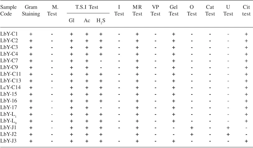
Table 1. Morphological, physiological and biochemical test for identification of isolated Lactobacilli sp
Sample Gram M. T.S.I Test I M R VP Gel O Cat U Cit
Code Staining Test Test Test Test Test Test Test Test test
Gl Ac H2S
LbY-C1 + - + + + - + - + - - - +
LbY-C2 + - + + + - + - + - - - +
LbY-C3 + - + + + - + - + - - - +
LbY-C4 + - + + + - + - + - - - +
LbY-C7 + - + + - - + - + - - - +
LbY-C9 + - + + - - + - + - - - +
LbY-C11 + - + + + - + - + - - - +
LbY-C13 + - + + + - + - + - - - +
LcY-C14 + - + + + - + - + - - - +
LbY-15 + - + + + - + - + - - - +
LbY-16 + - + + + - + - + - - - +
LbY-17 + - + + - - + - + - - - +
LbY-L1 + - + + + - + - + - - - +
LbY-L9 + - + + + - + - + - - - +
LbY-J1 + - + + + - + - - + - +
-LbY-J2 + - + + + + - - + - +
-LbY-J3 + - + + + - + - + - - - +
Table 2. Physiological and Biochemical test for identification of isolated Lactobacilli sp
Growth response at different pH, Temperature &NaCl concentration (%) for Lactobacilli and Lactococcus sp. are given below:
Sample Growth at different Growth at different Growth at different code Temperature pH Nacl conc. (%)
27°C 37°C 45°C 3 3.5 4 4.5 3 4 6
LbY-C1 ++ ++ + + + ++ +++ ++ ++ +
LbY-C2 ++ ++ + + + ++ +++ ++ ++ +
LbY-C3 ++ ++ + + + ++ +++ ++ ++ +
LbY-C4 ++ ++ + + + ++ +++ ++ ++ +
LbY-C7 ++ + - + + ++ ++ + -
-LbY-C9 ++ + - + + ++ ++ + + +
LbY-C11 ++ ++ + + + ++ +++ ++ ++ +
LbY-C13 ++ ++ + + + ++ +++ ++ ++ +
LbY-C14 ++ ++ + + + ++ +++ ++ ++ +
LbY-15 ++ ++ + + + ++ +++ ++ ++ +
LbY-16 ++ ++ + + + ++ +++ ++ ++ +
LbY-17 ++ ++ - + + ++ ++ +
-LbY-L1 ++ ++ + + + ++ +++ ++ ++ +
LbY-L9 ++ ++ + + + ++ +++ ++ ++ +
LbY-J1 +++ +++ ++ - + + ++ +++ ++ +
LbY-J2 +++ +++ ++ - + + ++ +++ +++ +
LbY-J3 +++ +++ + - + + ++ +++ +++ +
Note: Good=+, Moderate= ++, Excellent= +++
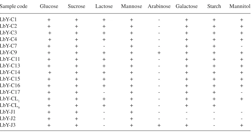
Table 3. Physiological and Biochemical test for identification of isolated Lactobacilli sp
Sample code Glucose Sucrose Lactose Mannose Arabinose Galactose Starch Mannitol
LbY-C1 + + + + - + + +
LbY-C2 + + + + - + + +
LbY-C3 + + + + - + + +
LbY-C4 + + + + - + + +
LbY-C7 + + - + - + +
-LbY-C9 + + + + + + + +
LbY-C11 + + + + - + + +
LbY-C13 + + + + - + + +
LbY-C14 + + + + - + + +
LbY-C15 + + + + - + + +
LbY-C16 + + + + - + + +
LbY-C17 + + - + - + +
-LbY-CL1 + + + + - + + +
LbY-CL9 + + + + - + +
LbY-J1 + + - + - - -
-LbY-J2 + + - + - - -
-LbY-J3 + + - + + + - +
Note: Positive = +, Negative =
-the culture filtrate was determined. The antimicrobial activity of the culture filtrate was also assayed by agar well diffusion method.
Determination of optimum Temperature for best antimicrobial activity
Table 4. Growth and final pH after incubation of the isolates at different pH
Isolates Parameters Initial medium pH
PH 3.0 PH 3.5 PH 4.0 PH 4.5
Lactobacillus delbrueckii Growth 0.327 0.421 0.489 0.89
(LbD) Final pH 3.28 3.60 4.30 4.69
Lactobacillus plantarum Growth 0.320 0.396 0.770 0.96
(LbP) Final pH 3.28 3.80 4.50 4.8
Table 5. Growth and final pH after incubation of the isolates at different temperature
Isolates Parameters Incubation Temperature
27°C 37°C 45°C
Lactobacillus delbrueckii Growth 0.195 0.210 0.177
(LbD) Final pH 1.09 1.08 1.13
Lactobacillus plantarum Growth 0.206 0.215 0.183
(LbP) Final pH 1.04 1.03 1.05
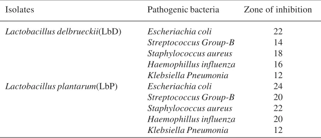
Table 6. Assay of Bacteriocin activity produced by the selected isolates given below
Isolates Pathogenic bacteria Zone of inhibition
Lactobacillus delbrueckii(LbD) Escheriachia coli 22
Streptococcus Group-B 14
Staphylococcus aureus 18
Haemophillus influenza 16
Klebsiella Pneumonia 12
Lactobacillus plantarum(LbP) Escheriachia coli 24
Streptococcus Group-B 20
Staphylococcus aureus 22
Haemophillus influenza 20
Klebsiella Pneumonia 12
on growth and the production of antimicrobial substances, the isolates were grown in broth medium with different temperature .The growth of the organisms after incubation was measured spectrophotometrically at 560 nm and final temperature of the culture filtrate was determined. The antimicrobial activity of the culture filtrate was also assayed by agar well diffusion method.
Determination of Bacteriocin Production
Zone of inhibition produced by all five isolates, indicated that all isolates are able to produce bacteriocin in our experiment. Zone of inhibition was found illustrated in the (Figure 1)
Effect of proteolytic enzymes on the antimicrobial activity of crude bacteriocin (Papain and Trypsin)
After treating Bacteriocin with Proteolytic enzymes papain, we have found that there were no
zone of inhibition indicated that the zone oh inhibition occurred by bacteriocin. The results are shown in the below (figure 2)
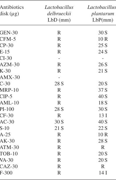
Assay of antibiotic susceptibility
Antibiotic sensitivity pattern of the selected isolates were also determined to observe the inhibitory effect of any antibiotics against them. Combination of this type of antibiotic with probiotic organism may destroy the therapeutic activity of the probiotic. The Antibiogram obtained is shown in table-7.
Fig. 1. Determination of Bacteriocin Production Capability of the Lab Isolates
Fig. 2. Effect of Proteolytic Enzymes on the
Antimicrobial Activity of Crude Bacteriocin (Papain and Trypsin)
Fig. 3. Percentage of Antibiotic Resistance, 21 for
Lactobacilli sp.)
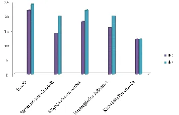
Fig. 4. Zone of inhibition produced produced by isolate LbD and LbP against 05 pathogenic organisms
Fig. 5-6. Zone of inhibition against (5) E.coli and (6) Streptococcus Group B
5 6
Fig. 7-8. Antibiotic sensitivity and resistant of (7) Lactobacillus delbrueckii (8) Lactobacillus plantarum
Assay of antimicrobial activity for bacteriocin
Bacteriocin an antimicrobial substance produced by most lactic acid bacteria. The activity of Bacteriocin produced by the selected isolates was also determined and the effectively of the substance was measured by the zone of inhibition (mm) which given following table-6.
In the Graph, LbD and LbP produced zone of inhibition (mm) respectively 22 and 24 mm against pathogenic bacteria E. coli., and 14, 20 mm against pathogenic bacteria Streptococcus Group-B., and 18, 22 mm against pathogenic bacteria
Staphylococcus aureus., and 16, 20 mm against
pathogenic bacteria Haemophillus influenza., and 13, 12 mm against pathogenic bacteria Klebsiella pneumonia.
Bile salt tolerance
As probiotic organism has to tolerate different bile salt concentration in the intestinal tract or stomach for their survival, the isolates were assayed for their sensitivity to different bile salt
concentration (i.e. 1%, 2% and 3%). The activities they showed were tabulated-12 in the following. In the graph showed that when bile salt concentration is increased, the No. of colony is decreased. Every isolate tolerate up to 3% bile salt but1% bile salt gives the best no. of colony (CFU/ 0.1ml) for all isolate.
DISCUSSION
For isolation of lactic acid bacteria, yoghurt samples were collected from different places of Chittagong & Jessore region. The identification of isolates were determined according to the Bergey,s Manual of Systematic Bacteriology
(2009).pH is an important factor which can dramatically affect bacterial growth. In our experimental design we have observed the growth of our isolated lactobacillus in various pH values ranges (3 to 4.5). The result shown that all the isolates grew best at pH 4.5 used range value (Table-5). Growth increases with the increasing of pH from pH-3 to pH-4.5 but growth decrease when it exceeds the neutral range of pH. It was found that Lactobacillus delbrueckii subsp. Bulgaricus,
Lactococcus lactis subsp. Lactis, Lactococcus
raffinolactis, Enterococcus faecium,
Pediococcuspentosaceuscan tolerate pH up to 5.0
9. The present results are closely related to their
findings. It was reported that Lactobacillus
plantarum could produce lactic acid and reduce
the pH to values lower than 4.0 (15). After growth at different temperature i.e. 27°C, 37°C and 45°C,it was found that the isolates grew best at 37°C but
Lactobacillus delbrueckii and Lactobacillus
plantarum can grew at 450 C and show suitable for
best antimicrobial activity (Table-7). The growth decreases with the increasing temperature (i.e.; 45°C). NaCl is an inhibitory substance which may inhibit growth of certain types of bacteria. The current results showed that Lactobacillus
delbrueckii and Lactobacillus plantarum were
able to tolerate 3%, 4%, & 6% NaCl and good growth was observed at 4% NaCl (Table-8). It was found that 56% of Lactococci tested were able to grow on 6.5% NaCl 14. It was reported that
Lactococcus lactis was the most tolerant to high NaCl concentration compared to the other isolates. The table shows the number of colonies of the isolates on agar medium after incubating them in
Fig. 10. Number of colony count for isolates against different bile salt concentration (%)
In the graph showed that when bile salt concentration is increased, the No. of colony is decreased. Every isolate tolerate up to 3% bile salt but1% bile salt gives the best no. of colony (CFU/0.1ml) for all isolate.
Fig. 9. Antibiotic sensitivity and resistant of
MRS broth medium containing varying concentration of bile salt i.e. 1.0 %, 2.0 %, 3.0%. In the Table-8, it is seen that at lower concentration the colony numbers were very high but gradually
Table 7. Antibiotic Sensitivity pattern of the selected isolates given below
Antibiotics Lactobacillus Lactobacillus
disk (µg) delbrueckii plantarum
LbD (mm) LbP(mm) Sensitive, GEN = Gentamicin, AZM = Azithromycin, E= Erythromycin, CI = Ceftriaxome, K= Kanamycin, CP= Cephalexin, CFM= Cefixime, S = Streptomycin, MRP = Meropenem, CF = Cefaclor, AML = Amoxycillin, C = Chloramphenical, AC= Amoxyclav, CIP = Ciprofloxacin, PI = Piperacillin, A = Ampicillin, AK = Amikacin, ATM = Aztreonam, TOB = Tobramycin, VA = Vanomycin, CAZ = Ceftazidime, F = Nitrofurantoin.
Table 8. Bile salt tolerance (at different concentrations) of the selected isolates given below
Isolates No. of colony grown on the
plates (CFU/0.1 ml)
1.0% 2.0% 3.0%
Lactobacillus delbrueckii (LbD) 360 220 180
Lactobacillus plantarum( LbP) 510 450 350
the numbers decreased with the increasing of bile salt concentration. The zone of inhibition in the Table-6 indicates that the isolates secrete Bacteriocin and it has bactericidal activities. The table-6 shows that Zone of inhibition (mm) produced by L. delbrueckii, L. plantarum, against pathogenic bacteria E. coli. Streptococcus Group-B, Staphylococcus aureus, Haemophillus influenza, Klebsiella pneumonia, Salmonella paratyphi, Bacillus subtillis, and Pseudomonas aeroginosawere satisfactory. Inhibition by isolates and further treated with papain indicate that our isolates inhibited the growth of pathogenic microorganisms by producing Bacteriocins like substances. Our study showed similarity with other research such results have shown L. plantarum
higher antimicrobial activities against E. coli and
Staph. aureusthan the others. The zone of
inhibition by L. plantarum against E. coli were 12.33 mm while zone of inhibition against Staph. aureus were 14.33 mm but it showed L. plantarum
was able to inhibit the growth of the Gram negative tested bacteria with an average inhibition zone of 18 and 26 mm in diameter against Salmonella sp. and E. coli strains respectively17.It was reported
Lactococcus lactis ssp. lactis antagonistic
behavior was demonstrated by the production of 14 mm against Salmonella sp. and 20 mm inhibition zones against E. coli strains which supports our findings16.
Lactic acid bacteria (LAB) from fermented products may act as a reservoir of antimicrobial-resistance genes7. Lactobacillus delbrueckii were
found sensitive to Chloramphenicol, Piperacillin, Amoxyclav and Streptomycin but resistant to remaining 17 antibiotics. Our results were similar to a study3 Lactobacillus plantarum is Sensitive
isolates were resistant to Ampicillin and one resistant to tetracycline. Lactobacilli isolated from milk and milk products were resistant to erythromycin, Ampicillin and tetracycline and were sensitive in 100 % to Gentamicin which is in agreement with our findings. Such resistance to a wide spectrum of antibiotic therapy may be helpful in faster recovery of the patients due to rapid establishment of desirable microbial flora. This study supports the use of selected probiotic agents for the prevention of antimicrobial-associated diarrhea. Resistance of the probiotic strains to some antibiotics could be used both preventive and therapeutic purpose in controlling intestinal infections4.
In conclusion, the experimental results showed that isolated two selected species were able to tolerate inhibitory substance bile salt at 1-3% where best at 1% and 3-6% NaCl. The suitable temperature for their growth is showed 370C and
pH-4.5, but Lactobacillus delbrueckii and
Lactobacillus plantarum grew at temperature 450C.
Growth inhibition against selected pathogens
(Escherichia coli ATCC 25922, Streptococcus
Group-B ATCC 12401, Staphylococcus aureus ATCC 10832, Haemophilus influenzae ATCC 49766, Klebsiella pneumoniae ATCC 13883, Salmonella paratyphi ATCC 9150, Bacillus subtilis ATCC 21332, and Pseudomonas aeruginosa ATCC 15442 and Neisseria
meninzitidis ATCC 35561) and resistance against
various antibiotics suggest that our identified Lactobacilli (Lactobacillus delbrueckii and
Lactobacillus plantarum). On the other hand
Lactobacillus delbrueckii was resistant to 17
antibiotics used against them but Lactobacillus
plantarum were found resistant only against 04
antibiotics (Cefixime, Ampicillin, Aztreonam and Ceftazidime). Finally, it can be said that the selected
lactobacillus delbrueckii found in our study
showed potentially to be used as a probiotics in near future. In spite of the problems with dosage and viability of probiotic strains, lack of industry standardization and potential safety issues, there is obviously considerable potential for the benefits of probiotics over a wide range of clinical conditions. Further study on these isolates will help to detect the genes responsible for therapeutic activities and this type of research will help to design more probiotic agents to control numerous
diseases and at the same time it will ensure the safe and healthy human civilization.
ACKNOWLEDGMENTS
We thank Bangladesh Council of Scientific and Industrial Research (BCSIR) for their logistic support and collaboration.
Conflict of Interest
We hereby declare that we have no conflict of interest regarding this paper
REFERENCES
1. Bacha, K., Mehari, T. & Ashenafi, M. In-vitro probiotic potential of lactic acid bacteria isolated from ‘Wakalim’, a traditional Ethiopian fermented beef sausage. Ethiop J Health Sci,
2009; 19: 21-29.
2. Bauer, A., Kirby, W., Sherris, J. C., Turck & Turck, M., Antibiotic susceptibility testing by a standardized single disk method. American journal of clinical pathology, 1966; 45: 493. 3. Belletti, N., Gatti, M., Bottari, B., Neviani, E.,
Tabanelli, G. & Gardini, F., Antibiotic resistance of lactobacilli isolated from two Italian hard cheeses. Journal of Food Protection®, 2009; 72:
2162-2169.
4. Bhutada, S. & Tambekar, D., An evaluation of probiotic potential of Lactobacillus sp. from milk of domestic animals and commercial available probiotic preparations in prevention of enteric bacterial infections. Recent Research in Science and Technology, 2010; 2.
5. Collins, C. H., Microbiological methods.
Microbiological methods, 1964.
6. Diop, M., Dubois Dauphin, R., Dortu, C., Destain, J., Tine, E. & Thonart, P., In vitro detection and characterization of bacteriocin-like inhibitory activity of lactic acid bacteria (LAB) isolated from Senegalese local food products.
African Journal of Microbiology Research [= AJMR], 2008; 2.
7. Flórez, A. B., Delgado, S. & Mayo, B., Antimicrobial susceptibility of lactic acid bacteria isolated from a cheese environment.
Canadian journal of microbiology, 2005; 51:
51-58.
8. Garrity, G. M., Bell, J. A. & Lilburn, T. G., Taxonomic outline of the prokaryotes. Bergey’s manual of systematic bacteriology. Springer, New York, Berlin, Heidelberg, 2004.
acid bacteria isolated from traditional fermented milk ‘Dahi’in Bangladesh. Pakistan J Nutr, 2007;
6: 647-652.
10. Heczko, P. B., Strus, M. & Kochan, P., Critical evaluation of probiotic activity of lactic acid bacteria and their effects. J Physiol Pharmacol,
2006; 57(9): 5-12.
11. Kumar, M., Kumar, A., Nagpal, R., Mohania, D., Behare, P., Verma, V., Kumar, P., Poddar, D., Aggarwal, P. K., Henry, C. J., Jain, S. & Yadav, H., Cancer-preventing attributes of probiotics: an update. Int J Food Sci Nutr, 2010;
61:473-96.
12. Liu, C. F., Tseng, K. C., Chiang, S. S., Lee, B. H., Hsu, W. H. & Pan, T. M., Immuno-modulatory and antioxidant potential of
Lactobacillus exopolysaccharides. Journal of the Science of Food and Agriculture, 2011; 91: 2284-2291.
13. Murray, B. E., The life and times of the Enterococcus. Clinical microbiology reviews,
1990; 3: 46-65.
14. Parente, E., Brienza, C., Moles, M. & Ricciardi,
A., A comparison of methods for the measurement of bacteriocin activity. Journal of microbiological methods, 1995; 22: 95-108. 15. Rao, M. S., Pintado, J., Stevens, W. F. & Guyot,
J. P., Kinetic growth parameters of different amylolytic and non-amylolytic Lactobacillus strains under various salt and pH conditions.
Bioresour Technol, 2004; 94: 331-7.
16. Yateem, A., Balba, M., surrayai, T., Al-mutairi, B. & Al-daher, R., Isolation of Lactic Acid Bacteria with Probiotic Potential from Camel Milk. International Journal of Dairy Science, 2008; 3.
17. Yang, E., Fan, L., Jiang, Y., Doucette, C. & Fillmore, S, Antimicrobial activity of bacteriocin-producing lactic acid bacteria isolated from cheeses and yogurts. AMB Express, 2012; 2: 1-12.