Physalis minima Leaves Extract Induces Re-Endothelialization in
Deoxycorticosterone Acetate-Salt-Induced Endothelial Dysfunction in Rats
Dian Nugrahenny1, Nur Permatasari1, Mohammad Saifur Rohman2 1 Department of Pharmacology, Faculty of Medicine, University of Brawijaya.
2 Department of Cardiovascular and Vascular Medicine, Faculty of Medicine, University of Brawijaya.
Email: [email protected], [email protected]
ABSTRACT
The administration of deoxy-corticosterone acetate (DOCA)-salt can induce oxidative stress leading to decrease the bioavailability of nitric oxide (NO), increase senescence of circulating endothelial progenitor cells (EPCs), thus contributing to endothelial dysfunction. This study was aimed to investigate the effects of Physalis minima L. leaves extract on serum NO levels, circulating EPCs number, and histopathology of tail artery endothelial cells in DOCA-salt-induced endothelial dysfunction in rats. Twenty-five male Wistar rats were randomly divided into five groups: rats without any treatment (normal), rats treated with DOCA (10 mg/kgBW s.c. twice weekly) and given 0.9% NaCl to drink ad libitum for 6 weeks, and DOCA-salt-induced rats orally supplemented with P. minima leaves extract at doses of 500, 1500, or 2500 mg/kgBW for 4 weeks. Serum NO levels were measured by colorimetry. The number of circulating EPCs (CD34+/CD133+ cells) was determined by flow cytometry. The tail artery sections were histologically processed with hematoxylin-eosin staining. DOCA-salt-induced rats showed significantly (p<0.05) decrease in serum NO levels and circulating EPCs number compared to the normal. There was also more detached tail artery endothelial cells in DOCA-salt-induced rats. P. minima leaves extract at a dose of 500 mg/kgBW significantly (p<0.05) increased serum NO level and circulating EPCs number, and also induced an optimal
re-endothelialization in DOCA-salt-induced rats. P. minima leave extract dose-dependently increases NO bioavailability contributing to enhanced EPCs mobilization, thereby promoting re-endothelialization in DOCA-salt-induced endothelial dysfunction in rats.
Keywords: Physalis minima; DOCA-salt-induced rats; Re-endothelialization; Nitric oxide; Endothelial progenitor cells.
INTRODUCTION
Oxidative stress, an increase in the production of reactive oxygen species (ROS), may play a role in the pathogenesis of hypertension. Studies have demonstrated that oxidative stress is markedly increased in patients with hypertension and renovascular disease (Minuz et al., 2002; Thuillez et al., 2005; Ceriello, 2008). Oxidative stress promotes functional deficiency of nitric oxide (NO), the factor released by the endothelium that causes vascular relaxation. The reduced NO levels in hypertension may result either from a decrease in NO production or an increase in NO degradation. The decrease in NO production may result from uncoupled endothelial NO synthase (eNOS) and deficiency of cofactor tetrahydrobiopterin (BH4) while the increase in NO degradation may result from interaction with enhanced superoxide production (Thuillez et al., 2005).
The NO-mediated signaling pathways are essential for endothelial progenitor cells (EPCs) mobilization and function (Aicher et al., 2003). Circulating EPCs are involved in endogenous injured endothelial recovery and play a critical role in maintaining endothelial integrity following arterial damage (Hill et al., 2003). Accumulating evidence shows that a fall in function and number of circulating EPCs (Giannotti et al., 2010; Liu et al., 2013), and an accelerated senescence of circulating EPCs are responsible for impaired EPCs-mediated re-endothelialization in patients with hypertension (Zhou et al., 2010).
Deoxycorticosterone acetate (DOCA) is a precursor of aldosterone. Increased aldosterone levels lead to enhanced reabsorption of sodium ions and water from epithelial cells in the distal nephron of the kidney. This condition concomitant with high salt intake promotes volume-dependent hypertension, leading to
oxidative stress (Zhou et al., 2008; Tomaschitz et al., 2010). DOCA-salt induction in this study was mainly intended to induce oxidative stress, leading to functional deficiency of nitric oxide (NO), decreased function and number of circulating EPCs, increased senescence of circulating EPCs, and impaired EPCs-mediated re-endothelialization.
Physalis minima L. of Solanaceae family is commonly known as wild gooseberry, ground-cherry, sunberry (English), popti (Gujarat), pokok letup-letup (Malaysia), or ceplukan (Indonesia). It is found throughout India, Afghanistan, Tropical Africa, Singapore, Malaysia, Indonesia, and Australia, and is reported as one of the principal medicinal plants in Indian Traditional System of Medicines (Chothani et al., 2012). Water in which the plant is boiled is traditionally taken to help ensure normal urination and also to lower hypertension (Muhamad & Mustafa, 2010). Our previous studies showed that P. minima leave extract enhances eNOS expression and preserves NO levels in endothelial cells, and also decreases oxidative stress and induces re-endothelialization in ovariectomized rats (unpublished observation). In the present study, we tested the hypothesis that P. minima preserve NO levels and EPCs number, thus promotes re-endothelialization in deoxycorticosterone acetate (DOCA)-salt-induced rats.
MATERIALS AND METHODS
Plant Materials.
stored at -20°C in airtight containers wrapped in aluminum foil.
Extraction.
The leaves powder (200 g) was mechanically stirred with 1000 mL of 80% (v/v) ethanol at RT for 24 h. The residue was extracted thrice; then the extract was filtered. Subsequently, the filtrate was evaporated to dryness using vacuum distillation and rotary evaporator at 50°C. The crude ethanol extract was then stored at -20°C in airtight containers until application.
Animals.
Twenty-five male Wistar rats, ten weeks of age, weighing 170–200 gram, were used for this study. The rats were housed in a clean wire cage and maintained under standard laboratory conditions at a temperature of 25 ± 2°C and dark/light cycle 12/12 h. Standard diet and water were provided ad libitum. The rats were acclimatized to laboratory conditions for two weeks before the experiment. Animal care and experimental procedures were approved by the institutional ethics committee of Faculty of Medicine, University of Brawijaya, Malang, Indonesia. The rats were randomly divided into five groups as follows. One group was a normal control group (N). One group was a positive control group induced with DOCA-salt (H). Then, three groups were DOCA- DOCA-salt-induced rats orally administered with P. minima leavesextract at doses of 500 (PM1), 1500 (PM2), and 2500 mg/kgBW (PM3), for four weeks.
DOCA-salt Induction.
DOCA-salt induction was performed according to the method of Imenshahidi et al. (2010) and Bankar et al. (2011) with modification. Briefly, the rats were subcutaneously injected with DOCA (10 mg/kgBW, Sigma-Aldrich, St. Louis, MO,
USA) twice a week and given 0.9% NaCl in drinking water ad libitum for six consecutive weeks (Imenshahidi et al., 2010; Bankar et al., 2011).
Tissue Sampling.
At the end of the experiment, the rats were euthanized by anesthetizing with ketamine i.p. 40 mg/kgBW and exsanguinated by cardiac puncture. The blood samples were collected. Serum was obtained by centrifugation (3000 rpm, RT, 5 min), labeled, and stored at -40°C until analysis. The EDTA-blood was used to isolate the circulating EPCs. The tail artery was stored in 10% buffered formalin, then underwent tissue processing and hematoxylin-eosin staining.
Isolation of Circulating EPCs.
The isolation of circulating EPCs was performed according to a method of Yuwono et al. (2013) with modification. Mononuclear cells (MNCs) were isolated using Histopaque 1.083 g/ml (Sigma-Aldrich, St. Louis, MO, USA) density gradient centrifugation (1600 rpm, 20°C, 30 min), then washed three times with Hanks’ balanced salt solution (Sigma-Aldrich, St. Louis, MO, USA).
CD34+/CD133+ cells were evaluated by immunostaining with PE-conjugated anti-mouse CD34+ monoclonal antibody (BioLegend, London, UK) and FITC-conjugated rabbit anti-CD133 polyclonal antibody (Bioss Inc., Woburn, Massachusetts, USA), and then detected by flow cytometry (BD FACSCalibur™ Flow Cytometer; BD Biosciences, San Jose, CA, USA) (Setiawan et al., 2013).
Analysis of Serum Nitric Oxide Levels.
Statistical Analysis
Data are presented as mean ± SD, and the differences between groups were analyzed using Kruskal-Wallis and Mann-Whitney test with SPSS 22.0 statistical package for Windows. Only probability values of p<0.05 were considered
statistically significant.
RESULTS AND DISCUSSION
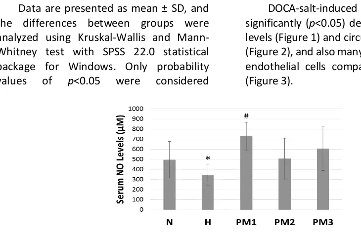
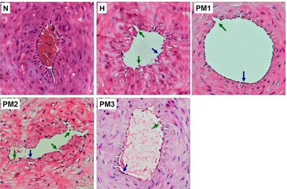
DOCA-salt-induced rats showed significantly (p<0.05) decrease in serum NO levels (Figure 1) and circulating EPCs number (Figure 2), and also many detached tail artery endothelial cells compared to the normal (Figure 3).
Figure 1: Serum NO Levels. Bar graph represents the mean + SD of serum NO levels; n = 5. DOCA-salt-induced rats showed a significant decrease in serum NO levels compared to the normal. P. minima leaves extract at a dose of 500 mg/kgBW significantly increased serum NO levels in DOCA-salt-induced rats. Normal control (N), DOCA-salt-induced control (H), DOCA-salt-induced rats administered with P. minima leaves extract at doses of 500 (PM1), 1500 (PM2), and 2500 mg/kgBW (PM3). *p<0.05 compared to normal control; #p<0.05
compared to DOCA-salt-induced control.
Figure 2. Circulating EPCs Number. DOCA-salt-induced rats showed a significant decrease in circulating EPCs number compared to the normal. P. minima leave extract at a dose of 500 mg/kgBW significantly increased EPCs number in DOCA-salt-induced rats. A. The isolated rats’ mononuclear cells were analyzed for the expression of CD34+ and CD133+ by flow cytometry. The ordinate and abscissa of representative flow cytometric graphs
show the fluorescence intensity of CD34+ and CD133+ mononuclear cells. The number in upper right
quadrant shows the percentage of CD34+/CD133+ double-positive cells. B. Bar graph represents the mean
percentage + SD of CD34+/CD133+ double-positive cells; n = 5. Normal control (N), DOCA-salt-induced
control (H), DOCA-salt-induced rats administered with P. minima leaves extract at doses of 500 (PM1), 1500 (PM2), and 2500 mg/kgBW (PM3). *p<0.05 compared to normal control; #p<0.05 compared to
Figure 3. Histopathological Examination of Hematoxylin-eosin-stained Rat Tail Artery Sections.
DOCA-salt-induced rats showed many endothelial cells detached from the basement membrane. P. minima leave
extract at a dose of 500 mg/kgBW promoted an optimal re-endothelialization in DOCA-salt-induced rats. The representative micrographs from 5 independent experiments in which the same results were obtained are shown. Blue arrow: intact endothelial cells, green arrow: detached endothelial cells. Magnification: 200x. Normal control (N), DOCA-salt-induced control (H), DOCA-salt-induced rats
administered with P. minima leaves extract at doses of 500 (PM1) and 1500 mg/kgBW (PM2).
The reduction of NO bioavailability in DOCA-salt-induced rats may be due to oxidative stress. The considerable evidence demonstrated that oxidative stress accompanies high blood pressure in salt-sensitive hypertension (Seifi et al., 2010). The increased aldosterone levels induce superoxide generation via mineralocorticoid receptor-mediated activation of NAD(P)H-oxidase and Rac1 in endothelial cells (Iwashima et al., 2008). Superoxide then reacts with NO to form peroxynitrite (ONOO¯), a potent oxidant, enhancing endothelial oxidative stress. Both superoxide and peroxynitrite reduce the endothelial cell cofactor BH4 levels by rapidly oxidizing BH4 to BH3, which further oxidized to BH2, resulting in eNOS uncoupling (Vasquez-Vivar et al., 2002; Kar & Kavdia, 2011). Studies have indicated that eNOS is uncoupled in DOCA-salt hypertension due to reduced BH4. The uncoupled eNOS then promotes the generation of superoxide rather than NO,
thus exacerbating oxidative stress (Du et al., 2008; Xie et al., 2008).
NO-mediated signaling pathways are essential for EPCs mobilization and function in blood vessel recovery by the processes of vasculogenesis or angiogenesis, or both (Aicher et al., 2003; Hill et al., 2003; George et al., 2011). During vasculogenesis, EPCs mobilize from bone marrow into circulation in response to various signals, and then home to the site of endothelial injury where they differentiate into mature endothelial cells, assisting in the ongoing new vessels formation. During angiogenesis, EPCs act in a paracrine fashion by secreting various angiogenic factors leading to endothelial cells activation. The activated endothelial cells then migrate and proliferate (sprouting) to form new blood vessels from pre-existing vessels (George et al., 2011).
decreased EPCs function in endothelial recovery following arterial damage. Our results are in line with recent studies indicated a reduction in number and function of circulating EPCs, and an acceleration of circulating EPCs senescence in patients with hypertension (Zhou et al., 2008; Giannotti et al., 2010; Liu et al., 2013). A study by Thum et al. (2007) has also shown that eNOS uncoupling impairs EPCs mobilization and function. Furthermore, a recent study has demonstrated that DOCA-salt hypertension features elevated endothelin (ET)-1 and oxidative stress. The ET-1 activation of ETA/NADPH oxidase pathway and diminished antioxidants critically contribute to EPCs reduction and dysfunction via increased oxidative stress in salt-sensitive hypertension (Chen et al., 2012).
The administration P. minima leaves extract at dose of 500 mg/kgBW significantly (p<0.05) increased serum NO levels (Figure 1) and circulating EPCs number (Figure 2), and also promoted an optimal re-endothelialization in DOCA-salt-induced rats (Figure 3), while the effects were much less with higher doses (1500 and 2500 mg/kg BW).
Our previous study showed that P. minima leave extract preserves NO levels and enhances eNOS expression in endothelial cells (unpublished observation), which is in line with this study results. This effect may be due to the antioxidant activity of flavonoids and polyphenols that are abundant in P. minima. A recent study has demonstrated that the ethanol extract of P. minima leaves exhibits potent antioxidant activity, as evidenced by 2, 2-diphenyl-1-picrylhydrazyl (DPPH) assay, Fe2+ chelating activity assay, and Fe3+ reducing power assay (Karpagasundari & Kulothungan, 2014). Moreover, our previous study showed that
P. minima leave extract enhances superoxide dismutase (SOD) activity (unpublished observation). SOD converts superoxide anion radicals to hydrogen peroxide, thereby reducing the likelihood of superoxide anion interacting with NO to form reactive peroxynitrite (Bais et al., 2012). This superoxide scavenging activity of antioxidants contained in P. minima promotes NO bioavailability and NO-mediated signaling pathways, thereby preserving EPCs mobilization and function in maintaining endothelial integrity following arterial damage.
CONCLUSION
P. minima leave extract at a dose of 500 mg/kgBW is useful to enhance NO bioavailability leading to increased EPCs mobilization from bone marrow, thereby promoting re-endothelialization in DOCA-salt-induced rats. However, the higher doses are less than optimal. For that reason, research on the toxicity of P. minima is needed to obtain the proper dosage to be used in hypertension aimed at reducing the incidence of hypertension-related cardiovascular diseases.
ACKNOWLEDGEMENTS
The authors acknowledge PHK-PKPD (Health Professional Education Quality-HPEQ), Faculty of Medicine, University of Brawijaya, for the financial support. Appreciation goes to Department of Pharmacology, Department of Pathology Anatomy, and also Central Laboratory of Biomedicine, Faculty of Medicine, University of Brawijaya for the laboratory facilities.
CONFLICT OF INTEREST
REFERENCES
Aicher A., Heeschen C., Mildner-Rihm C., Urbich C., Ihling C., Technau-Ihling K. et al. 2003. Essential role of endothelial nitric oxide synthase for mobilization of stem and progenitor cells. Nature Medicine 9: 1370-76. 2012. Evaluation of effects of rutin on oxidative stress in diabetic rat. International Journal of Pharmacy and Pharmaceutical Sciences Volume 4
(Suppl 5): 140-145.
http://www.ijppsjournal.com/Vol4Su
ppl5/4489.pdf.
Bankar G. R., Nayak P. G., Bansal P., Paul P., Pai K. S., Singla R. K. et al. 2011. Vasorelaxant and antihypertensive effect of Cocos nucifera Linn. endocarp on isolated rat thoracic aorta and DOCA salt-induced hypertensive rats. Journal of Ethnopharmacol 134 (1): 50-54.
https://linkinghub.elsevier.com/retrie
ve/pii/S0378-8741(10)00832-9.
Ceriello A. 2008. Possible role of oxidative stress in the pathogenesis of hypertension. Diabetes Care 31 (Suppl
2): S181-S184.
http://care.diabetesjournals.org/cont
ent/31/ Supplement_2/S181.
Chen D. D, Dong Y. G., Yuan H., and Chen A. F. 2012. Endothelin 1 activation of endothelin A receptor/NADPH oxidase pathway and diminished antioxidants critically contribute to endothelial progenitor cell reduction and dysfunction in salt-sensitive hypertension. Hypertension; 59: 1037-43.
http://hyper.ahajournals.org/content
/59/5/1037.short.
Chothani D. L. and Vaghasiya H. U. 2012. A phytopharmacological overview on Physalis minima Linn. Indian Journal of Natural Products and Resources
Volume 3 (4): 477-82.
http://nopr.niscair.res.in/bitstream/1 23456789/15571/1/IJNPR%203(4)%2
0477-482.pdf.
Deanfield J. E., Halcox J. P., Rabelink T. J. 2007. Endothelial function and dysfunction: testing and clinical relevance. Circulation; 115: 1285-95.
http://circ.ahajournals.org/content/1 specific GTP cyclohydrolase I overexpression attenuates blood pressure progression in salt-sensitive low-renin hypertension. Circulation;
117: 1045-54.
http://circ.ahajournals.org/cgi/pmidlo
okup?view=long&pmid=18268143.
George A. L, Bangalore-Prakash P., Rajoria S., Suriano R., Shanmugam A., Mittelman A. et al. 2011. Endothelial progenitor Mueller M. F., Bahlmann F. H., Horvàth T. et al. 2010. Impaired endothelial repair capacity of early endothelial progenitor cells in prehypertension: relation to endothelial dysfunction. Hypertension; 55 (6): 1389-97.
http://hyper.ahajournals.org/content
Hill J. M., Zalos G., Halcox J. P., Schenke W. H., Waclawiw M. A., Quyyumi A. A. et al. 2003. Circulating endothelial progenitor cells, vascular function, and cardiovascular risk. N Engl J Med; 13: 593-600.
https://www.ncbi.nlm.nih.gov/pubme d/12584367. DOI: 10.1056/NEJMoa 022287.
Imenshahidi M., Hosseinzadeh H., and Javadpour Y. 2010. Hypotensive effect of aqueous saffron extract (Crocus sativus L.) and its constituents, safranal and crocin, in normotensive and hypertensive rats. Phytother Res; 2008. Aldosterone induces superoxide generation via Rac1 activation in endothelial cells. Endocrinology; 149: 1009-14.
https://academic.oup.com/endo/artic
le-lookup/doi/10.1210/en.2007-0864.
Kar S. and Kavdia M. 2011. Modeling of biopterin-dependent pathways of eNOS for nitric oxide & superoxide production. Free Radic Biol Med; 51
(7): 1411-27.
https://www.ncbi.nlm.nih.gov/pmc/a
rticles/PMC3184605/.
Karpagasundari C. and Kulothungan S. 2014. Free radical scavenging activity of Physalis minima Linn. leaf extract 2005. Global burden of hypertension:
analysis of worldwide data. Lancet; improves endothelial repair capacity of endothelial progenitor cells from patients with essential hypertension. Int J Cardiol; 168: 3317-26.
http://www.sciencedirect.com/scienc
e/ article/pii/S0167527313006256.
Minuz P., Patrignani P., Gaino S., Degan M., Menapace L., Tommasoli R. et al. 2002. Increased oxidative stress and platelet activation in patients with hypertension and renovascular disease. Circulation; 106: 2800-05.
http://circ.ahajournals.org/content/1
06/22/2800.long.
Muhamad bin Zakaria and Mustafa Ali Mohd. 2010. Traditional Malay Medicinal Plants. Kuala Lumpur: Institut Terjemahan Negara Malaysia. Olives C., Myerson R., Mokdad A. H., Murray
Seifi B., Kadkhodaee M., Karimian S. M., Zahmatkesh M., Xu J., and Soleimani M. 2010. Evaluation of renal oxidative stress in the development of DOCA-salt-induced hypertension and its renal damage. Clin Exp Hypertens; 32: 90-97. D. et al. 2013. The effects of combined particulate matter 10 coal dust exposure and high-cholesterol diet on lipid profiles, endothelial damage, and hematopoietic stem cells in rats. J Exp Integr Med; 3 (3): 219-23.
http://www.scopemed.org/?mno=32 582.
Thuillez C. and Richard V. 2005. Targeting endothelial dysfunction in hypertensive subjects. J Hum Hypertens; 19: S21-S25. synthase uncoupling impairs endothelial progenitor cell mobilization and function in diabetes. Diabetes; 56: 666-74.
http://diabetes.diabetesjournals.org/
content/56/3/666.long.
Tomaschitz A., Pilz S., Ritz E., Pietsch B., and Pieber T. R. 2010. Aldosterone and arterial hypertension. Nat Rev Endocrinol; 6: 83-93.
https://www.nature.com/nrendo/jou
rnal/v6/n2/full/nrendo.2009.263.html
Tu K., Chen Z., and Lipscombe L. 2008. Prevalence and incidence of hypertension from 1995 to 2005: a
population-based study. CMAJ; 178 (11): 1429-35. https://www.ncbi. nlm.nih.gov/pmc/articles/PMC237487 0/.
Vasquez-Vivar J., Martasek P., Whitsett J., Joseph J., and Kalyanaraman B. 2002.
The ratio between
tetrahydrobiopterin and oxidized tetrahydrobiopterin analogs controls superoxide release from endothelial nitric oxide synthase: an EPR spin endothelial progenitor cell number and function via suppressing thrombospondin-1 expression in salt-sensitive hypertension. FASEB J; 22 (1, Suppl 964. 8).
http://www.fasebj.org/cgi/content/m eeting_abstract/22/1_MeetingAbstra cts/964.8.
Yuwono A., Permatasari N., Nugrahenny D., Sargowo D., Rudijsanto A., and Soeatmadji D. W. 2013. Caspase-3 expression and cell morphology of early endothelial progenitor cells exposed to N-epsilon-carboxymethyl lysine. Oxid Antioxid Med Sci; 2 (2): upregulation is required for early endothelial progenitor cell–mediated endothelial repair in patients with hypertension. Hypertension; 63: 383-89.
Zhou Y., Luo P., and Chang H. H. 2008. Clofibrate attenuates blood pressure and sodium retention in DOCA-salt hypertension. Kidney Int;74:1040-48.
https://www.ncbi.nlm.nih.gov/
pmc/articles/PMC2586931/.
Zhou Z., Peng J., Wang C. J., Li D., Li T. T., Hu C. P. et al. 2010. Accelerated
senescence of endothelial progenitor cells in hypertension is related to the reduction of calcitonin gene-related peptide. J Hypertens; 28 (5): 931-39.
http://insights.ovid.