Complete cDNA Sequences and Phylogenetic Analyses of the Th1 and Th2 Cytokines
of the Bactrian Camel (
Camelus bactrianus
)
Raadan ODBILEG1), Byambaa PUREVTSEREN2), Zayat BATSUKH2), Satoru KONNAI1), Kazuhiko OHASHI1) and
Misao ONUMA1)*
1)Department of Disease Control, Graduate School of Veterinary Medicine, Hokkaido University, Hokkaido 060–0818, Japan and 2)Immunological Research Center, Institute of Veterinary Medicine, Zaisan 210153, Ulaanbaatar, Mongolia
(Received 3 February 2006/Accepted 15 May 2006)
ABSTRACT. The complementary DNAs of the Th1 (IL-2, IL-12p35, and IFN-γ) and Th2 (IL-4, IL-10 and IL-13) cytokine genes of the
bactrian camel (Camelus bactrianus) were cloned, sequenced, and analyzed. IL-2, IL-4, IL-10, IL-12p35, IL-13, and IFN-γ were found to have 465, 402, 537, 669, 411, and 501 bp length open reading frames with 154, 133, 178, 222, 136, and 166 amino acid encodings, respectively. The homology ranged from 58.8% to 100% between the nucleotide sequences of the camel cytokine genes and the pub-lished sequences of other mammalian genes, including the llama, pig, cow, horse, human, and mouse. The cDNA had highest homology with orders Artiodactyla (pigs and cattle) and Perissodactyla (horses), especially to the recently cloned llama sequences.
KEYWORDS: camel cytokines, cDNA sequence, phylogenetic analysis, Th1, Th2.
J. Vet. Med. Sci. 68(9): 941–946, 2006
Camels are members of the Camelidae family, Tylopoda
suborder, Artiodactyla order, Mammalian class [2, 23]. Members of the camel family are among the principal large mammalian herbivores of arid habitats, and they make a cru-cial contribution to man’s existence and survival in desert environments. The animals of the Camelidae family are extremely important in the Puna of the Andes and Gobi desert and play a major role in the lives of people.
The knowledge of camel immunology has advanced rap-idly in the past several years. Studies on humoral immune response of camels and llamas had been dealt with to include findings on functional antibodies that formed with two heavy chains and no light chains [6, 10, 20, 26]. The camel is a comparatively hardy animal and is less suscepti-ble to many of the diseases that affect other livestock species in the same areas, such as trypanosomiasis and brucellosis [1, 14]. Camels have also been diagnosed with foot and mouth disease, but no naturally occurring cases seem to occur [25]. Although some cases of diseases have been reported, the resistance or susceptibility of the camel has not been elucidated. This makes it study of the immune system of Camelidae necessary.
The outcome of disease can be affected by cytokine responses, which play crucial roles in animal and human disease. In recent years, the central role of cytokines in the immune response has been widely studied. The T helper 1 (Th1) and Th2 cytokines are patterns of cytokines secreted by two different subpopulations of CD4+ T cells that
deter-mine the outcome of the antigenic response toward humoral or cell-mediated immunity (CMI) [7, 18]. In the recent past, a large variety of cytokines have been cloned for most important veterinary species, including the llama (Lama
glama). We have previously reported molecular cloning and phylogenetic analysis of IL-6 and TNF-α from this spe-cies [16]. However, camel cytokines have been studied to a little extent in the present time, and most of them are not well known.
In the present experiment, phylogenetic analysis was per-formed on the full-length nucleotide sequences of the known Th1 (IL-2, IL-12p35, and IFN-γ) and Th2 (4, IL-10, and IL-13) cytokine genes using the CLUSTALX pro-gram. Thus, the goal of the study reported here was cloning and sequence analysis of camel Th1 and Th2 cytokines and their comparison with those of other mammalian species including order Artiodactyla, Primates, Perissodactyla, Carnivora and Rodentia.
MATERIALS AND METHODS
Animals: Five healthy female bactrian camels (aged 4–9 years) were selected from a small herd in Erdene, Tuv Prov-ince, Mongolia, that was maintained by the Immunological Research Center, Institute of Veterinary Medicine, Ulaan-baatar, Mongolia.
Preparation of camel PBMCs, RNA isolation, and RT-PCR: In order to clone the Th1 and Th2 cytokines of the camels, blood samples were collected by jugular venipunc-ture and peripheral blood mononuclear cells (PBMCs) were isolated by density-gradient centrifugation using Percoll (Amersham-Pharmacia, Sweden). The sampling of prepara-tion was performed according to the protocols approved by the institutional committee for the use and care of animals. PBMCs were cultured in RPMI 1640 (Gibco BRL, USA) medium containing 10% heat-inactivated fetal bovine serum (FBS), 100 U/ml penicillin, and 100 ng/ml streptomycin. Cells (1 × 10⁷/ml) were grown in 24-well plates and
stimu-lated with Concanavalin A (Con A, 5 µg/ml) for 12 hr. * CORRESPONDENCE TO: Prof. ONUMA, M., Department of Disease
Total RNA was isolated from Con A-stimulated PBMCs using TRIzol reagent (Invitrogen, U.S.A.). An aliquot of the total RNA (5 µg) was reverse-transcribed using RAV2 reverse transcriptase (20 U/µl, TAKARA, Japan) and oligo-dT primer (0.5 mg/ml) in a total volume of 40 µl reaction mixture according to the manufacturer’s instructions. IL-2, IL-4, IL-10, IL-12p35, IL-13, and IFN-γ cDNAs were amplified from the mRNAs of the camel PBMCs by PCR using the primers used on bovine cytokine sequences pub-lished online [15, 17].
PCR was carried out in a total volume of 20 µl reaction buffer containing 10 mM Tris-HCl (pH 9.0), 50 mM KCl, 1.25 mM MgCl2, 0.2 mM dNTPs, 5 U of Taq polymerase
(Takara), 10 pmol each of the primers, and 2 µl of the cDNA sample. The cycling conditions for PCR were 35 cycles of 45s at 94°C, 45s at the annealing temperature depending on the cytokine to be amplified, and 1 min at 72°C. This was followed by a final extension for 5 min at 72°C. The result-ant PCR products were separated on 2% agarose gels con-taining ethidium bromide (10 mg/ml) and were visualized under a UV light.
Cloning and sequencing of camel Th1 and Th2 cytokine cDNAs: The amplified bands corresponding to cytokine cDNAs were excised from the gels and purified using a Geneclean kit (Bio 101, U.S.A.). The purified cDNA frag-ments were ligated into pGEM-T Easy Vector (Promega, U.S.A.), and the resultant recombinant plasmids were trans-formed into a competent E. coli strain, DH-5α. In each experiment, 10 plasmid clones containing cytokine cDNAs were sequenced using the BigDye Terminator Cycle Sequence kit (Applied Biosystems, Warrington, United Kingdom) and an automated DNA sequencer (PRISM 310
Genetic Analyzer, Applied Biosystems). The resulting sequences were identified by a BLAST search of the NCBI (National Center for Biotechnology Information) databases for homologous sequences.
Sequence and phylogenetic analysis: Sequence data anal-yses were performed using the BLAST search of the NCBI. The amino acid sequences of camel 2, 4, 10, IL-12p35, IL-13, and IFN-γ were deduced using genetic infor-mation processing software, (GENETYX MAC Ver.10.1.2, Software Development Co., Ltd., Japan). The sequence data herein have been submitted to Genbank and assigned Accession No. AB246671 for camel IL-2 cDNA, AB246673 for camel 4 cDNA, AB246674 for camel IL-10 cDNA, AB246672 for camel IL-12p35 cDNA, AB107658 for camel IL-13 cDNA, and AB107657 for camel IFN-γ cDNA.
Phylogenetic analysis was performed using the CLUST-ALX program [22]. The transition/transversion rates were calculated using the PUZZLE 4.0.2 program [21]. Boot-strapping values were calculated using the modules SEQ-BOOT (random number seed: 123; 100 replicates), DNADIST (distance estimation: maximum likelihood; anal-ysis of 100 data sets), NEIGHBOR (neighbor joining and UPGMA method; random number seed: 99; analysis of 100 data sets), and CONSENSE from the PHYLIP package (ver-sion 3.573) [9]. The phylogenetic trees were computed using the DNADIST and NEIGHBOR modules with the same parameters as above, and TREEVIEW version 1.6.0 [19] was used for visualization of the trees.
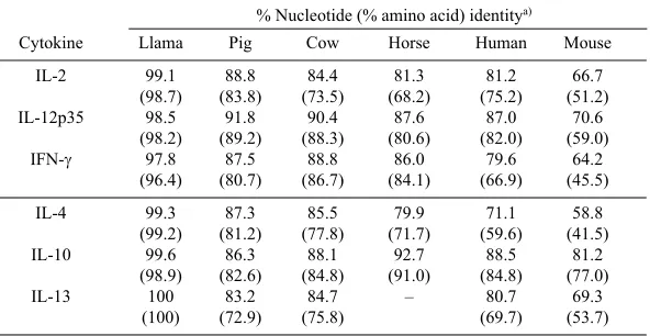
Table 1. Sequence identities between the llama, pig, cow, horse, human, and mouse cytokines
% Nucleotide (% amino acid) identitya)
Cytokine Llama Pig Cow Horse Human Mouse
IL-2 99.1 88.8 84.4 81.3 81.2 66.7
(98.7) (83.8) (73.5) (68.2) (75.2) (51.2)
IL-12p35 98.5 91.8 90.4 87.6 87.0 70.6
(98.2) (89.2) (88.3) (80.6) (82.0) (59.0)
IFN-γ 97.8 87.5 88.8 86.0 79.6 64.2
(96.4) (80.7) (86.7) (84.1) (66.9) (45.5)
IL-4 99.3 87.3 85.5 79.9 71.1 58.8
(99.2) (81.2) (77.8) (71.7) (59.6) (41.5)
IL-10 99.6 86.3 88.1 92.7 88.5 81.2
(98.9) (82.6) (84.8) (91.0) (84.8) (77.0)
IL-13 100 83.2 84.7 – 80.7 69.3
(100) (72.9) (75.8) (69.7) (53.7)
RESULTS
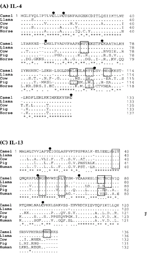
Cloning and sequence analysis of camel Th1 (2, IL-12p35, and IFN-γ) cytokines: To address the goals of this paper, we cloned and sequenced bactrian camel (Camelus bactrianus) IL-2, IL-12p35, and IFN-γ. The cDNAs of camel IL-2, IL-12p35, and IFN-γ were found to be 465, 669, and 501 bp in length, with open reading frames (ORF) encoding 154, 222, or 166 amino acids, respectively [Fig. 1. (A), (B), and (C)]. A high level of sequence conservation of the cloned cDNAs confirmed their identity. In most cases, most of the coding region of the cDNAs were obtained. The sequences were compared to the published llama, pig, cow, horse, human, and mouse sequences (Table 1). The deduced amino acid sequences of the cloned cytokine genes and their
alignment with the llama, cow, pig, and horse sequences are shown in Fig. 1. (A), (B), and (C). The cloned camel Th1 cytokine gene sequences showed the greatest homology to the published llama sequences, and the homology to llama of the camel sequences was significantly greater than the homology to the pig and cow sequences (Table 1).
Four cysteine (Cys) residues at positions 9, 78, 126, and 146 were conserved in all the analyzed mammalian species IL-2 sequences [(Fig. 1. (A), marked with a black dot]. Potential N-linked glycosylation site (as predicted by either NXT or NXS motifs) were found at residue position 111 in the IL-2 sequence (Fig. 1. (A), marked with an open bar). Therefore, eight Cys residues and one potential N-linked glycosylation site were conserved in all the analyzed mam-malian species IL-12p35 sequences. In addition, one
ble N-linked glycosylation site at position 174 was present in the llama and camel IL-12p35 sequences analyzed in this study [Fig. 1. (B)]. Finally, one Cys residue at position 13 and two potential N-linked glycosylation sites were found at residue positions 39 and 106 in all the analyzed IFN-γ sequences [Fig. 1. (C)].
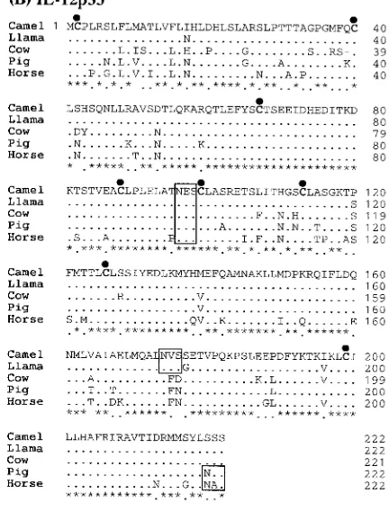
Cloning and sequence analysis of camel Th2 (IL-4, IL-10, and IL-13) cytokines: Full-length cDNAs of bactrian camel (Camelus bactrianus) IL-4, IL-10, and IL-13 were cloned and sequenced. The sequences of IL-4, IL-10, and IL-13 had 402, 537, and 411 bp nucleotides with ORF encodings of 133, 178, and 136 amino acids, respectively [Fig. 2. (A), (B) and (C)]. The identities of the nucleotide and deduced amino acid sequences of the camel and several mammalian species for IL-4, IL-10, and IL-13 as shown in Table 1. Most of the potential N-linked glycosylation sites and Cys residues were conserved among most of the species [Fig. 2.
(A), (B) and (C)]; marked with an open bar and a black dot]. The complete amino acid sequence of camel IL-4 and its comparison to llama, cattle, pig, and horse IL-4 are shown in Fig. 2. (A). The camel IL-4 deduced amino acid sequence includes four possible N-linked glycosylation sites (posi-tions 62, 96, 102, and 108) and six Cys residues (posi(posi-tions 13, 17, 48, 70, 105, and 133) [Fig. 2. (A)]. Camel IL-10 had four Cys residues (positions 30, 80, 126, and 132), with only one N-linked glycosylation site (position 134) at the same position as that of the llama, cow, pig and horse. The deduced amino-acid sequence of camel IL-13 bore six N-linked glycosylation sites (positions 38, 49, 57, 72, 75, and 131) and four Cys residues (positions 13, 48, 64, and 90). The identity (%) results were further confirmed by phyloge-netic analysis (Fig. 3).
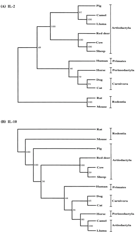
Phylogenetic analysis of camel Th1 and Th2 cytokines: In order to examine the phylogenetic relationships of camel Fig. 2. Alignment of the deduced amino acid sequences of the
Th1 and Th2 cytokines, we aligned mammalian those cDNA sequences with the respective sequences available in GenBank. As shown in Fig. 3, the mammalian species described in this study could be classified into the five major groups of Rodentia (mouse and rats), Primates (human),
Perissodactyla (horse), Artiodactyla (cow, sheep, pig, red deer, llama, and camel), and Carnivora (dog and cat). All the camel cytokine sequences reported here are most closely related to the llama, pig, cow, and horse sequences (Table 1). The homology results were further confirmed by phylo-genetic analysis (Fig. 3).
Our phylogenetic analyses indicated that camel cytokine genes are closely related to those of the order Artiodactyla, which includes the llama, pig, red deer, cow, and sheep. In the phylogenetic analysis, camel IL-2, IL-4, IL-12p35, IFN-γ and IL-13 were clustered with the llama, pig, cow, sheep, and red deer (Artiodactyla) with a high bootstrap value.
DISCUSSION
In this study, the camel Th1 (IL-2, IL-12p35, and IFN-γ) and Th2 (IL-4, IL-10, and IL-13) cytokines were cloned and sequenced for the first time. In general, the order Artiodac-tyla comprises three main suborders, Suiformes (pig, hog, and peccaries), Tylopoda (camel, llama, alpaca, vicuna, and guanaco) and Ruminantia (cow, sheep, red deer, antilope, buffalo, etc.), that are defined at the molecular level [2]. The camel Th1 and Th2 cytokine cDNAs showed a high degree of homology with respect to the cDNAs from the llama, pig, cow, horse, and other mammalian species, although there was relatively lower homology to the cDNA from the mouse. Therefore, these results revealed a closer phylogenetic relationship with the camel, llama, pig, cow and horse than with the mouse or rat. Interestingly, the nucleotide and amino acid sequences of camel (llama) IL-10 were closely related to those of the horse (Perissodactyla), human (Primates), cow (suborder Ruminantia) [11], and pig (suborder Suiformes) [3] [Table 1 and Fig. 3. (B)]. In addi-tion, phylogenetic analysis of IL-2, IL-4, IL-12p35, IFN-γ and IL-13 from the camels, and other species indicated that
Suiformes and Tylopoda are closely related to the order
Artiodactyla. These results were similar to those obtained from phylogenetic analysis of llama Th1 and Th2 cytokines, including IL-2, IL-4, IL-10, IL-12p35/p40, IL-13, and IFN-γ [15, 17]. The alignments of the deduced amino acid sequences of the Th1 and Th2 cytokines for several mam-malian species, including the camel, were similar in struc-ture to the cDNAs described for other species [4, 5, 8, 12, 13, 24]. In particular, most of the presumed Cys residues and N-linked glycosylation sites were found to be located in the same positions in all species. This result suggests that this region is highly conserved in the cytokine protein and may therefore play an important role in determining its ter-tiary structure and functional integrity. It also suggests that highly conserved amino acid residues are likely regulated in a similar manner and have an equally important role in the immune system of Camelidae.
Examination of the role of this cytokines in camel is essential for the understanding of Camelidae protective immune responses and disease development. An under-standing of biological properties of camel Th1 and Th2 cytokines will be important to study the CMI and humoral immune responses of camel, which has unique properties in heavy chain antibody. This cloning, sequencing, and phylo-genetic analyses of the camel Th1 and Th2 cytokines will be useful data for immunological study and formulation of dis-ease control strategies. Future studies are needed to take Fig. 3. Phylogenetic relationship based on nucleic acid sequences
advantage of these cytokines for enhancement of innate immune response in camels.
ACKNOWLEDGEMENT. This work was supported in part by Grant-in-Aids from the Ministry of Education, Cul-ture, Sports, Science and Technology of Japan.
REFERENCES
1. Abbas, B. and Agab, H. A review of camel brucellosis. 2002.
Vet. Med.55: 47–56.
2. Arnason, U. and Janke, A. 2002. Mitogenomic analyses of eutherian relationships. Cytogenet. Genome Res.96: 20–32. 3. Blancho, G., Gianello, P., Germana, S., Baetscher, M., Sachs,
D. H. and LeGuern, C. 1995. Molecular identification of por-cine interleukin 10: regulation of expression in a kidney allograft model. Proc. Natl. Acad. Sci. U. S. A.92: 2800–2804. 4. Cerretti, D. P., McKereghan, K., Larsen, A., Cantrell, M. A., Anderson, D., Gillis, S., Cosman, D. and Baker, P. E. 1986. Cloning, sequence, and expression of bovine interleukin 2.
Proc. Natl. Acad. Sci. U. S. A.83: 3223–3227.
5. Cerretti, D. P., McKereghan, K., Larsen, A., Cosman, D., Gil-lis, S. and Baker, P. E. 1986. Cloning, sequence, and expres-sion of bovine interferon-gamma. J. Immunol.136: 4561– 4565.
6. Conrath, K. E., Wernery, U., Muyldermans, S. and Nguyen, V. K. 2003. Emergence and evolution of functional heavy-chain antibodies in Camelidae. Dev. Comp. Immunol.27: 87–103. 7. Cousins, D. J., Lee, T. H. and Staynov, D. Z. 2002. Cytokine
coexpression during human Th1/Th2 cell differentiation: direct evidence for coordinated expression of Th2 cytokines. J. Immunol.169: 2498–2506.
8. Foss, D. L. and Murtaugh, M. P. 1997. Molecular cloning and mRNA expression of porcine interleukin-12. Vet. Immunol. Immunopathol.57: 121–134.
9. Felsenstein, J. 1989. PHYLIP: Phylogeny inference package (Version 3.2). Cladistics5: 164–166.
10. Hamers-Casterman, C., Atarhouch, T., Muyldermans, S., Rob-inson, G., Hamers, C., Songa, E. B., Bendahman, N. and Ham-ers, R. 1993. Naturally occurring antibodies devoid of light chains. Nature (Lond.)363: 446–448.
11. Hash, S. M., Brown, W. C. and Rice-Ficht, A. C. 1994. Char-acterization of a cDNA encoding bovine interleukin 10: kinet-ics of expression in bovine lymphocytes. Gene139: 257–261. 12. Heussler, V. T., Eichhorn, M. and Dobbelaere, D. A. 1992.
Cloning of a full-length cDNA encoding bovine interleukin 4 by the polymerase chain reaction. Gene114: 273–278.
13. McKenzie, A. N. 2000. Regulation of T helper type 2 cell immunity by interleukin-4 and interleukin-13. Pharmacol. Ther.88: 143–151.
14. Njiru, Z. K., Constantine, C. C., Ndung'u, J. M., Robertson, I., Okaye, S., Thompson, R. C. and Reid, S. A. 2004. Detection of Trypanosoma evansi in camels using PCR and CATT/T. evansi tests in Kenya. Vet. Parasitol. 124: 187–199.
15. Odbileg, R., Lee, S. I., Yoshida, R., Chang, K. S., Ohashi, K., Sugimoto, C. and Onuma, M. 2004. Cloning and sequence analysis of llama cytokines related to cell-mediated immunity.
Vet. Immunol. Immunopathol.99: 1–10.
16. Odbileg, R., Konnai, S., Ohashi, K. and Onuma, M. 2005. Molecular cloning and phylogenetic analysis of inflammatory cytokines of Camelidae (llama and camel). J. Vet. Med. Sci.
67: 921–925.
17. Odbileg, R., Lee, S. I., Ohashi, K. and Onuma, M. 2005. Clon-ing and sequence analysis of llama (lama glama) Th2 (IL-4, IL-10 and IL-13) cytokines. Vet. Immunol. Immunopathol.104: 145–153.
18. O’Garra, A. and Arai, N. 2000. The molecular basis of T helper 1 and T helper 2 cell differentiation. Trends Cell Biol.10: 542– 550.
19. Page, R. D. M. 1996. TREEVIEW: an application to display phylogenetic trees on personal computers. Comp. Appl. Biol. Sci. 12: 357–358.
20. Riechmann, L. and Muyldermans, S. 1999. Single domain anti-bodies: comparison of camel VH and camelised human VH domains. J. Immunol. Methods231: 25–38.
21. Strimmer, K. and von Haeseler, A. 1996. Quartet puzzling: a quartet maximum likelihood method for reconstructing tree topologies. Mol. Biol. Evol. 13: 964–969.
22. Thompson, J. D., Gibson, T. J., Plewniak, F., Jeanmougin, F. and Higgins, D. G. 1997. The CLUSTAL_X windows inter-face: flexible strategies for multiple sequence alignment aided by quality analysis tools. Nucleic Acids Res.24: 4876–4882. 23. Ursing, B. M., Slack, K. and Arnason, U. 2000. Subordinal
artiodactyl relationships in the light of phylogenetic analysis of complete mitochondrial genomes. Zoologica29: 83–88. 24. Vandergrifft, E. V., Swiderski, C. E. and Horohov, D. W.
1994. Molecular cloning and sequencing of equine interleukin 4. Vet. Immunol. Immunopathol.40: 379–384.
25. Wernery, U. and Kaaden, O. R. 2004. Foot-and-mouth disease in camelids, Vet. J.168: 134–142.