Brazilein in Combination with Cisplatin Inhibit Proliferation and
Migration on Highly Metastatic Cancer Cells, 4T1
Sri Handayani
2,3, Ratna Asmah Susidarti
1,2, Zalinar Udin
3, Edy Meiyanto
1,2,
Riris Istighfari Jenie
1,2,*1Faculty of Pharmacy, Universitas Gadjah Mada, Indonesia, 55281
2Cancer Chemoprevention Research Center, Faculty of Pharmacy, Universitas
Gadjah Mada, Indonesia, 55281
3Research Center for Chemistry, Indonesian Institute of Sciences (LIPI), Indonesia
Abstract
Brazilein performs anticancer activities on several cancer cells and potentially inhibits metastasis. The aims of this study is to observe the synergistic cytotoxic and migration inhibitory effect of brazilein combined with cisplatin on 4T1 breast cancer cells. Under MTT assay, we found that brazilein revealed cytotoxic effect on 4T1 cells in a dosedependent manner (IC50=50 ± 0.3 µM). Combination of brazilein and cisplatin showed synergistic effect (CI=0.72). Flowcytometry analysis on the cell cycle progression showed that single treatment of 25 µM brazilein induced G2/Mphase accumulation, 12.5 µM cisplatin induced Sphase accumulation, while combination of brazilein and cisplatin induced Sphase and G2/M phase accumulation. Combination of brazilein and cisplatin induced apoptosis higher than that of the single treatments. Based on wound healing assay, 12.5 µM brazilein and its combination with 6.25 µM cisplatin inhibited cells migration. Immunoblotting and gelatin zymography analysis showed that combination of brazilein and cisplatin inhibited the expression level of Rac1 and MMP9 proteins. Based on these results, we conclude that brazilein enhanced cytotoxic activity of cisplatin and inhibited migration on 4T1 cells and potentially can be developed as an enhancing cytotoxic and antimetastasis agent.
Keywords: Brazilein, cisplatin, combination treatment, cytotoxic effect, cells migration.
Introduction
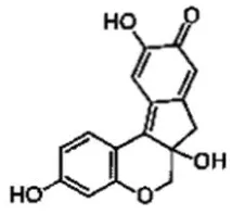
Brazilein (Figure 1), a major compound of Caesalpinia sappan L., performs potential antiproliferative effect on several cancer cells. In vitro studies have proven that brazilein exhibit cytotoxic activities and apoptosis induction on MCF7, T47D, and other breast cancer cell lines (Taoet al., 2011, 2013). These cytotoxic activities are mediated through aberration of some signal transduction, mainly on MAP Kinases signaling pathways (Hsieh et al., 2013). In many cancer cells, MAPKs involve on cancer cell growth and metastasis (Hanahan and Weinberg, 2011). Therefore, the physiologicallytargeted
mechanism of brazilein as anticancer on some specific cancer cells need to be explored.
Strategy on cancer treatment is not only focused on one target mechanism, but it should be developed on several target mechanism. On the other hand, cancer progression usually involve aberration of cell proliferation and cell migration as well as invasion (Kemperet al., 2014). The 4T1 cell is the example of metastatic breast cancer that *Corresponding author:
Riris Istighfari Jenie
Faculty of Pharmacy, Universitas Gadjah Mada, Indonesia, 55281
Email: [email protected]
39 performs active proliferation, migration and invasion (Kaur et al., 2012). Some growth signaling involve in the 4T1 cells proliferation including activation of NFκB transcription factor (Tsubaki et al., 2013). Our previous research performs that brazilein inhibits proliferation on MCF7 breast cancer cells via inhibition of NFκB activation (unpublished data). Hsieh et al., (2013) reported that brazilein inhibits NFκB activation leading to inhibition of migration on MDAMB231 cells. Since NFκB activation usually increase the side effect of chemotherapeutic agent such as cisplatin and doxorubicin, therefore the exploration of reduction of side effect through NFκB signaling is needed to be explored. Our previous study showed that brazilein inhibits doxorubicin resistantMCF7 cancer cells growth (Laksmiani et al., 2015). In this regards, Brazilein has a potential effect to be developed as cochemotherapeutic agent against cisplatin as well as doxorubicin.
Cell migration and invasion are indispensable of the metastasis process. Tumor metastasis is a complex process that allows the tumor cells grow on a variety of other tissues and organs. Metastasis occur when tumor cells shed from a primary tumor cells by damaging the extracellular tissues, migrate, invade surrounding tissue, enter the bloodstream and reach the target of a certain tissue to form secondary tumors (Brookset al., 2010). Cisplatin (CDDP) is one of the first line therapy in metastatic triple negative breast cancer (Zhanget al., 2015). Surprisingly, low concentration of cisplatin induces epithelialtomesenchymal transition (EMT) and followed by increasing instead of inhibiting of cancer metastasis (Latifiet al., 2011). On the other hand, brazilein suppresses cancer cells migration and invasion on MDAMB231 cells (Hsieh et al., 2013). It is interesting to know whether brazilein performs a potential effect to inhibit cisplatin inducedmigration and invasion on 4T1 cells. NFκB plays a role on cell proliferation and metastasis through the activation of targeted gene expression such as MMP9 and MMP2 (Hsiehet al., 2013; Kim, 2010). The 4T1 cells also performs such of this phenomenon
(Aminet al., 2015). Matrix metalloproteinases (MMPs) belong to a family of zincdependent endopeptidases, highly conserved and structurally related, capable of degrading many components of basement membrane and extracellular matrix (ECM). The active MMPs bind with membrane molecules and degrade ECM (Hua et al., 2011). Brazilein inhibits MMP2 protein in both its expression and activation on MDAMB231 cells (Hsieh et al., 2013). On the other hand, cisplatin induces migration mediated by inducing of Rac1 protein expression (Geng et al., 2016). The Rac1 protein is a member of RHO family of small GTPase that plays a role in neoplastic transformation, tumor progression and metastasis. Activation of p120/Vav2 and PI3K pathways activate Rac1 protein leading to lamellipodia formation through ASPfamily verprolinhomologous proteins (WAVEs) and p21activated kinases (PAK1) activation (Ridley, 2015; Valls et al., 2012). Our main purpose of this study is to explore the cytotoxic and antimetastasis potential of brazilein focusing on inhibiting of migration through inhibition of Rac1 and MMP9 protein expression on 4T1 cells.
Materials and Methods
Samples
Brazilein (Be) isolated fromCaesalpinia sappan L. was obtained from Cancer Chemoprevention Research Center (CCRC), Faculty of Pharmacy, Universitas Gadjah Mada. Cisplatin (CDDP) was purchased from Sigma (P4394).
Cell culture
4T1 metastatic cell line (ATCC CRL 2539) was kindly given by Prof. Dr. Mashashi Kawaichi (NAIST) and was cultured in Dulbecco's Modified Eagle Medium (DMEM) medium (Gibco) with 10% Fetal Bovine Serum (Gibco), 1.5% PenicillinStreptomycin (Gibco) and 0.5% fungizone (Gibco).
Cytotoxic assay
4T1 cells in 96well plates with 104
h incubation, culture medium was removed and cells were washed in PBS(Sigma). Then, cells were incubated with 100 µL culture medium and 10 µL MTT (Sigma) 5 mg/mL in every well for 4 h. MTT reaction was stopped by SDS reagent (10% Sodium dodecyl sulphate (Merck) in 0.01 M HCl (Merck)) and was incubated overnight. The absorbance was measured by ELISA reader (BioRad) at wave length of 595 nm.
Cell cycle assay
Propidium iodide (PI) staining kit (Becton Dickinson) was used to analyze DNA content. Cells were seeded in 24well plates with 5x10⁴within 24–48 h. Cells were treated
with various concentration of samples in single and combination. After a 24 h treatment, cells were harvested, and the cells were resuspended in PBS, fixed with 70% ethanol, labeled with PI (2 µg/mL), and incubated at room temperature in the dark for 10 min. DNA content was analyzed using a flow cytometry (Becton Dickinson) and followed by flowing software (version 2.5.1).
Apoptosis assay
Apoptotic population was determined by PIAnnexin V assay (Annexin VFITC Apoptosis Detection Kit Roche). Cells were seeded in 24well plates with 5x10⁴ within
24–48 h. Cells were treated with various concentration of samples in single and combination. After a 24 h treatment, cells were harvested, added with 1x binding buffer, labeled with PIAnnexin V, and incubated at room temperature in the dark for 5 min. Then the cells suspension was analyzed using a flow cytometry (Becton Dickinson).
Wound healing assay
Cells were seeded in each well of 24 well plate with 1×10⁵cells and incubated at
37°C in CO2incubator for 24 h. Thereafter cell were starved by using serum freemedium (containing 0.5% fetal bovine serum) for 24 h. The confluent cell monolayers were wounded by a yellowtip. After washing once with PBS, the cells were added with DMEM culture medium which contained of 1/4 IC50 of samples in single and combination. Time
lapse images were acquired at 18, 24 and 42 h. Percent of closured were analyzed by using ImageJ software.
Gelatin zymography assay
MMP9 activity was assayed by gelatin zymography. Cells were seeded in each well of 6well plate with 1×10⁶cells and incubate
at 37°C in CO2incubator for 24 h. Cells were incubated with 1/4 IC50of samples in single and combination in serum freemedium for 24 h. The supernatants were collected and subjected to gel electrophoresis on 10% running gel containing 0.1% gelatin. The gel were washed in renaturing solution containing 2.5% Triton x100 for 30 min, and followed by incubation for 20 h at 37°C with incubation buffer. The gels were stained for 30 min in 0.5% coomassie brilliant blue and then destained with destaining solution (10% v/v methanol and 5% v/v acetic acid). Gel were scanned and analyzed with imageJ software.
Immunoblotting assay
Cells were seeded in 10 cm cultured dish with 1×10⁶cells and incubated at 37°C
41 Statistical analysis
Parameter of inhibitory concentration
(IC50) and combinatory index (CI) were measured based on the previous reports (Mosmann, 1983; Reynolds and Maurer, 2005). The IC50 values of three independent experiments were expressed as mean ± standard deviation (SD). Analysis of significantly between control/untreated group and treated groups were analyzed by unpaired Student’sttest (Microsoft Excel 2013). Mean differences was significant at theP<0.05.
Results
Brazilein and cisplatin performed synergistic effect on 4T1 cells
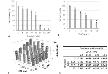
The confirmation of the cytotoxic activity of brazilein is important to be established as a basic anticancer performance and to be developed for the other anticancer activities. We used MTT assay as a standard method to measure the cytotoxic activity. After 24 h treatment, brazilein and cisplatin performed cytotoxic activities in a dose dependent manner with IC50values of 50 ±
0.3 µM and 26 ± 0.5 µM, respectively (Fig.2A–B). Furthermore, to confirm whether the compound affects on cytotoxic of cisplatin, we carried out combination treatment with series of concentrations under IC50value. The synergistic combination was measured by combination index (CI) parameter. The result showed that combination of 1/10, 1/8,¼, and½IC50of brazilein and cisplatin exhibited synergistic effect on inhibition of 4T1 cells growth with CI values less than 1 (Fig. 2C–D). The combination of½IC50of brazilein and cisplatin inhibited cell viability by 62%. These results showed that brazilein possessed cytotoxic activity on 4T1 cells with a middle intensity but potential to be developed as co chemotherapeutic agent. Whereas the combination treatment showed that brazilein increased the sensitivity of 4T1 cells upon cisplatin treatment. The cytotoxic activity of brazilein in single and combination with cisplatin may occur due to inhibition of cell cycle modulation or apoptosis induction. Thus, we conducted experiments to observe the cell cycle modulation and apoptosis event
upon treatment with brazilein and cisplatin.
Combination of brazilein and cisplatin induced cell cycle arrest on 4T1 cells.
To observe the specific mechanism of brazilein and its combination with cisplatin on inhibition of cell cycle progression, we analyze the cell cycle profiles after treatment of samples for 24 h by using flow cytometry. The results indicated that single treatment of
1/2 IC50 of brazilein caused subG1 and
G2/Mphase accumulation (Fig. 3B, 3E), while cisplatin caused Sphase accumulation (Fig. 2C, 2E) compared to untreated cells (Fig. 3A, 3E). The combination of brazilein and cisplatin (1/2 IC50) increased the cells accumulation in Sphase and G2/Mphase (Fig. 3D–E). Furthermore, these data showed that the cytotoxic activity of brazilein in single and in combination with cisplatin induced subG1. These phenomena suggested that the treatment of brazilein in single and in combination with cisplatin may induce cell death.
Combination of brazilein and cisplatin induced apoptosis on 4T1 cells.
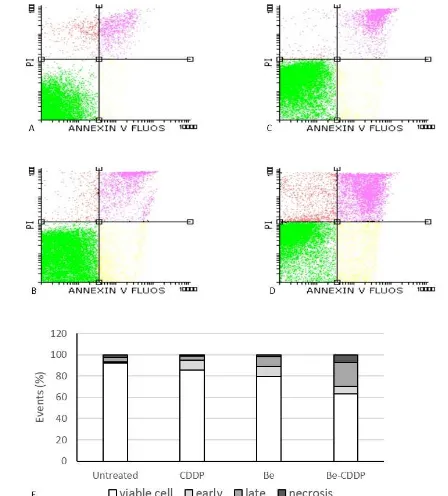
Next, we observed the effect of brazilein in single and in combination with cisplatin on apoptosis induction. Apoptosis is a programmed physiological event leading to cell death that involving the changing of membrane dynamic, nuclear fragmentation and usually become necrosis at the end (Ouyanget al., 2012). Therefore, to examine the phenomenon of brazilein and its combination with cisplatin on apoptosis induction we performed PIanexin V staining by using flow cytometry. The result showed that single treatment of 25 µM brazilein and 12.5 µM cisplatin exhibited total apoptosis (early and late apoptosis) up to19% and 13% respectively(Fig. 4B,4C,4), while combination of brazilein and cisplatin exhibited total apoptosis up to 30% (Fig. 4D–E) and increase the necrotic cells. These data suggested that combination of brazilein and cisplatin increased cells death compared to the single treatments on 4T1 cells.
There are internal and external molecular event in metastasis processes.
Internal molecular signaling during migration process involves actin polymerization and lamellipodia formation. This event mainly
Figure 3.The effect of single treatment of brazilein and its combination with cisplatin on cell cycle profiles on 4T1 cells. Cells were treated with vehicle(A), 25 µM brazilein (B),12.5 µM cisplatin (C), and combination of brazileincisplatin (D) for 24 h, stained with PI and analyzed by using flow cytometry according to the method and quantified by using flowing software (E).
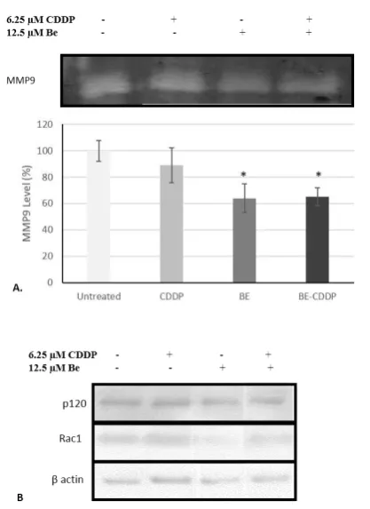
43 induce by the expression and activation of Rac1 and p120 (Vallset al., 2012). On the other hand, cancer progression is also marked by highly expression of proteinases in the microenvironment such as MMP9 to impair ECM (Huaet al., 2011). Therefore, to confirm whether the mechanism underlying migration inhibition of brazilein, we performed gelatin zymography to analyze the level of MMP9 in the environment and immunoblotting experiment to analyze the level of Rac1 and p120 protein in the cells. The level of MMP9 in the medium of 4T1 cells was decreased in response to treatment 12.5 µM brazilein and its combination with 6.25 µM cisplatin (Fig. 6A–B). Single treatment of 6.25 µM cisplatin did not significantly reduce the MMP9 level suggested that the decreasing of MMP9 level in the combination treatment was due to brazilein. Furthermore, brazilein and its
combination with cisplatin decreased Rac1
protein expression but not p120 in the 4T1 cells(Fig. 6B).
Discussion
The10 hallmarks of cancer noted that cell cycle modulation, apoptosis induction,
and inhibition of metastatic stages of cancer become important targets in cancer therapy development(Hanahan and Weinberg, 2011). In this report, we present the effect of brazilein on several physiological targets on cancer development, especially on metastatic breast cancer cells. We used 4T1 cells line that has been recognized as active cells in proliferation and migration (Kaur et al., 2012). Comprehensively, we found that brazilein exhibited middle or low cytotoxic effect, cell cycle modulation, apoptosis induction, and inhibition of cells migration. These results supported our previous data and other results
reported elsewhere (Hsieh et al., 2013;
Laksmianiet al.,2015; Taoet al.,2011,2013). Our data give additional information concerning on the physiological effects of brazilein in single and in combination with cisplatin on a progressive cell line, 4T1.
In this study, we showed that brazilein performed cytotoxic effect to 4T1 cells with IC50value of 50 ± 0.3 µM. This cytotoxic value is categorized as low level according to Larssonet al.(2006). Functional food such as
Caesalpinia sappanL. commonly safe and less
toxic on cells (Nirmal et al., 2015).
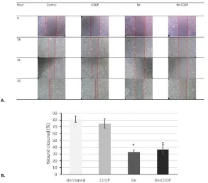
Figure 5.The effect of single treatment of brazilein and its combination with cisplatin on 4T1 cells migration. Cells were scratched according to wound healing assay, were treated with 12.5 µM brazilein and 6.25 µM cisplatin in single and in combination, were photographed at 0,18,24, and 42 h, then analyzed by using image J software. A. Wound closured, B. Relative wound closured after 42 h samples treatment. Error bars mean standard deviation (n
Nevertheless, we should consider when we consume this plant in high consumption because brazilein is the major compound in this plant. However, the synergistic effect with cisplatin in enhancing cytotoxicity on the cells gives insight for the use of brazilein to enhance the chemotherapeutic effect(the efficacy) and to reduce the side effect of cisplatin. Even we still do not know the molecular mechanism underlying this synergistic effect, but at least we have information that lower concentration of cisplatin gave the same cytotoxic effect to the cells when it combined with brazilein. This phenomenon also performed by brazilein in combination with doxorubicin which sensitized MCF7 resistant doxorubicin cancer cells (Laksmianiet al., 2015), thus, gives good prospect of brazilein to be developed as co chemotherapeutic agent. However, this data
are the in vitro report and should be confirmed with in vivo experiment.
The shift of cell cycle profile in combination treatment of brazileincisplatin is an interesting finding in this study. Based on the cell cycle profiles, brazilein showed different mechanism compare to cisplatin. Cisplatin is well known to induce cell cycle arrest at Sphase (Shenet al., 2013) and our finding showed the same phenomenon.
However, brazilein induced cell
accumulation in G2/M phase. This
phenomenon is similar with the effect of brazilin (the reduced form of brazilein) that induces cell accumulation in G2/M phase on
myeloma U266 cells (Kim et al., 2012).
Moreover, the different shifting of cell cycle profiles between cisplatin and brazilein indicated the differences targets of the two compounds, especially on the cell cycle regulation that should be investigated further. The differences of the targets mechanism are important to potentiate the cytotoxic effect to the cancer cells (Chou, 2006). In this regards, we confirmed that the combinational treatment of both compounds gave synergistic effect to inhibit cells growth by > 50% (Fig. 2C–D). Our data also showed that brazilein pulled the cells into subG1 phase indicated that this compound induces cell death or apoptosis. However, the combination treatment seemed to reduce the cell death(decrease the subG1 phase by 4%). This decreasing of the subG1 accumulation may be due to most of the apoptotic cells underwent necrosis. This phenomenon was confirmed by PIannexin V staining under flocytometry assay (Fig. 4D–E). Apoptosis
leading to necrosis is the normal
phenomenon in in vitro studies of cell death due to the absent of the phagocytic cells (Ouyanget al., 2012). Our result suggested that the combination treatment of brazilein and cisplatin enhanced cell cycle arrest and accelerated the apoptosis evidence.
Brazilein was reported to inhibit
migration and invasion in MDAMB231
breast cancer cells (Hsieh et al., 2013). Metastasis is a fatal state of cancer progression, which needs migration and
45 invasion to initiate the process. Targeted therapy to inhibit those two processes would be beneficially for triple negative cancer cells treatment that have characteristic of highly metastasis (FerrariAmorotti et al., 2014; Martinet al.,2013). The present data showed that brazilein, in single and in combination with cisplatin inhibited cells migration(Fig. 5) of 4T1 cells. Furthermore, we observed that single treatment of brazilein and its combination with cisplatin suppressed Rac1 protein expression(Fig. 6B). Rac1 is a member of RhoGTPase family which plays an important role in lamellipodium extension together with cdc42 and RhoA (Ridley, 2015). Previously, Chenet al.,(2014) reported that curcumin inhibits activation of PAK1, the downstream of Rac1, mediated by suppression of Rac1 protein expression. Flavonoid wogonin inhibits Rac1 expression as well as other Ras and activated ERK and PI3K/Akt pathway (Zhao et al., 2014). Upregulation of Rac1 activity is mediated
through several signaling pathways
including Wnt stimulation that involving binding of p120catenin with Rac1 and Vav2 (Vallset al., 2012). However, our finding (Fig. 6B) did not show any significance difference
of p120catenin expression among treated
and untreated cells. This phenomenon indicated that the cells migration inhibitory
effect of brazilein was due to other Rac1
activation mechanism such as PI3K kinase pathway. Kim et al. (2014) reported that rhapontigenin and the PI3K inhibitor wortmannin reduces Rac1 activation in MDAMB231 cells through inhibition of
PI3Kdependent Rac1 signaling pathway.
The MMPs family is important in the invasion step of metastasis to degrade the extra cellular matrix, and among them, MMP9 and MMP2 are known to be highly expressed in metastatic breast cancer cells (Hua et al., 2011). Our study demonstrated that brazilein and its combination with cisplatin decreased the gelatinolytic activity of MMP9 protein in 4T1 cells(Fig 5AB). It was reported by Hsieh
et al. (2013) that brazilein inhibits nuclear translocation of NFκB protein, thus,
suppressing the NFκB signaling pathway.
NFκB is constitutively active in most tumor cell lines and in tumor tissues derived from patients with various types of cancer including breast cancer NFκB is resides in the cytoplasm of all cells, however when NF κB is being activated it will be translocated in the nucleus and affected the downstream gene expression. MMP9 is one of many gene
products regulated by NFκB activation
(Aggarwal, 2004). Taken together, our findings indicated that brazilein and that
suppression of NFκB signaling may be
involved in the mechanism. However, further studies are needed to clarify the molecular mechanism underlies brazilein and its combination in modulating cell cycle, inducing apoptosis and inhibiting migration and invasion in metastatic breast cancer cells.
Conclusions
This study concludes that brazilein worked synergistically with cisplatin to inhibit proliferation and migration in 4T1 cancer cells through inhibition of MMP9 and Rac1 protein expression.
Acknowledgments
This work was supported by the grant of Penelitian Unggulan Perguruan Tinggi (PUPT) 2015–2016 from Indonesian Ministry of Research, Technology and High Education.
References
Aggarwal, B.B.2004. Nuclear factorkappaB: the enemy within. Cancer Cell, 6, 203–208. doi:10.1016/j.ccr.2004.09.003. Amin, H., Wani, N.A., Farooq, S., Nayak, D., Chakraborty, S., Shankar, S., Rasool, R. ur, Koul, S., Goswami, A., Rai, R. 2015. Inhibition of Invasion in Pancreatic Cancer Cells by Conjugate of EPA with β3,3PipOH via PI3K/Akt/NF kB Pathway. ACS Med. Chem. Lett., 6, 1071–1074. https://doi:10.1021/ acsmedchemlett.5b00257.
Baribeau, S., Chaudhry, P., Parent, S., Asselin, É. 2014. Resveratrol Inhibits Cisplatin
Induced EpithelialtoMesenchymal
Brooks, S.A., LomaxBrowne, H.J., Carter, T.M., Kinch, C.E., Hall, D.M.S. 2010. Molecular interactions in cancer cell metastasis. Acta Histochem., 112, 3–25. https://doi:10.1016/j.acthis.2008.11.02 2.
Chen, Q., Zheng, Y., Jiao, D., Chen, F., Hu, H., Wu, Y., Song, J., Yan, J., Wu, L., Lv, G. 2014. Curcumin inhibits lung cancer cell migration and invasion through Rac1dependent signaling pathway. J. Nutr. Biochem., 25,177–185. https:// doi:10.1016/j.jnutbio.2013.10.004. Chou, T.C. 2006. Theoretical Basis,
Experimental Design, and
Computerized Simulation of Synergism and Antagonism in Drug Combination Studies. Pharmacol. Rev., 58, 621–681. https://doi:10.1124/pr.58.3.10. FerrariAmorotti, G., Chiodoni, C., Shen, F.,
Cattelani, S., Soliera, A.R., Manzotti, G., Grisendi, G., Dominici, M., Rivasi, F., Colombo, M.P., Fatatis, A.,
Calabretta, B. 2014. Suppression of
Invasion and Metastasis of Triple Negative Breast Cancer Lines by Pharmacological or Genetic Inhibition
of Slug Activity. Neoplasia, 16,
1047–1058. https://doi:10.1016/ j.neo.2014.10.006.
Geng, S., Gu, L., Ju, F., Zhang, H., Wang, Y., Tang, H., Bi, Z., Yang, C. 2016.
MicroRNA224 promotes the
sensitivity of osteosarcoma cells to cisplatin by targeting Rac1. J. Cell. Mol.
Med., 20, 1611–1619. https://
doi:10.1111/jcmm.12852.
Hanahan, D., Weinberg, R.A. 2011. Hallmarks of Cancer: The Next Generation. Cell 144, 646–674. https://doi:10.1016/ j.cell.2011.02.013
Hsieh, C.Y., Tsai, P.C., Chu, C.L., Chang, F.R., Chang, L.S., Wu, Y.C., Lin, S. R. 2013. Brazilein suppresses migration and invasion of MDAMB231 breast cancer cells. Chem. Biol. Interact.,204,
105–115. https://doi:10.1016/
j.cbi.2013.05.005.
Hua, H., Li, M., Luo, T., Yin, Y., Jiang, Y.2011.
Matrix metalloproteinases in
tumorigenesis: an evolving paradigm. Cell. Mol. Life Sci., 68, 3853–3868. doi:10.1007/s000180110763x. Kaur, P., Nagaraja, G.M., Zheng, H.,
Gizachew, D., Galukande, M.,
Krishnan, S., Asea, A. 2012. A mouse model for triplenegative breast cancer tumorinitiating cells (TNBCTICs) exhibits similar aggressive phenotype to the human disease. BMC Cancer,12, 120. https://doi:10.1186/1471240712 120.
Kemper, K., Goeje, P.L. de, Peeper, D.S.,
Amerongen, R. van. 2014. Phenotype
Switching: Tumor Cell Plasticity as a Resistance Mechanism and Target for Therapy. Cancer Res., 74, 5937–5941. https://doi:10.1158/00085472.CAN 141174.
Kim, B.S. 2010. Brazilin Inhibits of TPA induced MMP9 Expression Via the
Suppression of NFκB Activation in
MCF7 Human Breast Carcinoma Cells. J. Food Hyg. Saf.
Kim, J.S., Kang, C.G., Kim, S.H., Lee, E.O. 2014. Rhapontigenin Suppresses Cell Migration and Invasion by Inhibiting the PI3KDependent Rac1 Signaling
Pathway in MDAMB231 Human
Breast Cancer Cells. J. Nat. Prod., 77, 1135–1139. doi:10.1021/np401078g. Laksmiani, N.P.L., Susidarti, R.A., Meiyanto,
E. 2015. Brazilein Increases The Sensitivity of Doxorubicin on MCF7 Resistant Doxorubicin (MCF7/DOX) Cells Through Inhibition of HER2 Activation. Int. J. Pharm. Pharm. Sci., 7, 525528.
Larsson, D.E., Lövborg, H., Rickardson, L., Larsson, R., Oberg, K., Granberg, D. 2006. Identification and evaluation of potential anticancer drugs on human
neuroendocrine tumor cell lines.
Anticancer Res., 26, 4125–4129. Latifi, A., Abubaker, K., Castrechini, N.,
Ward, A.C., Liongue, C., Dobill, F., Kumar, J., Thompson, E.W., Quinn, M.A., Findlay, J.K., Ahmed, N. 2011. Cisplatin treatment of primary and
47 carcinomas generates residual cells with mesenchymal stem celllike
profile. J. Cell. Biochem., 112,
2850–2864. doi:10.1002/jcb.23199. Martin, T.A., Ye, L., Sanders, A.J., Lane, J.,
Jiang, W.G.2013. Cancer Invasion and Metastasis: Molecular and Cellular Perspective. Landes Bioscience. Mosmann, T. 1983. Rapid colorimetric assay
for cellular growth and survival: application to proliferation and cytotoxicity assays. J. Immunol. Methods, 65, 55–63.
Nirmal, N.P., Rajput, M.S., Prasad, R.G.S.V.,
Ahmad, M. 2015. Brazilin from
Caesalpinia sappan heartwood and its pharmacological activities: A review. Asian Pac. J. Trop. Med., 8, 421–430. doi:10.1016/j.apjtm.2015.05.014. Ouyang, L., Shi, Z., Zhao, S., Wang, F.T.,
Zhou, T.T., Liu, B., Bao, J.K. 2012. Programmed cell death pathways in
cancer: a review of apoptosis,
autophagy and programmed necrosis. Cell Prolif., 45, 487–498. https:// doi:10.1111/j.13652184.2012.00845.x. Reynolds, C.P., Maurer, B.J. 2005. Evaluating
response to antineoplastic drug
combinations in tissue culture models.
Methods Mol. Med., 110, 173–183.
https://doi:10.1385/1592598692:173. Ridley, A.J.2015. Rho GTPase signalling in cell migration. Curr. Opin. Cell Biol., 36, 103–112. https://doi:10.1016/ j.ceb.2015.08.005.
Shen, H., Perez, R.E., Davaadelger, B., Maki, C.G.2013. Two 4N CellCycle Arrests Contribute to CisplatinResistance.
PLoS ONE, 8, e59848.
doi:10.1371/journal.pone.0059848. Tao, L., Li, J., Zhang, J. 2013. Brazilein, a
compound isolated from Caesalpinia sappanLinn., induced growth inhibition in breast cancer cells via involvement
of GSK3β/βCatenin/cyclin D1
pathway. Chem. Biol. Interact.,206,1–5. https://doi:10.1016/j.cbi.2013.07.015. Tao, L., Li, J., Zhang, J.2011. Brazilein Induced Cells Apoptosis in Human Breast Cancer MCF7 Cells and Its Action
Mechanism. J. Sun YatSen Univ. Med. Sci., 32, 449–453.
Tsubaki, M., Komai, M., Fujimoto, S., Itoh, T., Imano, M., Sakamoto, K., Shimaoka, H., Takeda, T., Ogawa, N., Mashimo, K., Fujiwara, D., Mukai, J., Sakaguchi,
K., Satou, T., Nishida, S. 2013.
Activation of NFκB by the
RANKL/RANK system upregulates snail and twist expressions and induces epithelialtomesenchymal transition in mammary tumor cell lines. J. Exp.
Clin. Cancer Res., CR 32, 62.
https://doi:10.1186/175699663262. Valls, G., Codina, M., Miller, R.K., VallePérez, B.D., Vinyoles, M., Caelles, C., McCrea, P.D., Herreros, A.G. de, Duñach, M.
2012. Upon Wnt stimulation, Rac1
activation requires Rac1 and Vav2 binding to p120catenin. J. Cell Sci.,125, 5288–5301. doi:10.1242/jcs.101030. Zhang, J., Wang, Z., Hu, X., Wang, B., Wang,
L., Yang, W., Liu, Y., Liu, G., Di, G., Hu, Z., Wu, J., Shao, Z.2015. Cisplatin and gemcitabine as the first line therapy in metastatic triple negative breast cancer.
Int. J. Cancer, 136, 204–211.
https://doi:10.1002/ijc.28966.
Zhao, K., Wei, L., Hui, H., Dai, Q., You, Q
D., Guo, QL., Lu, N. 2014. Wogonin
Suppresses Melanoma Cell B16F10 Invasion and Migration by Inhibiting
RasMedicated Pathways. PLoS ONE,