Corresponding author: [email protected]
Mammographic density and estrogen
receptor
a
gene polymorphism in Javanese
women
Lina Choridah1*, Teguh Aryandono2, Arif Faisal1, Ahmad Hamim Sadewa3, Dewajani Purnomosari4
1Department of Radiology, 2Department of Surgery, 3Department of Biochemistry, 4Department of Histology/Molecular Biology, Faculty of Medicine, Universitas Gadjah
Mada, Yogyakarta, Indonesia
ABSTRACT
Estrogen plays important roles in breast cancer as it binds its receptor in breast tissue. The most studied variants in estrogen receptor a encoded by ESR1 gene are the ESR1 PvuII and XbaI polymorphisms, which were associated with lower sensitivity to estrogen. We determined the proportion of ESR1 XbaI and PvuII polymorphisms in Javanese woman in Yogyakarta, Indonesia and analyzed the correlation between genetic variations with mammogram density. ESR1 XbaI and PvuII polymorphisms of 50 cases and 58 controls were identified using PCR-RFLP. Breast density was assessed based on digitizer mammograms. Quantitative analysis was performed using an interactive program based on cumulus of two thresholds. Mean of density and frequencies of SNPs were compared between cases and controls to identify the association between SNPs and cancer susceptibility. Mammographic density was significantly higher in cases (52%) than controls (0.41%) (p < 0.05). Women with one or two copies of the PvuII T allele and XbaI A allele had higher mammographic density compared with women with C and G alleles, respectively. The proportion between PP and TT genotype was not statistically significant (p > 0.05), while the proportion between AA and GG was significantly different (p < 0.05). Haplotype 2 (CG/PX) was associated with lower sensitivity to estrogen and reflects a decrease of mammographic density. These findings were consistent with other studies that showed that ESR1 polymorphisms may affect breast cancer risk through differences in breast density.
ABSTRAK
dibandingkan dengan wanita dengan alel C dan G, secara berurutan. Proporsi antara genotipe PP dan TT tidak signifikan secara statistik (p> 0,05), sedangkan proporsi antara AA dan GG berbeda secara signifikan (p <0,05). Haplotype 2 (CG / PX) berkaitan dengan sensitivitas yang lebih rendah terhadap estrogen dan mencerminkan penurunan kepadatan mamografi. Temuan ini konsisten dengan penelitian lain yang menunjukkan bahwa polimorfisme ESR1 dapat mempengaruhi risiko kanker payudara melalui perbedaan kepadatan payudara.
Keywords: breast cancer - DNA polymorphism - ESR1 PvuII - ESR1 XbaI - mammogram Digitizer
INTRODUCTION
Breast cancer (BC) is the highest ranked malignancy in women in the world and is particularly frequent in Yogyakarta, Java, Indonesia.1,2 Estrogen plays an important
role in the occurrence of BC through inducing proliferation and genotoxic effects by mechanisms involving estrogen binding to its receptor in breast tissue. However, the detailed mechanism of the induction of BC carcinogenesis is not fully understood.3 There
are two types of estrogen receptor, estrogen receptor alpha, which is the main estrogen receptor that is encoded by the ESR1 gene, and estrogen receptor beta which is encoded by ESR2 gene.4 ESR1 is one of the most
important mediators of hormonal response in estrogen-sensitive tissues such as the breast and plays a crucial role in breast growth and differentiation as well as in the development of cancer.5
Mammography density reflects the
number of stromal and epithelial cells and
is one of the strongest risk factors for BC. Some studies showed that women with a high density mammography pattern have a risk of BC 4–6 times higher than those with a low density pattern.6 An annual mammography
examination is currently recommended for women over the age of 40 years,1 but not for
women younger than 40 years, due to pain from compression techniques during the examination and the risk of radiation hazard.7
Many studies have demonstrated evidence for
the influence of estrogen on mammographic density.8-10 Women with hormone replacement
therapy showed increased breast density.8,9
Conversely, tamoxifen, a selective ER modulator and anti-estrogen, has been shown to decrease breast density and cancer risk.10 This suggests that the association of
mammographic density with BC risk may occur through an estrogenic mechanism.
Research on the relationship between
ESR1 polymorphism and mammographic density revealed different frequencies of
ESR1 polymorphisms in different ethnics.11,12
The most studied variants in the ESR1 gene are the PvuII and XbaI polymorphisms, which have been associated with lower sensitivity to estrogen.13 Determining the proportions of
ESR1 XbaI and PvuII polymorphisms in BC
cases in Yogyakarta may provide information that can be used as a preventive measure, especially for younger women who cannot be examined with mammography and as a reference for some interventions to reduce BC risk.
MATERIALS AND METHODS Subjects
diagnosed with BC and undergone mastectomy. The controls included women with no benign lesion based on breast examination, either using ultrasonography or mammography. All subjects agreed to participate in the study and provided signed informed consent. One hundred and eight blood samples from cases (n=50) and controls (n=58) were transported to the Molecular Biology Laboratory Faculty of Medicine Universitas Gadjah Mada for DNA extraction. Clinical data were obtained
from medical records and directly filling out
the questionnaire.
Density analysis
Density analysis was carried out after the
first mammogram scanning steps. The shape of a negative mammogram film is converted
into digital form using a mammogram digitizer. Density analysis was performed using a special program. Quantitative analysis was performed using base system computer-assisted methods of measurement with an interactive program based on two thresholds.
The first threshold distinguishes breast tissue
from the background in the mammogram. The second threshold is used to distinguish tissue in the breast itself.
Polymerase chain reaction-restriction fragment length polymorphism (PCR-RFLP)
Genomic DNA was extracted with saturated NaCl method. PCR was conducted in a total reaction volume of 25 µL, consisting of 100 ng genomic DNA, 2.5 µL 10 X PCR buffer, 0.75 µL MgCl 50 mm, 0.2 µL 25-mm dNTPs, 0.1 µL Platinum Taq (Invitrogen) and 10 pmol of each primer 1 µL forward- and reverse-coupled with DHPLC. The PCR reaction conditions were as follows: 95°C 5 min, followed by 35 cycles of 95°C 35 s, annealing temperatures of 15 s and 72°C for
25 s, followed by a final extension at 72°C
for 10 min. PCR products were digested with
XbaI and PvuII restriction enzymes (New England Biolabs) and visualized on 2.5–3% agarose gels containing ethidium bromide.
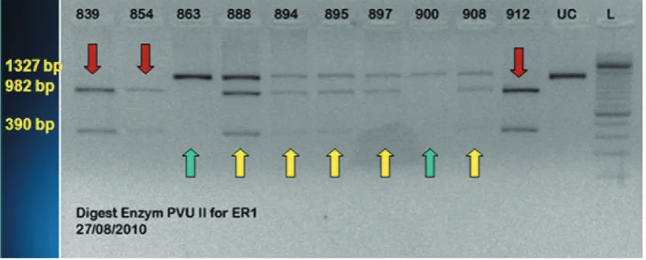
The homozygous mutant in ESR1 XbaI AA shows two bands sized 936 and 436 bp, the heterozygous AG produces three bands (1327, 936 and 436 bp), and the homozygous wildtype GG shows a single band size of 1327 bp.9 For ESR1 PvuII, the TT homozygous
mutant shows two bands sized 982 and 390 bp, the heterozygous CT produces three bands (1372, 982 and 390 bp), and the homozygous CC wildtype shows a single band of 1327 bp.14
Statistical analysis
Data were analyzed with student t-test statistical analyses. P-values less than 0.05
were considered significant.
RESULTS
This study included 108 BC research subjects in Dr. Sardjito General Hospital, Yogyakarta between 2009 and 2010. The average age of subjects was 50.6 years and average age of controls was 48.72 years. The youngest subject among them was 33 years old and the oldest was 68 years old. There
was no significant difference of age between
cases and controls (p > 0.05). The percent mammogram density of cases was 52% and controls was 41%%. Compare means with independent sample t-test between cases and
controls was significantly different (p < 0.05).
We next performed PCR-RFLP of all cases. The ESR1 PvuII TT homozygous mutant showes two bands of 982 and 390 bp, the heterozygous CT produces three bands of 1372, 982 and 390 bp, and the homozygous CC wildtype showes one band of 1327 bp (FIGURE 1).14 Furthermore the frequency
Green arrow: C/C, yellow arrow: C/T, red arrow: T/T
FIGURE 1. ESR1 PvuII polymorphism
TABLE 1. Frequency of ESR1 PvuII polymorphism
Allele FrequencyCase % FrequencyControl
%
TT 15 30 6 10.3
TC 28 56 33 56.9
CC 7 14 19 32.8
The homozygous mutant in the ESR1 XbaI AA shows two bands sized 936 and 436 bp, the heterozygous AG produces three bands (1327, 936 and 436 bp), and the homozygous
wildtype GG shows a band size of 1327 bp (FIGURE 2).14 Furthermore the frequency
of ESR1 XbaI polymorphism is presented in TABLE 2.
Green arrow: G/G, yellow arrow: G/A, red arrow: A/A
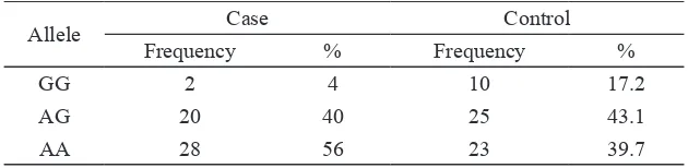
TABLE 2. Frequency of ESR1XbaI polymorphism
Allele Case Control
Frequency % Frequency %
GG 2 4 10 17.2
AG 20 40 25 43.1
AA 28 56 23 39.7
We also analyzed and compared the mammography density in all groups. The mean percentage density in women with one or two copies of the PvuII p allele (CT/Pp and TT/pp, 49% and 48%, respectively) was higher than in those with the CC/PP genotype (39%). However, this difference did not
show statistical significance (TT vs. CC, p >
0.05). Women with one or two copies of the
XbaI x allele had a higher mean percentage density (AG/Xx and AA/xx, 49% and 47%, respectively) than those with the GG/XX
genotype (32%), and we detected a significant
difference between AA and GG genotypes
(p < 0.05). The percentages of heterozygous
(CT) and homozygous (TT) PvuII in BC cases were higher (86%) than those in control (66%). The percentages of heterozygous (AG) and homozygous (AA) XbaI were also higher in BC cases (96%) compared with controls (82%).
DISCUSSION
The average age of the subjects in this study was 50.6 years. Age less than 50 years as many as 25 (50%), equal to or more than 50 years 25 subjects (50%). A study in the United Kingdom in 2007 reported that most GC patients are over 50 years of age (81%).15 These data reflect the high number of young
GC survivors in Indonesia, especially in Java, which is consistent with the pattern of GC in Asian females.16 In general, the frequency of
cancer incidence will increase in line with age.
This increased incidence can be explained by the accumulation of somatic mutations in the human body. Another factor that plays a role is the decline in immune competence that accompanies the aging process. Although very few cases of BC occur in women in their teens or early 20s, BC is the second most common cancer diagnosed in women under 35 years. Approximately 1,400 BC cases are diagnosed each year among women aged 35–39 years. BC incidence rates generally increase with age, with the largest rate of increase before menopause.1 The differences in rates between
patients after BC in Indonesia and in developed countries is probably because of the lower life expectancy in Indonesia and may also be due
to specific genetic factors.
Mammogram density
High breast density on mammography is one of the strongest risk factors for BC.
Breast density seen on a mammogram reflects
differences in the number of stromal and epithelial cells and fatty tissue in the breast. Stroma and epithelium are radiologically will provide the density, while fat provides a radioluscent.17 The average mammographic
density of BC cases is 52% with an average age 50.6%, but in control group 41% with an
average age 48.7 % (p < 0.05). A previous
average age of 55.95 years.18 Another study
reported an average density in BC patients with an average age of 57.4 years of 36.7%, while the control group showed 30.6% with an average age of 56.8 years.19 These
data showed that our study subjects had a higher average mammogram density than previous studies. Martin and Boyd proposed a hypothesis that the biological occurrence of mammogram density is determined by cell proliferation (mitogenesis) and cell damage caused by mutagens (mutagenesis). Both mitogenesis and mutagenesis are affected by many factors, including age, reproductive status, endogenous hormones and growth factors.20 The high-density mammogram
data in this study support the hypothesis that mammogram density is one of the risks for BC.
DNA polymorphisms XbaI and PvuII in
ESR1
Estrogen is a mitogen that affects physio-logical processes including cell growth. Estrogen is found in breast tissue and affects the regulation of cell growth. High estrogen levels are also reportedly associated with an increased risk of BC. Estrogen mostly exerts its cellular action through binding its receptor. Between the two forms of estrogen
receptor α and b, estrogen receptor α plays
an important role in BC because of its high prevalence in breast tissue.21 The ESR1
gene, also known as ESRα, is located on human chromosome 6q25. ESR1 is a core receptor that mediates the action of estrogen or other steroid hormones that regulate gene transcription, especially in estrogen-sensitive tissues such as breast, and plays a crucial role in the growth and differentiation of BC. Most research has focused on the ESR1PvuII (C/T) and XbaI (G/A) polymorphisms in intron 1.21 Both variants have implications in
affecting transcription and gene expression. Some studies also indicate an increased risk of BC with polymorphisms at allele A and T (homozygous mutant) of XbaI and PvuII.22
In this study, percentage of heterozygous (CT) and homozygous mutant (TT) of PvuII in breast cancer cases (86%) higher than controls (66%). The percentage of heterozygous (AG) and homozygous mutant (AA) of XbaI in breast cancer cases (96%) also higher than controls (82%). The average percentage density was higher in women with one or two copies of the PvuII p allele (CT/Pp and TT/pp, 49% and 48%, respectively) than in those with the CC/ PP genotype (39%). However, this difference
did not show statistical significance (TT vs.
CC). Women with one or two copies of the
XbaI x allele had higher mean percentage density (AG/Xx and AA/xx, 49% and 47%, respectively) than those with the GG/XX
genotype (32%), and we detected a significant
difference between AA and GG genotypes. The results of our study are consistent with those published by van Duijnhoven.23 Mammographic density was significantly
higher in women with one or two copies of p (T) allele in ESR1 PvuII. Haplotype ESR1
gene 1 (px/TA) was also associated with increased mammographic density while haplotype 2 (PX/CG) was associated with a decrease in density. On the whole subject is a breast cancer contained a high percentage and high mammographic density of heterozygous and homozygous mutant compared with homozygous wild type at ESR 1 XbaI and
differences.22 The existence of sex steroid
metabolic enzymes and ESRs in breast tissue caused an activation of local estrogen that could potentially lead to reactive metabolites in breast tissue and may have a role in the initiation and promotion of carcinogenesis.
CONCLUSION
Our study showed that haplotype 1 (px/ TA) of ESR1 gene is associated with high
sensitivity to estrogen and reflects an increase
of mammographic density. Haplotype 2 (CG/PX) is associated with lower sensitivity
to estrogen and reflects a decrease of mammographic density. Our findings support
the view that ESR1 polymorphisms may affect BC risk through differences in breast density.
ACKNOWLEDGEMENT
We would like to thank all patients who have participated in this study.
REFERENCES
1. American Cancer Society. Can breast cancer be found early? American Cancer Society, 2010.
2. Ghozali A. Registrasi kanker. Proceedings of Seminar Onkologi Yayasan Kanker Indonesia. 2009.
3. Cuzick J. Epidemiology of breast cancer-selected highlights. The Breast 2003; 12(6):405-11. http://dx.doi.org/10.1016/ S0960-9776(03)00144-9
4. Couse JF, Lindzey J, Grandien K, Gustafsson JA, Korach KS. Tissue distribution and
quantitative analysis of estrogen receptor-α (ERα) and estrogen receptor-β (ERβ)
messenger ribonucleic acid in the wild-type
and ERα-knockout mouse. Endocrinology
1997; 138(11):4613-21. http://dx.doi. org/10.1210/endo.138.11.5496
5. Gruber CJ, Tschugguel W, Schneeberger C, Huber JS. Production and actions of estrogens. N Engl J Med 2002; 346(5):340-52. http:// dx.doi.org/10.1056/ NEJMra000471
6. Boyd NF, Byng JW, Jong RA, Fishell EK, Little LE, Miller AB, et al. Quantitative
classification of mammographic densities and
breast cancer risk: results from the canadian national breast screening study. J Natl Cancer Inst 1995; 87(9):670-5. http://doi. org/10.1093/jnci/87.9.670
7. Hall F. Mammographic screening in younger women at high risk. Am J Roentgenol 2009; 193(4):1188. http://dx.doi.org/10.2214/ AJR.09.2753
8. Persson I, Thurfjell E, Holmberg L. Effect of estrogen and estrogen-progestin replacement regimens on mammographic breast parenchymal density. J Clin Oncol 1997; 15(10):3201-7. http://dx.doi.org/10.1200/ jco.1997.15.10.3201
9. Greendale GA, Reboussin BA, Slone S, Wasilauskas C, Pike MC, Ursin G. Postmenopausal hormone therapy and change in mammographic density. J Natl Cancer Inst 2003; 95(1):30-7. http://dx.doi.org/10.1093/ jnci/95.1.30
10. Cuzick J, Warwick J, Pinney E, Warren RM, Duffy SW. Tamoxifen and breast density in women at increased risk of breast cancer. J Natl Cancer Inst 2004; 96(8):621-28. http:// dx.doi.org/10.1093/jnci/djh106
11. Shimada N, Iwasaki M, Kasuga Y, Yokoyama S, Onuma H, Nishimura H et al. Genetic polymorphisms in estrogen metabolism and breast cancer risk in case–control studies in Japanese, Japanese Brazilians and non-Japanese Brazilians. J Hum Genet 2009; 54(4):209-15.
http://dx.doi.org/10.1038/jhg.2009.13
susceptibility: a meta-analysis involving 17,600 subjects. Breast Cancer Res Treat 2010; 122(2):521-5.
http://dx.doi.org/10.1007/s10549-009-0731-4 13. Ding H, Fu Y, Chen W, Wang Z. COMT
Val158Met polymorphism and breast cancer risk: evidence from 26 case-control studies. Breast Cancer Res Treat 2010; 123(1):265-70. http://dx.doi.org/10.1007/s10549-010-0759-5
14. Boroumand M, Ghaedi M, Mohammadtaghvaei N, Pourgholi L, Anvari MS, Davoodi G, et
al. Lipid profile and inflammatory markers
associated with estrogen receptor a PvuII and XbaI gene polymorphisms. Transl Res 2009; 153(6):288:95.
http://dx.doi.org/10.1016/j.trsl.2009.02.006 15. Cancer Research UK. Breast Cancer-UK.
2011.
from:http://www.cancerresearchuk.org/ cancer-info/cancerstats/types/breast/ incidence/uk-breast-cancer-incidence-statistics#age
16. Leong SP, Shen ZZ, Liu TJ, Agarwal G, Tajima T, Paik NS, et al. Is breast cancer the same disease in Asian and Western countries? World J Surg 2010; 34(10):2308-24. http:// dx.doi.org/10.1007/s00268-010-0683-1 17. Yaffe MJ. Measurement of mammographic
density. Breast Cancer Res 2008; 10(3):209-18. http://dx.doi.org/10.1186/bcr2102
18. Boyd NF, Guo H, Martin LJ, Sun L, Stone J, Fishell E, et al. Mammographic density and the risk and detection of breast cancer. N Engl J Med2007;356(3):227-36.
http://dx.doi.org/10.1056/NEJMoa062790
19. Takata Y, Mascarinec G, Le Marchand L. Breast density and polymorphisms in genes coding for CYPA2 and COMT: the Multiethnic Cohort. BMC Cancer 2007; 7:30. http://dx.doi.org/10.1186/1471-2407-7-30 20. Martin LJ, Boyd NF. Mammographic density,
potential mechanisms of breast cancer risk associated with mammographic density: hypotheses based on epidemiological evidence. Breast Cancer Res 2008; 10(1):201. http://dx.doi.org/10.1186/bcr1831
21. Slattery ML, Sweeney C, Herrick J, Wolff R, Baumgartner K, Giuliano A, et al. ESR1, AR, body size, and breast cancer risk in Hispanic and non-Hispanic white women living in the Southwestern United States. Breast Cancer Res Treat 2007; 105(3):327-35.
http://dx.doi.org/10.1007/s10549-006-9453-z 22. Crandal CJ, Sehl ME, Crawford SL, Gold
EB, Habel LA, Butler LM, et al. Sex steroid metabolism polymorphisms and mammographic density in pre- and early perimenopausal women. Breast Cancer Res 2009; 11(4):R5.
http://dx.doi.org/10.1186/bcr2340
23. van Duijnhoven FJ, Bezemer ID, Peeters PH, Roest M, Uitterlinden AG, Grobbee DE, et al. Polymophism in the estrogen receptor
α gene and mammographic density. Cancer
Epidemiol Biomarkers Prev 2005; 14(11 Pt 1):2655-60.