Microscopic Examination of Fecal Leukocytes as a
Simple Method to Detect Infective Colitis in Children
Nuraini I Susanti*, Reynaldo**, Aria Kekalih**,Anis Karuniawati***,
Badriul Hegar****
*Department of Child Health, Fatmawati General Hospital, Jakarta
**Department of Community Medicine, Faculty of Medicine, Universitas Indonesia, Jakarta ***Department of Microbiology, Faculty of Medicine, Universitas Indonesia, Jakarta
****Departement of Child Health, Universitas Indonesia/Dr. Cipto Mangunkusumo General National Hospital, Jakarta
Corresponding author:
Badriul Hegar. Division of Gastroentero-hepatology, Department of Child Health, Dr. Cipto Mangunkusumo General National Hospital. Jl. Diponegoro No.71 Jakarta Indonesia. Phone: +62-21-3907742; Facsimile: +62-21-3907743. E-mail:[email protected]
ABSTRACT
Background: Various pathogenic bacteria are reported as the cause of infectious colitis in children. Infectious colitis does not have a specific sign, therefore an accurate examination is required. The implementation of fecal cultures accompanied with drug resistance tests often have constraints, beside the relatively expensive costs, longer times are needed, and not all health care facilities have required instruments. On the other hand, this condition requires an immediate antibiotic therapy, so that the infection should not be continued. In daily practice, it is not uncommon to find diarrhea with the amount of fecal leukocyte < 10/hpf with pathogenic bacteria on the examination of the fecal culture.
Method: Cross-sectional study was conducted to observe the pattern of bacterial distribution in children’s fecal who have acute diarrhea and the correlation between the existence of pathogenic bacteria and the number of leukocytes in the fecal, as well as antibiotic resistance patterns. The population of this study is children with age of 6 months old - 18 years old who were suffering from acute diarrhea with the amount of fecal leucocyte ≥ 5/hpf, who recruited from polyclinic or patient admitted at Cipto Mangunkusumo Hospital and Fatmawati General Hospital, Jakarta.
Results: Based on examinations of fecal cultures and PCR, Salmonella sp and C. dificille were found
subsequently in 2 children (33.3%), Enterophatogenic E. Coli(EPEC) and Shigella were found subsequently in 1 child (16.7%). Based on the ROC curve, it was found that there was no intersection of maximum and minimal leukocyte value with the midline, whereas the best sensitivity and specificity value was found at the cut-off point of 8.5, hence the cut-off point of leukocytes was determined at < 8 and > 8. The sensitivity value was 83.3% and the specificity value was 45.1%.
Conclusion: The antibiotic sensitivity test showed that one child infected by EPEC was sensitive to
ciprofloxacin. Two children infected by Salmonella, were still sensitive to chloramphenicol, cotrimoxazole, cefixime, and ceftriaxone. Two children infected by C. Difficile were sensitive to ceftriaxone, and 1 child infected by Shigella was sensitive to cefixime, ceftriaxone and ciprofloksazine.
ABSTRAK
Latar belakang: Berbagai bakteri pathogen dilaporkan sebagai penyebab kolitis infeksi pada anak. Kolitis infeksi tidak mempunyai gambaran spesifik, sehingga diperlukan pemeriksaan penunjang akurat. Implementasi kultur tinja disertai uji resistensi obat seringkali mendapatkan kendala, selain biaya, memerlukan waktu cukup lama, dan tidak semua fasilitas pelayanan kesehatan memilikinya. Dilain pihak, kondisi ini memerlukan terapi antibiotika segera, agar infeksi tidak berkelanjutan. Dalam praktik sehari-hari, tidak jarang ditemukan diare dengan jumlah leukosit tinja < 10/LPB disertai bakteri patogen pada pemeriksaan kultur tinjanya.
Metode: Dilakukan penelitian potong lintang untuk melihat pola sebaran bakteri pada tinja anak yang
mengalami diare akut dan sekaligus ingin mengetahui hubungan keberadaan bakteri patogen dengan jumlah leukosit dalam tinja, serta pola resistensi antibiotika. Populasi penelitian adalah anak berumur 6 bulan - 18 tahun menderita diare akut dengan jumlah leukoit tinja ≥ 5/LPB yang berobat jalan di Poliklinik atau dirawat di Rumah Sakit Cipto Mangunkusumo, Jakarta Indonesia.
Hasil: Pemeriksaan kultur tinja dan PCR didapatkan Salmonella sp dan C. dificille masing-masing pada 2
anak (33,3%), Enterophatogenic E. Coli dan Shigella masing-masing pada 1 anak (16,7%). Pada kurva ROC tidak didapatkan perpotongan nilai leukosit maksimal dan minimal dengan garis tengah, sedangkan nilai sensitivitas dan spesifisitas terbaik terletak pada titik potong 8.5, maka ditentukan titik potong leukosit adalah < 8 dan > 8. Nilai sensitivitas didapatkan sebesar 83,3% dan nilai spesifisitas sebesar 45,1%.
Simpulan: Uji sensitivitas antibiotika memperlihatkan 1 anak terinfeksi EPEC sensitif terhadap ciprofloksazin.
Dua anak terinfeksi oleh Salmonela, masih sensitif terhadap Kloramfenikol, Kotrimoksazol, Cefiksim, dan Ceftriakson. Dua anak terinfeksi C. difficile sensitif terhadap ceftriakson, dan 1 anak terinfeksi Shigella sensitif terhadap cefiksim, ceftriakson, dan ciprofloksazin.
Kata kunci: kolitis infeksi, leukosit tinja, kultur tinja
INTRODUCTION
Infectious colitis is an inflammation process of the colon caused by pathogenic bacterial infection.1
Various pathogenic bacterias, such as Shigella, E Coli, Salmonella, Campylobacter are reported as causes of infectious colitis in children. The prevalence of acute diarrhea in children caused by pathogenic bacterial infection is approximately 16%.2 In addition
to diarrhea, infectious colitis does not have specific features, so an accurate investigation is required. Fecal culture with drug resistance test is a gold standard test to prove the cause of infection colitis by pathogenic bacteria. However, the daily implementation of fecal cultures accompanied with drug resistance tests often have constraints, beside the relatively expensive costs, longer times are needed, and not all health care facilities have required instruments. On the other hand, infectious colitis caused by pathogenic bacteria required immediate antibiotic therapy, so that the should not be continued.
World Health Organization (WHO) recommends the number of fecal leucocyte > 10/ hpf as an infection marker of pathogenic bacterial Shigella dysenteriae
in children with clinical dysentery. Considering the infection could get worse and life-threatening,
therefore WHO recommends administering empirical antibiotics based on epidemiological data in area where there are no facilities for fecal cultures examination. The recommendation also applies while waiting for results of a fecal culture examination that takes several days. 3
METHOD
This study is a descriptive cross-sectional study which aims to obtain fecal leukocyte values in infectious colitis bacterial. The study also conducted a diagnostic test by assessing the sensitivity of fecal leukocyte counts to diagnose bacteria of infectious colitis. The population of this study is children with age of 6 months old - 18 years old suffering from acute diarrhea with the amount of fecal leucocyte 5/hpf, treated as outpatient in polyclinic or admitted at Cipto Mangunkusumo Hospital and Fatmawati General Hospital Jakarta. Children with severe systemic infections (HIV-AIDS, sepsis), clinical symptoms of lactose intolerance other than diarrhea, or parents who refused to include their children in the study were excluded from the study. Based on a single sample formula for estimating the proportion of a population by using absolute accuracy, the sample formula for the diagnostic test with sensitivity output and calculating the 'drop out' of 10%, then a sample size of 88 was obtained.
Explanations given to parents are including the objective of the study and examination procedures which will be performed. Parents, who were willing to commit their children in this study, were required to sign a consent form for participation in study before the study was conducted.
The data obtained in this study are primary data, including interviews with subject parents through the completion of a questionaire of demographic data, history taking that included a history of diarrhea duration, fecal consistency, fever, abdominal pain, associated diseases, and previous antibiotics administration. Physical examination includes general condition, nutritional status, body temperature, and hydration status. Fecal macroscopic examination includes consistency, color, blood and mucus, whereas microscopic examination of fecal includes the number of leucocytes/hpf and erythrocytes/hpf. Fecal culture and antibiotic resistency tests were performed on children with fecal leukocyte count > 5/hpf.
Fecals were collected in plastic-coated diapers or directly put in a plastic bag. Parents took fecals using a spoon/scoop and put in 1 non-sterile pot and 1 sterile pot which had been prepared before. After the fecals collected, non-sterile pots were delivered to Clinical Pathology Laboratory Cipto Mangunkusumo Hospital to be examined microscopicly. If the microscopic examination obtained leucocyte count > 5/hpf, fecal sample in sterile pot will continue by fecal culture examination and resistance test in Microbiology Laboratory, Faculty of Medicine University of Indonesia. Fecal cultures were performed on Shigella spp, Salmonella spp,
Campylobacter spp, E Coli, Yersinia Enterocolica and
Aeromonas spp. Antibiotic resistance test performed on
Cefixim, Azithromycin, Ceftriaxone, Ciprofloksazin, Ciprimoxazole, and Chloramphenicol.
Determination of nutritional status was acomplished based on age, weight (W) and height (H) in a state without dehydration. Weight was scaled by using a digital scale which had a precision of 0.1 kg, length or height measured by using height measuring instrument with a precision of 0.1 cm. Nutritional status was assessed based on the weight and height score converted to a standardized value (Z-score) by using the WHO-2005 toddler anthropometric standard. Furthermore, based on the Z-score value of each indicator, nutritional status was determined according to weight/age index.
All data obtained recorded in a research report form that had been prepared for subsequent incorporation into the computer using SPSS program version 15.0. Descriptive data would be presented in textile and tabular. This study was approved to pass the ethical review of the Health Research Ethics Committee, Faculty of Medicine, Universitas Indonesia, Cipto Mangunkusumo Hospital.
RESULTS
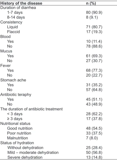
The subjects consisted of 48 (54.5%) girls and 40 (45.5%) boys with the age range from 6 to 216 months. Most children (65.9%) were < 2 years old and only 8% aged over 6 years. The diarrhea duration of 1-7 days when examined was obtained in 80 children (90.9%) with consistency of fluid fecals in 71 children (80.7%). Fecals containing blood was obtained in 10 children (11.4%) and mucus in 61 children (69.3%). Based on the history,fever found in children (77.3%) and abdominal pain in 31 children (35.2%). A total of 45 children (51.1%) had received antibiotics when the fecal sample was examined with a mean duration of 3 days. 48 children (54.5%) with good nutritional status and poor nutritional status in 7 children (8%). Based on dehydration status assessment, it was found that 13 children (14.8%) were severe dehydration and 25 (28,4%) children were without dehydration (Table 1).
Fecal culture examination conducted on 88 children
obtained E. Coli on 34 children (48.5%). Then PCR
examination was conducted to observe the strain of pathogenic E. coli, and found Enterophatogenic E. coli
on 1 child (16.7%). Other pathogenic bacteria found in the fecal culture examination were Salmonella sp
and C. dificille respectively in 2 children (33.3%), and
Based on the ROC curve, it was found that there was no intersection of maximum and minimal leukocyte value with the midline, whereas the best sensitivity and specificity value was found at the cut-off point of 8.5, hence the cut-off point of leucocytes was determined at < 8 and > 8. The sensitivity value was 83.3% and the specificity value was 45.1%.
Table 2. Results of fecal culture examination
No Culture results Pathogenic bacterial n (%)
1.
Table 1. Duration of diarrhea, fecal characteristics, complaints, nutritional status, and dehydration
History of the disease n (%)
Duration of diarrhea
The duration of antibiotic treatment < 3 days
Figure 1. ROC curve of fecal leukocytes on microscopic examination
Of the 88 children which fecal being examined, it was found that 38 children with microscopic leukocyte leukocyte < 8/hpf and 1 child (2.6%) with pathogenic bacteria in their culture examination. Of the other 50 children with microscopic fecal leukocytes > 8/hpf, 5 children (10%) was being found with pathogenic bacteria on fecal culture examination (Table 3). Sensitivity value of 83.3% and specificity value of 45.1% were obtained. Based on the assessment using Pearson Chi-square, value 1.845, df 1, and p 0.174 were obtained. This study showed no significant correlation between leucocyte count with pathogenic bacteria cause of diarrhea.
Based 6 children who had pathogenic bacteria on their fecals, it was followed by examination of antibiotic sensitivity test. The sensitivity test was performed on antibiotics which were often used as the preferred treatment for diarrhea caused by pathogenic bacterial infection, namely Trimethoprim (Trimethoprim-Sulfametoksazol), Chloramphenicol, Cefixime, Ceftriaxone, Ciprofloksazin, and Azithromycin. The antibiotic sensitivity test for 6 children infected by pathogenic bacteria showed that
Table 4. Correlation between fecal culture with antibiotic resistance test
No Culture results
Chloram: Chloramphenicol; Cotrim: Cotrimoxazole; Ceftri: Ceftriaxone; Cipro: Ciprofloksazine; Azithr: Azithromycin; S: Sensitive; I: Intermediate; R: Resistant
Table 3. Correlation betwen microscopic fecal leukocytes and
fecal cultures examination using the cut-off point value of 8 in
fecal leukocytes
Total leukocyte/hpf Culture results Total Positive (%) Negative (%)
> 8 5 (10) 45 (90) 50
≤ 8 1 (2.6) 37 (97.4) 38
Total 6 82 88
Pearson Chi-square obtained value 1.845 , df 1, and P 0.174, sensitivity
1 child was infected by EPEC and was sensitive to ciprofloksazin. Two children infected by Salmonella, were still sensitive to chloramphenicol, cotrimoxazole, cefixime, and ceftriaxone. Two children infected by C. difficile were sensitive to ceftriaxone, and 1 child infected
by Shigella was still sensitive to cefoxime, ceftriaxone,
and ciprofloksazine (Table 4).
DISCUSSION
This study showed no difference between boys and girls for diarrhea, which are 48 (54.5%) and 40 (45.5%) respectively. Likewise, based on WHO data that the most common age group affected by diarrhea due to infection was age below 2 years, similar tothe results of this study, that was equal to 65.9%.3
Pathogenic bacteria on fecal cultures were obtained in 6 children (6.8%).The results of this study were slightly lower than the results of surveillance of pathogenic bacteria in diarrheal disease in Indonesia (Oyofo et al). Oyofo et al included 6760 patients found 9% of them having pathogenic bacteria, namely Shigella flexneri (39%), Salmonella spp (26%), Vibrio spp
(17%), S.sonnei (7%), Campylobacter jejuni (4.4%),
Salmonella typhi (3%) and S.dysenteriae (2.3%).4 The
study by Bodhidatta et al of 623 Thai children who having dysentery with leukocyte count > 10/hpf, found
Campylobacter as the most pathogenic bacteria (28%),
followed by Salmonella spp (18%), Shigella spp (9%),
and E. coli ETEC (6%).5 An increase in the number
of fecal leukocytes was also found by Huicho et al in 36% of children infected with Salmonella-Shigella-Campylobacter, 16% children infected by Rotavirus or enterotoxigenic E. colli, and 18% children are infected with Cryptosporidial diarrhea.6 In our study, we found
that Salmonella spp and C.difficile respectively of 33.3%
and EPEC and Shigella spp respectively of 16.67%, neither Campylobacter nor E.Coli strain other than
EPEC were found.
Our study found E. coli in 34 children, after
isolation and identification, and only one 13-month-old child infected by pathogenic E. coli strain (2.9%) ie Enterophatogenic E. Coli (EPEC). The results of this
study were smaller than the previous studies conducted on children aged < 5 years who experienced diarrhea in Cairo. Of the 134 E. coli samples examined, EPEC
isolates were obtained in 7 samples (5.2%) and most at age < 2 years.7
Klebsilla pneumonia was found in 43.2% of fecal
samples examined. Until this time, Klebsiella spp
family Enterobacteriaceae regarded as normal flora,
although Klebsiella spp. was obtained as the only
species in fecal cultures. In recent years, many studies on the role of some pathogenic bacteria previously regarded as normal or commensal flora. One of the most studied bacteria was Klebsiella spp along with K. pneumoniae species. The results of the study showed
that K. pneumoniae, which had been considered as
commensal bacteria could turn out to be a pathogen in the human intestine. The K. pneumonia bacteria
themselves were influenced by several factors, namely capsule, fimbriae (villi), serum lipopolysaccharide resistance, K. pneumoniae virulence, hemolysin and siderophores production, and toxin.8
Klebsilla pneumonia enteropathogen was
commonly reported as the cause of acute diarrhea in children, even some circumstances caused nosocomial infections, resulting in food-borne outbreaks occured.9
Several K. pneumonia strains produced thermolable or
thermostable enterotoxins which caused diarrhea with a mechanism similar to E. coli. Klebsiella sp. could be
the pathogenic bacteria which caused diarrhea resulted from the transfer of plasmid of virulent E.coli strain and Klebsiella in gastrointestinal tract. Janczura et al using
PCR found the lth gene as a heat-labileenterotoxin
code as much as 8.5% in plasmid of Klebsiella spp
strain, and 77% of which caused cytopathic effects on the cell. Even parts of cells that did not contain the lth gene also caused cytopathic effects. This indicated that
Klebsiella spp considered as normal flora and unable
caused diarrhea, could become cause of diarrhea.8
In this study, it was found that there was relatively large variation in the number of leukocyte from fecal with pathogenic bacteria culture results. Therefore, calculation for estimation of minimum and maximum value of fecal leucocytes was conducted by using ROC coordinate curve. In line with ROC curve, there was no intersection of minimal and maximal value with the center line. Considering best sensitivity and specificity value were 8.5, then the cut-off point of leukocyte was determined as many as < 8 or > 8. The sensitivity value was 83.3% and the specificity value was 54.9%. From 6 fecal samples found with pathogenic bacteria, 5 of them had leukocyte values > 8/hpf, whereas from 82 samples with no pathogenic bacteria found, 45 showed a leukocyte count > 8/hpf. However, these results did not provide statistically significant differences. Huicho L et al stated that the number of leukocytes > 5/lpb as a cut-off point for pathogenic bacterial infection in their study of 446 children suffering from diarrhea, which found almonella, Shigella, Campylobacter
enterotoxigenic and rotavirus respectively 16%.6
DeWitt also determined bacterial infection when fecal leukocyte count > 5/hpf was found.13 Children
having diarrhea with fecal leukocytes > 10/hpf and mixed with blood, strongly correlated with E coli
infection.7 Number of fecal leucocytes > 10/hpf used
by WHO as a marker of Shigella disentriae bacterial infection in children with clinical dysentery, so it was permissible to administer antibiotics without fecal culture examination first.3 The number of leukocytes
> 8/hpf in this study indicated that there were bacterial infections.
Half of children (51.1%) were or had received antibiotics and 62.2% of them had received antibiotics for more than 3 days. This situation is indeed require a separate assessment to determine the effect of antibiotics toward the results of fecal culture. The antibiotic sensitivity test in this study revealed only Ciprofloksazin which was sensitive to E.coli strain
EPEC. Antibiotics such as Chloramphenicol, Cefixime, and Ceftriaxone were sensitive to Salmonella spp. In the 2 infected children, most antibiotics were resistant
to C.difficile, except Ceftriaxone and Ciprofloksazin
were resistant to 1 of 2 infected children. Cefixime, Ceftriaxone, and Ciprofloksazin were among those antibiotic that were still sensitive toward Shigella spp.
Several previous studies had shown that E.coli was
still sensitive to Cotrimoxazole and Ciprofloksazin. In this study, it was found a child infected by E coli, EPEC
isolates had been resistant to Cotrimoxazole. Two antibiotics namely Cotrimoxazole and Ciprofloksazin were resistant to one Salmonella bacteria. Our study showed Cotrimoxazol and Chloramphenicol were resistant to Shigella spp. This result was similar to study conducted by Dharmawan BS et al. which found 8.6% of EPEC isolates and 7.1% Salmonella. Provision of Cotrimoxazole compared to placebo did not indicate significant differences in duration and frequency of diarrhea. Cotrimoxazole was resistant to all EPEC isolates and 3 of 5 Salmonella.10 This result may caused
by uncontrolled use of Cotrimoxazole resulting in an altered bacterial resistance pattern.11
Another study by Nafianti S et al showed that Trimethoprim-Cotrimoxazole and placebo administered to 2-24 months old children suffering bloody diarrhea and subsequently the amount of fecal leukocytes were examined, it did not indicate any significant difference. Thesefact proved that bloody diarrhea was not always caused by bacteria. Furthermore, in infants aged 6 months age or less, need to be thought other cause factors, including cow milk or soy allergy.12
Study conducted by Gogate A at al showed that
C. difficile was only sensitive to Ceftriaxone and Ciprofloksazin. C. difficile infections commonly
occured in children who were receiving broad-spectrum antibiotics over long periods, resulting in Antibiotic Associate Diarrhea. This study conducted in India of
250 children, 5-12 year old with severe diarrhea, fluid fecal with blood and mucus, with leukocyte counts > 5 hpf, found that 18% of children were infected
by C.difficile and 68.9% children has improved after
provisions of antibiotics were discontinued, while others improved after treatment with Metronidazole.13
The pattern of bacterial sensitivity to antibiotics was always changing and different in each area, so survey on pattern of antibiotic resistance should be conducted which could be used as a guide of therapy in an area. The relatively high intensity of antibiotic used may lead to bacterial resistance problems. According to Antimicrobial Resistant in Indonesia (AMRIN-Study) study on 2494 individuals in the community, it was found 43% E coli resistant to various antibiotics,
among others Ampicilin (34%), Cotrimoxazole (29%) and Chloramphenicol (25%). In order to optimize the used of antibiotics wisely (prudent use of antibiotics), general guidelines for the use of antibiotics in hospital and health fasilities is needed to be prepared. This guidelines is expected to be applied as a reference in developing antibiotic policies and guidelines for hospitals and other healthcare facilities.14
CONCLUSION
In conclusion, the prevalence of pathogenic bacterial infectious colitis in children with acute diarrhea was 6.82% with Salmonella sp. dominance. Microscopic examination of fecal leukocytes and fecal cultures examination showed no significant association in the diagnosis of bacterial infectious colitis. Most of the pathogenic bacteria were resistant to Cotrimoxazole.
REFERENCES
1. Riset Kesehatan Dasar (Riskesdas). Jakarta: Badan Penelitian dan Pengembangan Kesehatan Indonesia: Departemen Kesehatan Republik Indonesia;2007.
2. Guandalini S. Acute Diarrhea. In: Pediatric Gastrointestinal Disease. 4th ed. United States: BC Decker Inc 2004;h:166-76.
3. World Health Organization. Diarrhoeal Treatment Guidelines. Geneva: World Health Organization 2009;h:1-46.
5. Bodhidatta l, Vithayasari N, Eimpokalarp B. Bacterial Enteric Pathogens in Children with Acute Dysentery in Thailand: Increasing Importance of Quinolone Resistant Campylobacter.
Southeast Asian J Trop Med Public Health 2002;33:752-7. 6. Huicho L, Sanchez D, Contreras M, Paredes M. Occult
blood and fecal leucocytes as screening test in childhood infectious diarrhea: an old problem revisited. Pediatr Infect Dis J 1993;12:474-7.
7. Mercado EH, Ochoa TJ, Ecker L, Cabello M, Durand D. Fecal leukocytes in children infected with Diarrheagenic Escherichia coli. J Clin Microbiol 2011;49:1376-81.
8. Janczura A, Smutnicks, Junka A, Gosciniak G. The detection and expression of enterotoxin encoding lthgene
among Klebsiella spp. Isolated from diarrhea. C Eur J Biol
2013;8:121-9.
9. Gilquin J. Bloody diarrhea caused by Klebsiella pneumonia : A new mechanism of bacterial virulence? CID 1998;27:648-9. 10. Dharmawan BS, Firmansyah A, Chair I. The benefit of Co-trimoxazole treatment in the management of acute watery diarrhea caused by invasive bacterial infection. Paediatr. Indones 2007;47:104-8
11. Guerrant RL, Gilder TV, Steiner TS, Thielman NM, et al. Practice Guidelines for The Management of Infectious Diarrhea. IDSA Guidelines. CID 2001:32:331-50.
12. Nafianti S, Ramayani OR, Daulay DG, Supriatmo. The efficacy of Trimethoprim- Sulfamethoxazole treatment in children with acute bloody diarrhea. Paediatrica Indonesiana 2007:47;17-20
13. Gogate A, De A, Nanivadekar R, Mathur M. Diagnostic role of fecal culture and toxin detection in antibiotic associated diarrhea due to Clostridium difficile in children. Indian J Med Res 2005;Dec:518-24.