Vol. 41. No. 3 July–September 2008
The management of oral candidosis in diabetic patient with
maxillary Herpes Zoster
Kus harijanti1, dwi Setyaningtyas2, and isidora KS2
1 Departement of Oral Medicine Faculty of Dentistry Airlangga University 2 Oral Medicine Clinic of Surabaya Navy Hospital
Surabaya - Indonesia
abStract
background: Oral candidosis is an infection caused by mainly Candida albicans. Candida species are common normal flora in Oral candidosis is an infection caused by mainly Candida albicans. Candida species are common normal flora in the oral cavity and have been reported to be present in 40% to 60% of the population. Candida is predominantly an opportunistic infectious agent. Infection frequency has increased because of the presence of both local and systemic risk factors. The elderly age and diabetes mellitus may decrease the amount of saliva (xerostomia) and potentially increase the risk of colonization and secondary infection by Candida. Herpes Zoster (HZ) is a manifestation of the reactivation of latent varicella zoster virus. It is characterized by unilateral, painful, vesicular rash with a dermatomal distribution. The clinical manifestations of this disease can erupt to the skin and mucous membrane. If maxillary nerve is involved, the lesion can appear on unilateral facial skin and oral mucous membrane.
Purpose: The purpose of this paper is to report and discuss the difficulties in managing the oral candidosis in elderly patient (57 The purpose of this paper is to report and discuss the difficulties in managing the oral candidosis in elderly patient (57 year old male) who suffered from maxillary Herpes Zoster and diabetes mellitus. case management: At first, the patient was treated At first, the patient was treated with 2% chlorhexidine gluconate and mycostatin oral suspension as topical antimycotic and reffered to dermathology clinic for viral infection treatment, however the oral candidosis did not improved. Subsequently, ketokonazole tablet was given three times daily for three weeks and regulated blood glucose level. In systemic antifungi (ketokonazole) treatment the oral candidosis disappeared.
conclusion: In this case, it is conclude that the management of oral candidosis are adequate, antiviral, blood glucose level regulating In this case, it is conclude that the management of oral candidosis are adequate, antiviral, blood glucose level regulating and systemic antifungal therapy.
Key words: Herpes Zoster, oral candidosis
Correspondence: Kus Harijanti, c/o: Departemen Ilmu Penyakit Mulut, Fakultas Kedokteran Gigi Universitas Airlangga. Jl. Mayjend. Prof. Dr. Moestopo no. 47 Surabaya 60132, Indonesia. Email: [email protected]
introduction
Candida is a yeast-like fungus. Candida species are common in the normal flora and have been reported to be present in 40% to 60% of the population.1 It is harmless, but it might become opportunistic pathogen when there is oral immune decreasing as a result of the oral microorganism ecosystem changes. Candidosis is an infection caused by Candida, mainly C. albicans. Infection frequency by Candida species has increased because of the presence of both local and systemic risk factors. The systemic risk factors are treatment with broad spectrum antibiotic, diabetes mellitus and immunosuppressive illness.1,2
The symptoms of candidosis may include burning sensation, sensitivity, altered taste and smell. Clinical
intra oral sign may vary from a red form (erythematous) to psedomembranous form (white, thrush); simultaneous red and white changes are common. Oral candidal overgrowth is almost superficial and rarely penetrates deeper than epithelial cells surfaces. The pseudomembranous form is the colonies of organisms that attach to the surface and can be removed with rubbing, frequently leaving red or bleeding sites.1
Varicella Zoster Virus (VZV) is DNA virus that causes chicken pox in children as primary infection and herpes zoster (shingles) in adult as subsequent reactivation. This disease can affect the skin and oral mucous membrane. Chicken pox is caused by droplet infection or direct contact with patient, usually benign illness that occurs in epidemic among susceptible children. Although, the clinical
symptom on skin or mucous membrane disappear, zoster virus still resides in dorsal-root ganglia and subsequent reactivation can occur.3,4 Virus reactivation does not always give the clinical symptoms, because virus-cell mediated immunity can protect the host. The risk of shingle is increased by declining cell mediated immune responses, which occur naturally as a result of aging or induced by immunosuppressive illness or medical treatments.3,5
At an incidence of 2/1000, about 500,000 cases annually would occur in the United States and about half this number in the United Kingdom. The incidence rises steeply with age, being less than 1 per 1000 person years in children and as many as 12 per 1000 person years in those over 65 years old.5 Seventy percent of herpes zoster occurred on persons over 50 year old.4
The initial symptoms are pain and tenderness in the affected area and prodromal severe pain of herpes zoster. Characteristic of herpes zoster are multiple vesicles, which distributed in appropriate sensory nerve region. When the maxillary nerve is involved, the lesion can appear on unilateral facial skin and oral mucous membrane, this is about 15% of the cases.4 The patient suffers from tooth-ache in the nerve region which is involved and frequently it can not be distinguished with trigeminal zoster.3,6
When this disease is not treated, vesicles on skin and ulcers in oral mucous membrane will disappear in 2-4 weeks or more. Crusts appears on the skin and might form scarring. Although there are several serious complications of zoster (ophthalmic, splanchnic, cerebral motor), in immunocompromise adult the most common case is post herpetic neuralgia- pain. Postherpetic neuralgia is a condition in which the patient suffers from anesthesia, paraesthesia and severe pain in the involved nerve region. The pain sensation is still persisted for prolong time until years after clinical sign disappears. Both the incidence and the duration of postherpetic neuralgia are directly correlated with the patient’s age.3,4,5
This article reported a case of oral candidosis on herpes zoster which involved the maxillary nerve. The patient was 57 years old and also suffered from diabetes mellitus.
caSe
In October 17, 2006, a 57 years old male came to the dental clinic at Dr. Soetomo General Hospital Surabaya, the patient complained of severe left tooth-ache in 5 days. Initially, the patient came to the public medical center for treating his teeth, but the pain didn’t reduce although the teeth was already filled, even the left facial skin was oedem and erythemathous, followed by fever, malaise and weakness. Then, the patient went to general practitioner and was treated with antibiotic and analgesic/anti-inflammation. The patient still suffered from severe teeth-ache on the maxillary left-region. Formerly, the patient suffered from localized abnormal skin sensations, ranging from itching
or tingling on the left facial-skin and followed by the appearance of clusters of clear vesicles.
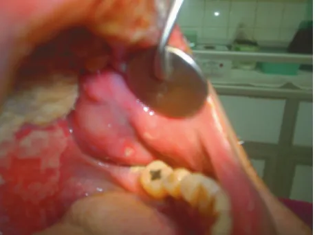
Extra oral examination showed pustulation, ulceration, crusting and oedema on the left facial skin (Figure 1). Several lesions on the left lower lid caused lid edema and ectropion, involved left nose and upper lip. Left submandibular lymph nodes were enlarged and tender. On intra oral examination (Figure 2 & 3), white plaque was revealed on hard palate, thick and soft as creamy milk with irregular border and form, surrounded by erythemathous and can be removed with rubbing. Erythemathous and multiple ulcers with 2-4mm diameter were found on maxillary left buccal-fold and left buccal mucosa, the center of ulcers were yellowish and painful. Erythemathous mucosa and white thin-plaque, irregular form and scrapable were found on the left upper lip.
caSe management
The patient previously came to the public medical center with chief complaint left upper teeth-ache. In fact, the figure 1. First visit: Extra oral view.
figure 3. First visit: Palatal view.
lesions on the skin had emerged but the patient neglected and only focused on his tooth-ache.
In the dental clinic, the patient was treated with benzidamine-HCl rinse 4 times daily and multivitamin plus Zn one caplet daily. Because of weak general condition and chief complaint of severe pain on the facial skin, the patient referred to dermatology clinic and had to control a week later.
Control first (October 30, 2006): According to the anamnesis, the patient said that he had been hospitalized for 4 days in dermatology clinic and given erythromycin 3 × 500 mg/daily, acyclovir 5 × 800 mg/daily, amytriptilin 2 × 25 mg/daily, mefenamic acid 3 × 1/daily, bioneuron 1 × 1/daily and eye drops and skin ointment. The patient suffered from severe pain, liked tingling sensation on the left-facial skin, with pain-duration about 15 seconds continually almost everyday. Extra oral examination showed left submandibular lymph nodes were enlarged, springy and pain. Dark brown-crusting surrounded by erythemathous on left facial skin, and desquamation
followed yellowish by crusting on upper lips. Intra oral examination showed that ulcers and erythemathous on the left buccal mucosa and buccal-fold disappeared, but white plaque on the left palate still persisted. Chlorhexidine gluconate 0, 2% oral rinse and multivitamin plus Zn caplet one daily were administered.
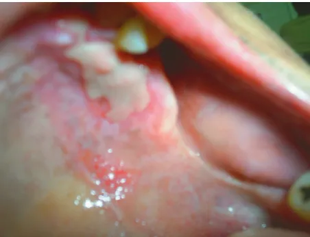
Control second (November 07, 2006): Pain sensation on the left facial skin and teeth-ache still persisted, as well as the left ear region although aulin tablet had been given. Left submandibular lymph nodes did not change, left facial skin revealed hyper-pigmentation. White plaque on the left palate was thinner and difficult to be scraped (Figure 4). Oral rinse and multivitamin plus Zn caplet were repeated and mycostatin oral suspention was added. Therefore, the patient referred to mycological laboratory and ENT (Ear Nose Throat) clinic.
Control third (November 14, 2006): Pain sensation on the left ear had gone, but still existed on the facial skin and teeth. Left submandibular lymph nodes were normal; hyperpigmention on the facial skin was thinner. White plaque on the left palate changed to be smaller and thinner, left upper teeth region were unstable grade three. Radiographic examination showed the resorbtion of alveolar bone. This data supported that the patient has diabetes mellitus.
Mycological finding showed positive Candida infection, therefore final diagnosis on the palatum was oral candidosis (thrush). The mycostatin oral suspension and chlorhexidine gluconate 0.2% oral rinse treatment was continued. In fact, the third control (four weeks later) showed that oral candidosis did not change; therefore the patient was given ketokonazole tablets three times daily. Then, the patient was referred to clinic of diabetes mellitus and neurology.
Control fourth (November 21, 2006): From neurology clinic, the patient was given amitriptilyn, carbamazepin, neurobion and pondex, that were regularly taken for reducing pain. Actually, pain sensation on the left facial skin and left upper teeth region were reduced, but the patient complained of xerostomia.
Hyperpigmentation on the left facial skin still existed although it was thinner. White plaque on left palate was smaller and slighter, on upper and lower buccal mucosa were painful. There was white plaque on dorsal tongue, thick and scrapable, surrounded by erythematous. Oral rinse was repeated and ketokonazole tablet was continued.
Control fifth (November 28, 2006): Laboratory result (November 23, 2006) showed that fasting blood glucose level was 109mg/dl and 2 hours postprandial was 162mg/dl, it proved that the patient suffered from diabetes mellitus. According to the anamnesis, 5 days before control the patient got spontaneous loss of two of the upper left teeth (canine and first premolar).
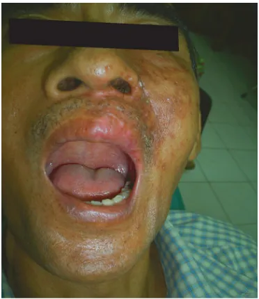
Extra oral and intra oral examination showed that hyperpigmentation of the left facial skin was thinner and sometime less pain sensation (Figure 5 and 6). The upper lip was dry, desquamative and fissured. Dry socket was found in the region of post spontaneous teeth loss. White plaque on
the left palate disappeared, but on the dorsum of the tongue still existed. Therefore, ketokonazole tablet was repeated, and vitamin C tablet as supporting treatment, mefenamic acid and benzidamine HCl oral rinse for reducing pain was added and alvogyl fibre was topically given in the dry socket region. The patient was referred to diabetes clinic for blood regulation controlling. If the blood regulation was normal, the left upper teeth which unstable grade three would be extracted.
Control sixth (December 05, 2006): Although the patient was given medicines for diabetes mellitus, the blood glucose level was still higher than normal, consequently the patient’s pain-labile-teeth could not be extracted, therefore the patient had to wait for normal condition. The
brown-macula presented on the left facial skin and there was no pain sensation, thinner white plaque was presented on the dorsum of the tongue. Ketokonazole tablets was continued, and the patient was given vitamin C and B complex tablets for supporting treatment. The patient suggested to control one month later.
Control seventh (January 9, 2007): A week before control, the patient got spontaneous loss of the front of the upper teeth (left second incisor). The brown macula on the left facial skin was thinner, and intra oral examination showed white plaque disappeared. The fasting blood glucose level was 78 mg/dl and 2 hours postprandial blood glucose level was 136mg/dl, it indicated the normal condition was reached. Then, the patient referred to oral surgery clinic for extracting of the front upper teeth which unstable grade three (the left upper central incisor and canine). Blood glucose level decreasing would improve the patient’s general condition.
diScuSSion
Base on the general condition, anamnesis and extra oral examination, the clinical diagnosis of this case was herpes zoster which involved trigeminal nerve. The appearance of herpes zoster is sufficiently distinctive that a clinical diagnosis is usually accurate.7 This case involved left facial skin and limited on the left lower lid caused lid edema and ectropion, involved left nose and upper lip. The present unilateral ulceration or skin lesions limited in area supplied by one division of the trigeminal nerve suggests a diagnosis of zoster.3
The maxillary division of the trigeminal nerve is entirely sensory, supplying the skin of the middle portion of the face, lower eyelids, side of the nose, upper lip, mucous membranes of the nasopharynx, maxillary sinus, soft palate, and teeth. Near its origin it branches off to form the middle meningeal nerve, which supplies the ipsilateral middle meningeal artery and branches to durameter. One of its terminal branches is the greater palatine nerve, which supplies the hard palate, a portion of the maxillary gingival, the uvula, and a portion of the soft palate. The other important branch is the superior alveolar nerve, which supplies the maxillary gingival, teeth, and mucous membranes of the cheek.8
If the maxillary or mandibulary division of the trigeminal nerve were involved, the patient may experience toothache-like pain for several days prior to the onset of more characteristic cutaneous lesions.7 The patient suffers from tooth-ache in the nerve region which is involved and frequently can not be distinguished with trigeminal zoster.3,6
On intra oral examination, erythemathous and multiple ulcers were found on maxillary left buccal-fold mucosa, left buccal and upper lip mucosa, uvula and left soft palate. On left hard palate there was white plaque, thick and soft as
figure 5. Post treatment: extra oral view.
creamy milk, irregular border and form, can be removed with rubbing and surrounded by erythemathous. Based on intra oral examination, trigeminal nerve which involved in this case was maxillary division.
Thrush as secondary infection was suspected on the left hard palate and upper lip mucosa. It occurred because the patient took antibiotic from private physician and there were multiple ulcers caused by maxillary herpes zoster infection.
Acyclovir 800 mg five times daily will inhibit the replication of varicella zoster virus by shortening the duration of viral shedding, halting the formation of new lesions more quickly, accelerating the rate of healing, and reducing the severity of acute pain. Attenuation to the severity of the acute infection and the neural damage will reduce the likelihood of post herpetic neuralgia.3,5The pain of post herpetic neuralgia can be reduced by a number of medications. Tricyclic antidepressant medications such as amitriptyline (Elavil), as well as anti-seizure medications such as gabapentin (Neurontin) and carbamazipine (Tegretol), have been used to relieve the pain associated with herpetic neuralgia. Finally, capsaicin cream (Zostrix), a derivative of hot chili peppers, can be used topically on the area after all the blisters have healed, to reduce the pain.9
Until the 13th days, the patient still suffered from severe pain on the left facial skin although a number of medications was given to reduce pain. It proved that, the patient suffered acute herpetic neuralgia. According to Johnson and Dworkin,5 acute herpetic neuralgia defines as pain within 30 days from rash onset.
At the second control, white plaque at he left palate was thinner but difficult to be scrapped, therefore chlorhexidine gluconate 2% (and multivitamin plus Zn caplet) were repeated and mycostatin oral suspension as antimycotic topical was added.
Intra oral examination at the third control revealed that left upper teeth region were unstable grade three. Radiographic examination showed resorbtion of alveolar bone. This data supported that the patient suffered from diabetes mellitus. Mycological finding showed positive Candida infection, thus final diagnosis on the palatum was oral candidosis (thrush). Antimycotic topical was continued and the patient was given antimycotic systemic (ketokonazole tablets three times daily).
Laboratory finding (November 23, 2006) showed that fasting blood glucose level was 109 mg/dl and 2 hours postprandial was 162 mg/dl, it is really proved that the patient suffered from diabetes mellitus. Diabetes mellitus on this patient is caused by poor condition, xerostomia, leading to unstable upper left-teeth on the third degree and followed by spontaneous loss of teeth. This disease also caused oral candidosis which was difficult to treat. Blood glucose level decreasing would improve the patient’s general condition.
Since the patient not only suffered from herpes zoster in the maxillary nerve but also oral candidosis and diabetes mellitus, he was classified as immunocompromised patient. Oral candidosis of immuno-compromised patients should be treated with systemic antifungals.10
The involvement of oral candidosis, herpes zoster virus infection and diabetes mellitus, made this case complicated and multidisciplinary approached is needed. It is concluded that the management of in this case, it oral candidosis are adequate antiviral, blood glucose level regulating and systemic antifungal therapy.
referenceS
1. Silverman S, Epstein JB. Oral fungal infection on Burket’s of oral medicine. diagnosis and treatment. 10th ed. Ontario: BC Decker Inc; 2003 p. 170–9.
2. Scully C. Candidosis, mucosal. Available at: eMedicine.com, Inc. Accessed January 24, 2002.
3. Gnan JW, Whitley RJ. Herpes Zoster. The New England Journal of Medicine 2002; 347(5): 340–6.
4. Field A, Longman L. Tyldesley’s oral medicine. 5th ed. New York: Oxford University Press; 2003. p. 42–43.
5. Johnson RW, Dworkin RH. Treatment of Herpes Zoster and post herpetic neuralgia. Br Med J 2003; 326:748–50.
6. Cawson RA, Odel EW. Oral pathology and oral medicine. 7th ed. Churchill Livingstone. London: Churchill Livingstone; 2005. p. 181-2, 185–90.
7. Lamey PJ, Lewis MAO. Oral medicine and practice: Viral infection. Br Dent J 1989;. 167:269–74.
8. Okeson JP. Orofacial pain. Chicago: Quintessence Publishing Co, Inc; 1996. p. 3–5.
9. Cunningham AL, Breuer J, Dwyer DE, Gronow DW, Helme RD, Litt JC, et al. The prevention and management of Herpes zoster. Clinical Update MJA 2008 February; 188 (3): 171–6.