EDITOR-IN-CHIEF
CHRISTINE RAINES
!
University of Essex, Colchester, United Kingdom
[email protected] — Photosynthesis — Carbon fixation — Transgenic plants — Molecular biology — Plant physiology
Webpage
ASSOCIATE EDITORS
KARL-JOSEF DIETZ
!
Bielefeld University, Bielefeld, Germany
[email protected] — Abiotic stress
— Antioxidant
— Enzyme activity and regulation — Gene regulation
— Metal toxicity — Organelle signalling — Photosynthesis
— Protein translation and assembly — Redox signalling
— Vacuole
Webpage
HOWARD GRIFFITHS
!
University of Cambridge, Cambridge, United Kingdom [email protected]
— Acclimation
— Crops and tropical epiphytes — Photoinhibition
— Photosynthesis and water use in trees
— Photosynthetic CO2
concentrating mechanisms in terrestrial and aquatic plants — Plant and soil water relations — Regulation of Crassulacean Acid
Metabolism — Stable isotopes
Webpage
journal of
experimental
botany
Editorial Board
journal of
experimental
botany
jxb.oxfordjournals.org
KEY TO EDITOR’S EXPERTISE
PHOTOSYNTHESIS AND METABOLISM
CROP MOLECULAR GENETICS
PLANT GROWTH AND DIFFERENTIATION
CHRIS HAWES
!
Oxford Brookes University, Oxford, United Kingdom
[email protected] — Plant cell biology — Endomembrane systems — Golgi
— Confocal and electron microscopy
Webpage
LARS HENNIG
Flowering Newsletter Editor
!
Swedish University of Agricultural Sciences, Uppsala, Sweden [email protected]
— Chromatin — Epigenetics
— Flower Development — Flowering time — Epigenomics
Webpage
GERHARD LEUBNER
!
Royal Holloway, London, United Kingdom
[email protected] — Seed, fruit and seedling biology — Abiotic stress responses (seeds/
seedlings)
— Crops, weeds, climate change, food security
— Molecular physiology of seed germination
— Seed biomechanics and water relations
— Seed and bud dormancy
— Plant hormones, natural products — Embryo growth mechanisms — Cell-wall hydrolyses and
apoplastic ROS — Evolution seed/fruit trait
Webpage 1 and 2
JOHN LUNN
!
Max Planck Institute of Molecular Plant Physiology, Potsdam, Germany
[email protected] — C3 photosynthesis
— C4 photosynthesis
— Carbohydrate metabolism — CO2 fixation
— Evolution of metabolism/enzymes — Protein purification
— Protein-protein interactions — Sugar signalling
— Sulphur metabolism
Webpage
JIM MURRAY
!
Cardiff School of Biosciences, Cardiff, United Kingdom [email protected] — Synthetic biology — Biotechnology — Cell cycle — Development
— Gene regulatory networks — Systems biology
Webpage
GRAHAM NOCTOR
!
Université de Paris-Sud,Orsay, France
[email protected] — Stress/oxidative stress — Antioxidants
— Leaf metabolism/metabolomics — Redox homeostasis
DON ORT
Darwin Reviews Editor
!
University of Illinois, Urbana, IL, United States
[email protected] — Photosynthesis — Global Change — FACE
— Environmental/abiotic Stress
Webpage
CRISTOBAL UAUY
!
John Innes Centre, Norwich, United Kingdom
[email protected] — Wheat
— Genomics — Yellow rust — Senescence — Grain development — Genetics
— QTL — Breeding
Webpage
JIANHUA ZHANG
!
Chinese University of Hong Kong, Hong Kong
[email protected] — Crop physiology — Water stress
— Water-saving crop production — ABA
— Drought tolerance
Webpage
HANDLING EDITORS
LISA AINSWORTH
!
University of Illinois, Urbana, IL, United States
[email protected] — Climate change — Photosynthesis — Ozone
— Source-sink balance — Plant molecular physiology
Webpage
TOM BEECKMAN
!
Ghent University, Ghent, Belgium [email protected] — Cell cycle
— Gene expression — Plant anatomy — Plant development — Plant morphogenesis — Root architecture — Root development
CHRISTINE BEVERIDGE
!
University of Queensland, Brisbane, Australia
[email protected] — Auxin
— Plant Development — Shoot branching — Strigolactone
Webpage
PETER BOZHKOV
!
Swedish University of Agricultural Sciences and Linnean Center for Plant Biology, Uppsala, Sweden [email protected]
— Cell death — Ageing — Proteases — Autophagy — Development — Catabolism
Webpage
TIM COLMER
!
University of Western Australia, Crawley, Australia
[email protected] — Aerenchyma and oxygen
transport — Anoxia tolerance — Flooding tolerance — Halophytes — Salinity tolerance — Submergence tolerance
Webpage
KATHERINE DENBY
!
University of Warwick, Coventry, United Kingdom
[email protected] — Plant disease resistance — Systems biology
— Gene regulatory networks — Environmental stress responses — Host-pathogen interactions
Webpage
IAN DODD
!
Lancaster University, Lancaster, United Kingdom
[email protected] — Water deficit
— Root-to-shoot signalling — Plant-growth promoting
rhizobacteria — Phytohormones — Stomatal physiology
Webpage
CHRISTINE FOYER
!
University of Leeds, Leeds, United Kingdom
— Redox regulation and signalling (ROS, antioxidants)
— Abiotic stress (drought, cold, high light)
— Photosynthesis — Respiration
— Carbon nitrogen interactions — Aphid resistance
Contents
FOCUS PAPERS: PHOTOSYNTHESIS IN VARIABLE ENVIRONMENTS
Preface
Johnson GN, Lawson, T, Murchie EH, Raines C. Photosynthesis in variable environments 2371
Review papers
Schöttler MA, Tóth SZ, Boulouis A and Kahlau S. Photosynthetic complex stoichiometry dynamics in higher plants: biogenesis,
function, and turnover of ATP synthase and the cytochrome b6 f complex 2373
Dietz K-J. Eficient high light acclimation involves rapid processes at multiple mechanistic levels 2401 Kaiser E, Morales A, Harbinson J, Kromdijk J, Heuvelink E and Marcelis LFM. Dynamic photosynthesis in different environmental
conditions 2415 Allahverdiyeva Y, Suorsa M, Tikkanen M and Aro E-M. Photoprotection of photosystems in luctuating light intensities 2427
Research paper
Retkute R, Smith-Unna SE, Smith RW, Burgess AJ, Jensen OE, Johnson GN, Preston SP and Murchie EH. Exploiting heterogeneous
environments: does photosynthetic acclimation optimize carbon gain in luctuating light? 2437
RESEARCH PAPERS
Sun H, Li J, Song W, Tao J, Huang S, Chen S, Hou M, Xu G and Zhang Y. Nitric oxide generated by nitrate reductase increases nitrogen
uptake capacity by inducing lateral root formation and inorganic nitrogen uptake under partial nitrate nutrition in rice 2449 Girondé A, Poret M, Etienne P, Trouverie J, Bouchereau A, Le Cahérec F, Leport L, Orsel M, Niogret M-F, Deleu C and Avice J-C.
A proiling approach of the natural variability of foliar N remobilization at the rosette stage gives clues to understand the limiting
processes involved in the low N use eficiency of winter oilseed rape 2461
O’Brien M, Kaplan-Levy RN, Quon T, Sappl PG and Smyth DR. PETAL LOSS, a trihelix transcription factor that represses
growth in Arabidopsis thaliana, binds the energy-sensing SnRK1 kinase AKIN10 2475 Onoda Y, Schieving F and Anten NPR. A novel method of measuring leaf epidermis and mesophyll stiffness shows the ubiquitous
nature of the sandwich structure of leaf laminas in broad-leaved angiosperm species 2487
Jost R, Pharmawati M, Lapis-Gaza HR, Rossig C, Berkowitz O, Lambers H and Finnegan PM. Differentiating phosphate-dependent and phosphate-independent systemic phosphate-starvation response networks in Arabidopsis thaliana through the application of
phosphite 2501 Li Q, Cao C, Zhang C, Zheng S, Wang Z, Wang L and Ren Z. The identiication of Cucumis sativus Glabrous 1 (CsGL1) required for
the formation of trichomes uncovers a novel function for the homeodomain-leucine zipper I gene 2515
Rasmann S, Chassin E, Bilat J, Glauser G. and Reymond P. Trade-off between constitutive and inducible resistance against
herbivores is only partially explained by gene expression and glucosinolate production 2527 Zhang Y, Wang Y, Taylor JL, Jiang Z, Zhang S, Mei F, Wu Y, Wu P and Ni J. Aequorin-based luminescence imaging reveals differential
calcium signalling responses to salt and reactive oxygen species in rice roots 2535
Riach AC, Perera MVL, Florance HV, Penield SD and Hill JK. Analysis of plant leaf metabolites reveals no common response to
insect herbivory by Pieris rapae in three related host-plant species 2547
Jin C, Fang C, Yuan H, Wang S, Wu Y, Liu X, Zhang Y and Luo J. Interaction between carbon metabolism and phosphate accumulation
is revealed by a mutation of a cellulose synthase-like protein, CSLF6 2557
Barbier F, Péron T, Lecerf M, Perez-Garcia M-D, Barrière Q, Rolčík J, Boutet-Mercey S, Citerne S, Lemoine R, Porcheron B, Roman H, Leduc N, Le Gourrierec J, Bertheloot J and Sakr S. Sucrose is an early modulator of the key hormonal mechanisms controlling bud
outgrowth in Rosa hybrida 2569
Walter S, Kahla A, Arunachalam C, Perochon A, Khan MR, Scoield SR and Doohan FM. A wheat ABC transporter contributes to
both grain formation and mycotoxin tolerance 2583
Chai G, Kong Y, Zhu M, Yu L, Qi G, Tang X, Wang Z, Cao Y, Yu C and Zhou G. Arabidopsis C3H14 and C3H15 have overlapping
roles in the regulation of secondary wall thickening and anther development 2595
Xu C, Liu Y, Li Y, Xu X, Xu C, Li X, Xiao J and Zhang Q. Differential expression of GS5 regulates grain size in rice 2611 Boyer JS. Turgor and the transport of CO2 and water across the cuticle (epidermis) of leaves 2625
Dong L, Cheng Y, Wu J, Cheng Q, Li W, Fan S, Jiang L, Xu Z, Kong F, Zhang D, Xu P and Zhang S. Overexpression of GmERF5, a
new member of the soybean EAR motif-containing ERF transcription factor, enhances resistance to Phytophthora sojae in soybean 2635 Chateigner-Boutin A-L, Suliman M, Bouchet B, Alvarado C, Lollier V, Rogniaux H, Guillon F and Larré C. Endomembrane proteomics
reveals putative enzymes involved in cell wall metabolism in wheat grain outer layers 2649
Porto DD, Bruneau M, Perini P, Anzanello R, Renou J-P, Santos HPD, Fialho FB and Revers LF. Transcription proiling of the chilling
requirement for bud break in apples: a putative role for FLC-like genes 2659
Rojas-González JA, Soto-Súarez M, García-Díaz Á, Romero-Puertas MC, Sandalio LM, Mérida Á, Thormählen I, Geigenberger P, Serrato AJ and Sahrawy M. Disruption of both chloroplastic and cytosolic FBPase genes results in a dwarf phenotype and
important starch and metabolite changes in Arabidopsis thaliana 2673
Carrie C, Venne AS, Zahedi RP and Soll J. Identiication of cleavage sites and substrate proteins for two mitochondrial intermediate
peptidases in Arabidopsis thaliana 2691
Zhang X, Wu Q, Cui S, Ren J, Qian W, Yang Y, He S, Chu J, Sun X, Yan C, Yu X and An C. Hijacking of the jasmonate pathway by the
mycotoxin fumonisin B1 (FB1) to initiate programmed cell death in Arabidopsis is modulated by RGLG3 and RGLG4 2709 Li J, Han Y, Liu L, Chen Y, Du Y, Zhang J, Sun H and Zhao Q. qRT9, a quantitative trait locus controlling root thickness and root
length in upland rice 2723
Rosas-Santiago P, Lagunas-Gómez D, Barkla BJ, Vera-Estrella R, Lalonde S, Jones A, Frommer WB, Zimmermannova O, Sychrová H
and Pantoja O. Identiication of rice cornichon as a possible cargo receptor for the Golgi-localized sodium transporter OsHKT1;3 2733 Wang B, Li G and Zhang W-H. Brassinosteroids are involved in Fe homeostasis in rice (Oryza sativa L.) 2749 Lu Y, Sasaki Y, Li X, Mori IC, Matsuura T, Hirayama T, Sato T and Yamaguchi J. ABI1 regulates carbon/nitrogen-nutrient signal
transduction independent of ABA biosynthesis and canonical ABA signalling pathways in Arabidopsis 2763 Deshpande GM, Ramakrishna K, Chongloi GL and Vijayraghavan U. Functions for rice RFL in vegetative axillary meristem
speciication and outgrowth 2773
Jahan SN, Åsman AKM, Corcoran P, Fogelqvist J, Vetukuri RR and Dixelius C. Plant-mediated gene silencing restricts growth of the
potato late blight pathogen Phytophthora infestans 2785
Piazza A, Zimaro T, Garavaglia BS, Ficarra FA, Thomas L, Marondedze C, Feil R, Lunn JE, Gehring C, Ottado J and Gottig N. The dual nature of trehalose in citrus canker disease: a virulence factor for Xanthomonas citri subsp. citri and a trigger for plant
defence responses 2795
Abu-Abied M, Rogovoy (Stelmakh) O, Mordehaev I, Grumberg M, Elbaum R, Wasteneys GO and Sadot E. Dissecting the contribution
of microtubule behaviour in adventitious root induction 2813
Journal of Experimental Botany, Vol. 66, No. 9 pp. 2501–2514, 2015 doi:10.1093/jxb/erv025 Advance Access publication 19 February 2015
This paper is available online free of all access charges (see http://jxb.oxfordjournals.org/open_access.html for further details)
RESEARCH PAPER
Differentiating dependent and
phosphate-independent systemic phosphate-starvation response
networks in
Arabidopsis thaliana
through the application of
phosphite
Ricarda Jost1,*, Made Pharmawati1,2, Hazel R. Lapis-Gaza1, Claudia Rossig1, Oliver Berkowitz1,3,†, Hans Lambers1,4 and Patrick M. Finnegan1,4
1 School of Plant Biology, The University of Western Australia, Crawley (Perth), Western Australia, Australia
2 Biology Department, Faculty of Mathematics and Natural Sciences, Bukit Jimbaran Campus, Udayana University, Bali, Indonesia 3 School of Veterinary and Life Sciences, Murdoch University, Murdoch, Western Australia, Australia
4 Institute of Agriculture, The University of Western Australia, Crawley (Perth), Western Australia, Australia
*To whom correspondence should be addressed. E-mail: [email protected]
† Present address: Australian Research Council Centre of Excellence in Plant Energy Biology, University of Western Australia, Crawley
(Perth), Western Australia, Australia.
Received 18 November 2014; Revised 28 December 2014; Accepted 9 January 2015
Abstract
Phosphite is a less oxidized form of phosphorus than phosphate. Phosphite is considered to be taken up by the plant through phosphate transporters. It can mimic phosphate to some extent, but it is not metabolized into organophos-phates. Phosphite could therefore interfere with phosphorus signalling networks. Typical physiological and transcriptional responses to low phosphate availability were investigated and the short-term kinetics of their reversion by phosphite, com-pared with phosphate, were determined in both roots and shoots of Arabidopsis thaliana. Phosphite treatment resulted in a
strong growth arrest. It mimicked phosphate in causing a reduction in leaf anthocyanins and in the expression of a subset of the phosphate-starvation-responsive genes. However, the kinetics of the response were slower than for phosphate, which may be due to discrimination against phosphite by phosphate transporters PHT1;8 and PHT1;9 causing delayed shoot accumulation of phosphite. Transcripts encoding PHT1;7, lipid-remodelling enzymes such as SQD2, and phospho-choline-producing NMT3 were highly responsive to phosphite, suggesting their regulation by a direct phosphate-sensing network. Genes encoding components associated with the ‘PHO regulon’ in plants, such as At4, IPS1, and PHO1;H1,
generally responded more slowly to phosphite than to phosphate, except for SPX1 in roots and MIR399d in shoots. Two
uncharacterized phosphate-responsive E3 ligase genes, PUB35 and C3HC4, were also highly phosphite responsive. These
results show that phosphite is a valuable tool to identify network components directly responsive to phosphate.
Key words: Arabidopsis thaliana, phosphate-starvation response, phosphate transport, phosphite, phosphonate, phosphorous acid, phosphorus signalling networks, PSR genes, transcriptional regulation.
Introduction
Phosphite (H2PO3–, Phi) is a less oxidized form of phospho-rus (P) than phosphate (H2PO4–, Pi). Phi is highly water solu-ble and less prone than Pi to adsorb to soil particles, which
makes it more accessible to plants (Ruthbaum and Baille, 1964). Phi competes with the essential macronutrient Pi for uptake by plants, most probably through both high- and
This is an Open Access article distributed under the terms of the Creative Commons Attribution License (http://creativecommons.org/licenses/by/3.0/), which permits unrestricted reuse, distribution, and reproduction in any medium, provided the original work is properly cited.
© The Author 2015. Published by Oxford University Press on behalf of the Society for Experimental Biology.
Abbreviations: P, phosphorus; Phi, phosphite; Pi, inorganic phosphorus/phosphate/H2PO4–, PSR, phosphate-starvation-responsive.
by guest on August 6, 2015
http://jxb.oxfordjournals.org/
2502 | Jost et al.
low-afinity transport systems (d’arcy-Lameta and Bompeix, 1991; Danova-Alt et al., 2008). Phi uptake is strongly and competitively inhibited in the presence of Pi (Pratt et al., 2009). Within the plant, Phi can be translocated, and it pref-erentially accumulates in sink tissues (Nartvaranant et al., 2004).
Phosphite was once abundant in the oceans, but it has been oxidized over time (Pasek et al., 2013). Many microbes have retained the ability to oxidize Phi to Pi, and even use it as a reducing agent, namely for sulphate reduction (Poehlein et al., 2013). Plants, however, are not able to metabolize Phi (McDonald et al., 2001). Instead, P-limited plants are highly sensitive to Phi and display toxicity symptoms such as leaf chlorosis and stunted growth (McDonald et al., 2001; Ratjen and Gerendas, 2009; Thao and Yamakawa, 2009). Other detrimental effects caused by Phi are the arrest of primary root growth, yellowing of the leaf lamina of young leaves, and a patchy accumulation of anthocyanins in older leaves (Varadarajan et al., 2002). Pratt et al. (2009) also showed that respiration rates declined upon Phi treatment of P-limited sycamore cells. It was recently found that the accumulation of Phi impacts on metabolism in Arabidopsis thaliana, leading to changes in the levels of several central metabolites (Berkowitz et al., 2013).
Phi also triggers broad-spectrum resistance against patho-gens with a (hemi)biotrophic lifestyle, such as oomycetes, fungi, and nematodes (Smillie et al., 1989; Hofgaard et al., 2010; Dias-Arieira et al., 2013; Percival and Banks, 2014). Phi has been suggested to act as a priming agent of plant defence responses in a number of plant–pathogen interactions (Machinandiarena et al., 2012; Massoud et al., 2012; Dalio et al., 2014). However, it is unclear how the primary recogni-tion of Phi takes place, and which molecular pathways are altered within the plant subsequently to induce this primed state of heightened defence. Given that Phi is transported by Pi transporters, these primary molecular interactions could trigger changes in signal perception (Schothorst et al., 2013).
Phi accumulates in both the cytosol and organelles, while the presence of Pi enhances Phi sequestration in the vacuole (Danova-Alt et al., 2008). This is probably why plants with an adequate P status can tolerate moderate Phi exposure with-out visible toxicity symptoms (Thao and Yamakawa, 2009). Conversely, Phi inhibits the eflux of Pi from the vacuole, which could exacerbate Pi-starvation symptoms (Pratt et al., 2009) and lead to accelerated plant death (Singh et al., 2003). Interestingly, the combined concentrations of Phi plus Pi within roots and shoots of A. thaliana were remarkably con-stant, regardless of their ratio in the growth medium, dem-onstrating that plants sense both Pi and Phi and adjust their uptake and allocation accordingly (Berkowitz et al., 2013).
Due to its physical similarity to Pi and non-metaboliz-able nature, Phi has been used as a tool to understand Pi -dependent signalling networks in plants. In several studies, Phi in fact seemed to mimic Pi effectively. Brassica nigra seedlings germinated on low-Pi media in the presence of high (1–10 mM) Phi concentrations had reduced activation of Pi -starvation-induced phosphoenolpyruvate phosphatase and pyrophosphate-dependent phosphofructokinase compared
with P-limited control plants (Carswell et al., 1996). While Phi did not affect the total adenylate pool in P-limited Brassica napus suspension cells in the same way as Pi, it did cause changes in the in vivo phosphorylation status of a number of proteins (Carswell et al., 1997). In A. thaliana, Ticconi et al. (2001) observed that Phi prevented the induction of tran-scripts from the Pi-starvation-responsive (PSR) genes ACP5,
At4, and PT2 upon 14 d exposure of P-suficient seedlings to a medium lacking Pi, but containing high concentrations of Phi. The same plants showed reduced in vitro activities of PSR ribonucleases RNS1 and RNS2 and of an acid phosphatase. Within 1 d of transfer of P-suficient A. thaliana seedlings to a medium lacking Pi, Phi suppressed the typical root hair formation and transcript accumulation of purple acid phos-phatase PAP1 and Pi transporters PT1 and PT2 that occur upon Pi withdrawal (Varadarajan et al., 2002). Exposure of A. thaliana to Phi prevented not only PSR MGD2 and
MGD3 expression, but also changes in glycerolipid proiles that accompany P-limited growth (Kobayashi et al., 2006). In P-limited tomato seedlings, Phi mimicked Pi in promot-ing proteolytic turnover of purple acid phosphatases (Bozzo et al., 2004). In rice, long-term exposure (5–7 d) to Phi sup-pressed the Pi-starvation-induced expression of OsIPS1 and
OsIPS2 (Hou et al., 2005). In tobacco BY-2 cells, Phi caused the reversion of autophagic protein turnover triggered by Pi deprivation (Tasaki et al., 2014).
The irst evidence suggesting that Phi and Pi have dis-crete effects on P signalling networks came from work by Stefanovic et al. (2007), who showed that transcripts of
PHO1 and its close paralogue PHO1;H1 differentially accumulated in plants treated with Pi or Phi. The PHR1-dependent induction of PHO1;H1 under P-limiting condi-tions was attenuated by Phi, while the PHR1-independent induction of PHO1 was not. This effect does not directly depend on the MYB transcription factor PHR1, because, unlike for PHO1;H1, the induction of another PHR1-regulated paralogue, PHO1;H10, was not affected by Phi (Ribot et al., 2008). Interestingly, both PHO1 and PHO1;H1
transcripts were less abundant in the P-limited pho2 mutant and more strongly induced in the P-limited pdr2 mutant compared with those in the wild type (Stefanovic et al., 2007). Disruption of the gene encoding endoplasmic retic-ulum (ER)-resident P5-type ATPase PDR2 affected local Pi-sensing networks and heightened the sensitivity and amplitude of metabolic responses to P limitation (Ticconi et al., 2004). The conditional pdr2 short-root phenotype was reversible by Phi. These observations strongly suggest that Phi mimics Pi in local signalling networks, irrespective of the plant’s P status.
Studies have so far addressed the question of whether Phi can prevent the long-term accumulation of PSR gene tran-scripts. In this study, the question of whether the shorter term kinetics of Phi suppression were similar to those of Pi was addressed (Müller et al., 2004; Morcuende et al., 2007). Organ-level accumulation of both Pi and Phi in P-limited seedlings in A. thaliana accession Col-0 and three PHT1 trans-porter mutants was therefore determined. Root growth and anthocyanin accumulation as well as gene expression proiles
by guest on August 6, 2015
http://jxb.oxfordjournals.org/
Short-term phosphite effects on phosphate signalling networks | 2503
in response to Phi treatment or Pi resupply were monitored in P-limited Col-0 seedlings over a time-course from 1 d to 7 d.
Materials and methods
Plant material and growth conditions
Seeds of A. thaliana (L.) Heynh. Col-0 and homozygous T-DNA
insertion lines for pht1;1–2 (SALK 088568C) (Shin et al., 2004),
pht1;8 (SALK 056529, Lapis-Gaza et al., 2014), and pht1;9-1 (SALK 050730) (Remy et al., 2012) were surface-sterilized for 2 min in 70% (v/v) ethanol and 5 min in 5% (v/v) NaOCl, before being rinsed ive times in sterile water. Seeds were resuspended in sterile 0.1% (w/v) agar and stratiied in the dark for 24–48 h at 4 °C. Seedlings (12 per plate) were grown vertically on 10 × 10 cm plates containing 50 ml of
nutrient solution [1 mM Ca(NO3)2, 2 mM KNO3, 0.5 mM MgSO4,
0.25 mM KH2PO4, 40 μM Fe-EDTA, 25 μM H3BO3, 2 μM MnCl2,
2 μM ZnSO4, 0.5 μM CuSO4, 0.075 μM (NH4)6Mo7O24, 0.15 μM
CoCl2, 50 μM KCl, pH 5.8] with 0.5% (w/v) 2-(N-morpholino)
ethanesulphonic acid and 1% (w/v) sucrose, and solidiied with 0.7% (w/v) agar (Plant TC Agar, cat.#A111, PhytoTechnology Laboratories, Shawnee Mission, KS, USA). Plates were sealed with 3M™ Micropore medical tape (Intouch Direct, Springwood, Australia). Seedlings were grown in a 10/14 h day/night cycle with
200 μmol m–2 s–1 photosynthetically active radiation (PAR) at
21 °C (day), 19 °C (night), and 65% relative humidity. The
plant-available Pi present in the agar added another 5 μM to the medium.
This amount is within the range of Pi concentrations across gelling
agents (Jain et al., 2009). Preliminary experiments showed that
con-centrations of Pi ranging from 250 μM to 1 mM do not limit
seed-ling growth in this system (data not shown). For the experiment,
seedlings grown on a medium with 250 μM Pi for 5 d were grown
for 4 d on plates without Pi supplementation (containing 250 μM
KCl instead) before being transferred to plates containing minimal Pi (5 μM residual Pi in agar), or equimolar concentrations (250 μM)
of either Pi or Phi. The Phi solution was prepared from a fresh batch
of phosphorous acid (99%, Sigma Aldrich, Castle Hill, Australia) as a ilter-sterilized 250 mM stock. The pH was adjusted to pH 5.8 with KOH. There was <0.1% oxidation of Phi to Pi in this solution during
1 month storage at 4 °C.
At harvest, the 12 seedlings on each plate were pooled into one sample. Roots were rinsed in MilliQ water for 5 min. Roots and shoots were blotted dry and shock-frozen in liquid nitrogen. Harvesting started 3 h after the beginning of the light period in syn-chrony with the experimental time-course to ensure that plants were at a comparable physiological state.
Root growth analysis and microscopy
After emergence of the radicle or transfer to a new plate, the posi-tion of the primary root tip was marked at 24 h intervals. Prior to transfer or harvest, the seedlings were scanned at 600 dpi resolution to determine root and root hair length, growth rate, and lateral root number (LSM Image Browser v4.2; Carl Zeiss Microscopy GmbH, Jena, Germany).
For microscopy (Axioplan Universal microscope; Carl Zeiss Microscopy GmbH), roots were mounted onto slides in water under glass cover slips. Images were electronically processed (AxioVision4; Carl Zeiss Microscopy GmbH).
Metabolite quantiication
Fifteen volumes of 1% (v/v) acetic acid were added to frozen plant powder (30–50 mg) and homogenized for three cycles of 45 s at 5000 rpm in the presence of two ceramic beads (ø 2 mm, Precellys 24 Tissue Disruptor; Bertin Technologies, Montigny-le-Bretonneux, France). After incubation for 15 min on ice, the homogenization process was repeated once. Cleared supernatants were used to
determine organ Pi concentrations via the reduction of a
phospho-molybdate complex by ascorbic acid (Ames, 1966). Phi
concentra-tions were determined using the same extracts in a high-throughput enzymatic luorescence assay (Berkowitz et al., 2011).
Anthocyanins in leaf samples were determined using a
pH-dif-ferential method (Wrolstad et al., 2005). Concentrations were
cal-culated using the molar absorptivity of cyanidin-3-glucoside (ε=26
900 l mol–1 cm–1), the predominant anthocyanin in A. thaliana leaves
(Tohge et al., 2005).
Relative quantiication of transcript abundance
mRNA was captured from tissue homogenates using oligo(dT)25
-coated magnetic beads (Dynabeads, Life Technologies Australia Pty Ltd, Mulgrave, Australia) and converted to cDNA as previously described (Jost et al., 2007). Aliquots of 0.5 ng of cDNA were
ampli-ied in a 10 μl reaction volume containing 0.3 μM of each primer
and PCR master mix (Power SYBR® Green, Applied Biosystems,
Scoresby, Australia). Quantitative PCR and threshold cycle (Ct)
determination were performed using a luorescence baseline setting of 0.3 (7500 FAST Real-Time PCR System, Applied Biosystems,
Scoresby, Australia). Data were normalized against PP2AA3
(for-merly PDF2) and UBC9 reference genes (Czechowski et al., 2005).
PCR eficiencies for each primer pair were determined using the
LinReg algorithm (Ruijter et al., 2009) (Supplementary Table S1
available at JXB online). Data were expressed either relative to
nor-malized Ct values in control samples (ΔΔCt) or as 40–ΔCt values that
correlate with the relative transcript expression of the gene of inter-est (Bari et al., 2006). The detection limit of the assay was calculated to be a 40–ΔCt value of 25.7 ± 0.1.
Statistical analysis
Statistically signiicant differences between treatments were
deter-mined using analysis of variance (ANOVA) and deined as P≤0.05
(SigmaStat v. 12.3, Systat Software Inc., San Jose, CA, USA). Two-way ANOVA followed by Tukey’s post-hoc test was used to separate means. Hierarchical clustering was performed using squared Euclidean distance and complete linkage [J-Express 2012, Norwegian Bioinformatics Platform and Norwegian Microarray
Consortium (http://www.molmine.com)] http://jexpress.bioinfo.no/
site/ (last accessed 27 January 2015) (Dysvik and Jonassen, 2001).
Results
Phosphite strongly reduced plant biomass production
A vertical growth system was used for A. thaliana Col-0 seed-lings that allowed a direct comparison of the effects of Phi versus Pi on the repression of Pi-starvation responses with-out the confounding effects of competition between Pi and Phi. Using this system, P-limited seedlings were subjected to continued Pi deprivation, Pi resupply, or Phi treatment. Plant biomass did not differ signiicantly among the treat-ments within the irst 2 d of transfer (Fig. 1). After 3 d of treatment, both the P-limited seedlings and those resupplied with Pi had greater root and shoot biomass than seedlings at days 1 and 2, while the biomass of the Phi-treated seedlings was unchanged. Over the next 4 d, seedlings resupplied with Pi recovered from P limitation with a proportional increase in both root and shoot biomass that maintained the root-to-shoot ratio at 0.30 ± 0.01. P-limited seedlings preferentially allocated resources to roots over shoots, leading to a inal root-to-shoot ratio of 0.47 ± 0.03. Despite the greater partitioning of biomass to roots, the root biomass after 7 d of further P
by guest on August 6, 2015
http://jxb.oxfordjournals.org/
2504 | Jost et al.
limitation was only 84% of that in the Pi-resupplied seedlings. The shoot biomass of the P-limited seedlings was only 53% of that of the Pi-resupplied seedlings. By contrast, the Phi treat-ment slowed seedling growth much more severely. After 7 d exposure to Phi, the inal root and shoot biomass of seedlings was only 26% and 38%, respectively, of that in Pi-resupplied seedlings. While there was a 55% increase in shoot biomass over time, the root biomass of Phi-treated seedlings did not change. Since this severe inhibition of root growth contrasted with both P-limited and P-suficient seedling growth, the kinetics of root elongation were examined in more detail.
Phosphite strongly inhibited primary root elongation
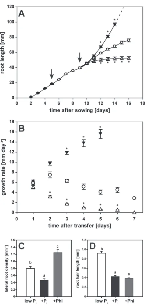
Seedlings germinated on high-Pi media showed steady primary root growth from 2 d after sowing (Fig. 2A). Root growth was initially maintained when the seedlings were transferred to a Pi-deicient medium 5 d after sowing. Imposing Pi resupply or Phi treatments after 4 d of Pi withdrawal did not affect root elongation during the irst day (Fig. 2A). Two days after the transfer to the inal medium (day 11), roots of both P-limited and Pi-resupplied seedlings grew at similar rates (Fig. 2B). In contrast, primary roots of Phi-treated seedlings showed much lower growth rates during this period, and elongation ceased completely within the next 48 h. Root growth in Pi-resupplied seedlings accelerated exponentially during this same time period (Fig. 2B), with roots reaching the bottom of the 10-cm plate by 6 d after imposing the treatment. Root growth in P-limited seed-lings decelerated by 2%, resulting in a inal total root length that was almost 30% shorter than in Pi-resupplied seedlings. These results show that, unlike Pi resupply, Phi treatment accentuated the reduction in root growth caused by Pi depletion.
Phosphite altered seedling root architecture
At the end of the time-course experiment, high-resolution scans of primary root segments initiated on day 3 after the inal transfer were used to analyse the effects of the three treat-ments on root development (Fig. 2C, D). The chosen root seg-ment was proximal to the root apex, at the beginning of the root branching zone (Dubrovsky and Forde, 2012). The short-root phenotype caused by Phi resulted in an almost 2-fold greater lateral root density than in P-limited seedlings in this newly formed section of the root (Fig. 2C). Remarkably, the number of lateral roots per segment in Phi-treated seedlings (2.3 ± 0.2) was 2-fold lower than that in Pi-limited (4.7 ± 0.3) and Pi-resupplied (5.0 ± 0.5) segments. Primary root growth in Pi-resupplied seedlings decreased lateral root density by nearly 2-fold compared with P-limited seedlings. While lateral roots elongated similarly under both Pi limitation and Pi resupply, emergence of lateral roots was inhibited in the presence of Phi. This phenomenon was also observed in a hydroponics growth system, where transfer to different nutrient solutions is less damaging to roots (Supplementary Fig. S1 at JXB online; note that in order to compensate for slower uptake of Phi over Pi, 1 mM Phi was used in this experiment).
Root hairs of Phi-treated seedlings were 57% shorter than in P-limited seedlings (Fig. 2D). This shortening was similar to the 52% reduction observed for seedlings resupplied with Pi. A concomitant reduction in root hair density by 38% for Phi-treated seedlings (14 ± 1 mm–1) compared with P-limited seedlings (22 ± 1 mm–1) was also very similar to the 44% reduction observed in Pi-resupplied seedlings (13 ± 1 mm–1). Hence this local response to Pi resulting in fewer and shorter root hairs appears to be mimicked by Phi.
Anthocyanin accumulation in P-limited seedlings was repressed by both Pi and Phi
Anthocyanins accumulated to signiicant levels in leaves of seedlings after a total of 11 d of growth on minimal Pi media (4 d Pi withdrawal+7 d treatment; Fig. 3). This slow accumu-lation indicates that the seedlings were not highly stressed by the Pi deprivation imposed during the early stage of the experi-ment, and were probably accessing and gradually depleting P reserves that accumulated during the initial 5-d growth on Pi -containing medium. Seedlings resupplied with Pi after a star-vation period of 4 d had lower levels of anthocyanins within 2 d of treatment. Phi-treated seedlings also had reduced leaf anthocyanin levels within the irst 2 d of treatment, but not as low as in Pi-resupplied seedlings. In Phi-treated seedlings, the leaf anthocyanin concentration was higher at day 7 than in Pi -supplied seedlings, but was 72% lower than in P-limited seed-lings. Therefore, Phi attenuated anthocyanin accumulation in P-limited plants that was completely suppressed by Pi resupply.
Root-to-shoot transport favoured Pi over Phi
To appreciate fully the differences in the physiological and molecular responses to Phi compared with Pi, the accumula-tion of both anions in roots and shoots was determined over time. While roots accumulated both Pi and Phi equally within
Fig. 1. Accumulation of root and shoot biomass. Seeds were germinated on media containing 250 μM phosphate (Pi) on vertical plates as described
in the Materials and methods. Five-day-old seedlings were transferred to a low-Pi medium for 4 d before being transferred to plates containing
minimal Pi (5 μM, white bars), high Pi (250 μM, black bars), or phosphite
(250 μM, grey bars) media. Root and shoot biomass was determined at 1, 2, 3, and 7 d after transfer (mean ±SE, n=4 replicates with 12 seedlings each). Statistically signiicant differences between time points as determined by Tukey’s HSD for each treatment at P<0.001 are indicated by an asterisk. Differences between treatments were signiicant for both organs only at 7 d after transfer (P<0.001).
by guest on August 6, 2015
http://jxb.oxfordjournals.org/
Short-term phosphite effects on phosphate signalling networks | 2505
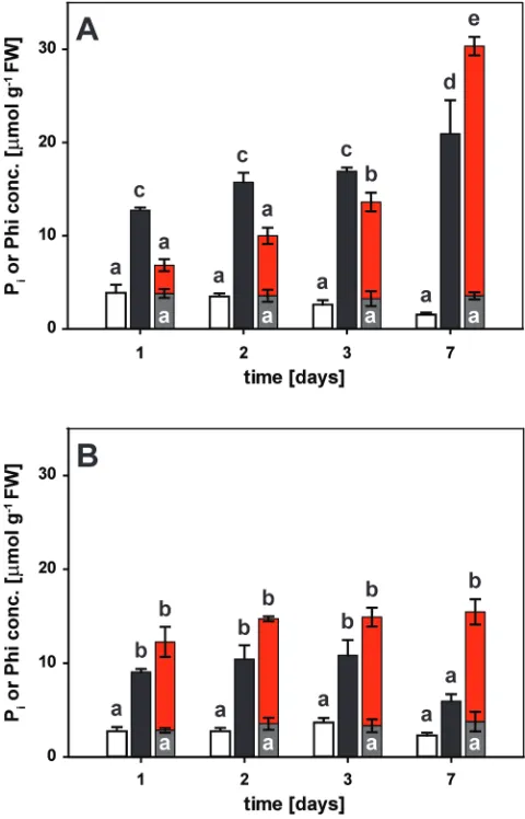
1 d of exposure, there was a delay in the accumulation of Phi relative to that of Pi in the shoot (Fig. 4). Shoot Pi concentra-tions reached ~13 μmol g–1 fresh weight (FW) within 1 d of resupply (Fig. 4A) which was greater than the level of free Pi in seedlings continuously grown on a suficient Pi supply (~5 μmol g–1 FW). The shoot P
i concentration nearly doubled over the next 6 d to a inal concentration of ~21 μmol g–1 FW. In roots, Pi levels increased to 9 μmol g–1 FW within 1 d of resupply, matching the Pi concentration found in roots of seedlings continuously receiving Pi (~10 μmol g–1 FW). Pi concentrations remained at this level for several days, before dropping to 6 μmol g–1 FW by day 7 (Fig. 4B). The drop in Pi was probably due to a combination of depletion from the medium, continued export to the shoot, conversion to organic P compounds, and internal dilution by root growth. In roots of Phi-treated seedlings, Phi accumulated to similar levels as Pi within 1 d, and remained high for the 7 d of the experiment, with a inal concentration of 12 μmol g–1 FW. In shoots of Phi-treated seedlings, Phi concentrations were lower than the Pi concentrations in Pi-resupplied seedlings at the two earliest time points (3 μmol g–1 FW; Fig. 4A). After 3 d, the shoot Phi concentration of 10 μmol g–1 FW caught up with the shoot Pi concentration found after only 1 d of Pi resupply. At the inal harvest, the shoot Phi concentration of 27 μmol g–1 FW in Phi-treated seedlings was higher than that of the free Pi concentration in resupplied seedlings, probably due to metabolic conversion of Pi but not Phi into organic compounds. In shoots of P-limited seedlings, the Pi concen-tration tended to decline over the course of the experiment to a inal concentration of 1.5 μmol g–1 FW. The P
i concentra-tion in roots of P-limited plants (2 μmol g–1 FW) was con-stant over the time-course. In roots and shoots of Phi-treated seedlings, the Pi concentration (3 μmol and 4 μmol Pi g–1 FW, respectively) was constant over time, and Pi concentrations at
Fig. 2. Changes in root architecture in response to phosphate (Pi)
resupply and phosphite (Phi) treatment. (A) Primary root growth over the course of the experiment. Seedlings were germinated on media containing 250 μM Pi (illed circles). After 5 d, they were transferred to media
containing minimal Pi (5 μM, open circles) before being transferred to
plates with minimal Pi (5 μM, open circles), high Pi (250 μM, illed triangles),
or Phi (250 μM, open triangles). Arrows indicate transfer to new plates. (B) Root growth rates in response to treatments. Symbols are the same as in (A). Shown in (A, B) are means ±SE, n=16 (four seedling roots each were measured individually from four separate plates). (C) Lateral root density in seedlings harvested 7 d after transfer to minimal Pi (white bars), high
Pi (black bars), or 250 μM Phi (grey bars). Emerging lateral roots were
counted in root segments that were formed 3 d after transfer. Shown are means ±SE, n=10 (ive seedlings each from two plates). (D) Root hair length of seedlings harvested 7 d after transfer to minimal Pi (white
bars), high Pi (black bars), or 250 μM Phi (grey bars). Shown are means
±SE, n=30 (3 root hairs×5 seedlings×2 plates). Statistically signiicant differences across time points in (A, B) were determined by Tukey’s HSD for treatments relative to Pi-limited seedlings at P<0.001. In (C) and (D),
pairwise multiple comparisons between treatments identiied statistically signiicant differences at P<0.005.
Fig. 3. Anthocyanin accumulation in leaves of phosphorus-limited seedlings. Five-day-old seedlings were grown on low-phosphate (Pi)
medium for 4 d before being transferred to plates with minimal Pi- (5 μM,
white bars), high Pi- (250 μM, black bars), or phosphite- (250 μM, grey
bars) containing media. Leaf anthocyanin concentrations were determined at day 1, 2, 3, and 7 after transfer (mean ±SE, n=3 replicates with 12 seedlings each). Statistically signiicant differences between time points and treatments were determined by Tukey’s HSD at P<0.001. Differences within the low-Pi series and between the low-Pi and the other two
treatments were signiicant only at 7 d after transfer (P<0.001).
by guest on August 6, 2015
http://jxb.oxfordjournals.org/
2506 | Jost et al.
the inal harvest were higher than those in P-limited organs, most probably due to Phi-induced Pi retention in the vacuole (Pratt et al., 2009).
Phosphite tissue accumulation was differentially affected among a set of pht1 mutants
To gather direct evidence that Phi is transported by Pi trans-porters of the PHT1 family, the Phi accumulation in roots and shoots of homozygous T-DNA insertion lines was ana-lysed in the Col-0 background lacking either PHT1;1, one of the major Pi transporters at the root–soil interface (Shin et al., 2004), PHT1;8, or PHT1;9. The latter two PHT1 trans-porters are involved in translocation of Pi to the shoot ( Lapis-Gaza et al., 2014). Seedlings were grown on vertical plates and depleted of Pi as described above, before supplying them with either 250 μM Pi or 250 μM Phi for 24 h prior to harvest.
Pi starvation led to similar residual organ Pi concentrations across genotypes (Fig. 5). Compared with the corresponding wild-type Col-0, the pht1;1–2 mutant accumulated 58% less Pi in roots and 22% less Pi in shoots of Pi-resupplied seed-lings over the 24-h period (Fig. 5). The effect of this mutation on Phi uptake by P-limited seedlings was signiicantly more pronounced, leading to 71% less Phi in roots and 84% less Phi in shoots of the mutant than in the wild type. Knocking out PHT1;8 or PHT1;9 had no effect on either root or shoot Pi accumulation. In contrast to pht1;1–2, Phi concentrations in roots of both pht1;8 and pht1;9-1 were similar to those in the wild type, but Phi accumulation in shoots was reduced by 76% for pht1;8 and by 60% for pht1;9-1 compared with Col-0, the same extent as seen in pht1;1–2. The basal organ Pi concentrations in Phi-treated seedlings were similar across mutants. The same trends in organ Pi and Phi concentrations were observed after 2 d of treatment, although differences between Col-0 and the three mutants were diminished by day 7 (Supplementary Fig. S2 at JXB online). Throughout the time-course, root and shoot biomass accumulation was
Fig. 5. Phosphate (Pi) and phosphite (Phi) accumulation in P-limited
Col-0 and pht1 mutant organs after 1 d of Pi resupply or Phi treatment.
Five-day-old seedlings were depleted of Pi for 4 d before being treated
as indicated. (A) Shoot and (B) root accumulation of Pi in P-limited (white
bars), Pi-resupplied (black bars), and Phi-treated (grey bars) seedlings and
accumulation of Phi (red bars). Shown are means ±SE, n=3 replicates with 12 seedlings each. Genotypes and treatments with a letter in common are not signiicantly different according to Tukey’s HSD at P<0.05.
Fig. 4. Kinetics of phosphate (Pi) and phosphite (Phi) accumulation in
seedling organs. Five-day-old seedlings were depleted of Pi for 4 d before
being transferred to plates for the different treatments as indicated. (A) Shoot and (B) root accumulation of Pi in phosphorus-limited seedlings
(white bars), upon Pi resupply (black bars), or with Phi treatment (grey
bars). Phi accumulation in Phi-treated seedlings is shown as red bars. Shown are means ±SE, n=3 or n=4 replicates with 12 seedlings each. Statistically signiicant differences between time and treatments are indicated by different letters according to Tukey’s HSD at P<0.001.
by guest on August 6, 2015
http://jxb.oxfordjournals.org/
Short-term phosphite effects on phosphate signalling networks | 2507
largely unaffected by the lack of individual PHT1 proteins (Supplementary Fig. S3).
Phosphite altered transcript accumulation for a subset of Pi-responsive genes
The short-term effect of Phi on PSR gene expression was assessed by quantitative reverse-transcriptase PCR (qRT-PCR) for a set of well-documented PSR genes representing various metabolic and regulatory steps within plant P sig-nalling networks (Hammond et al., 2003; Wu et al., 2003; Misson et al., 2005; Morcuende et al., 2007; Woo et al., 2012). If Phi was a true Pi analogue and sensed in the same way as Pi by as yet unidentiied cellular signalling components, one would expect the effect of the two chemicals on transcript proiles to be similar; that is, lower transcript levels for Pi -starvation-induced genes and higher transcript abundance for genes involved in organophosphate biosynthesis or encoding negative regulators such as the E2 ubiquitin conjugase PHO2 (Aung et al., 2006; Bari et al., 2006) or the F-box protein FBX2 and transcription factor BHLH32 (Chen et al., 2008).
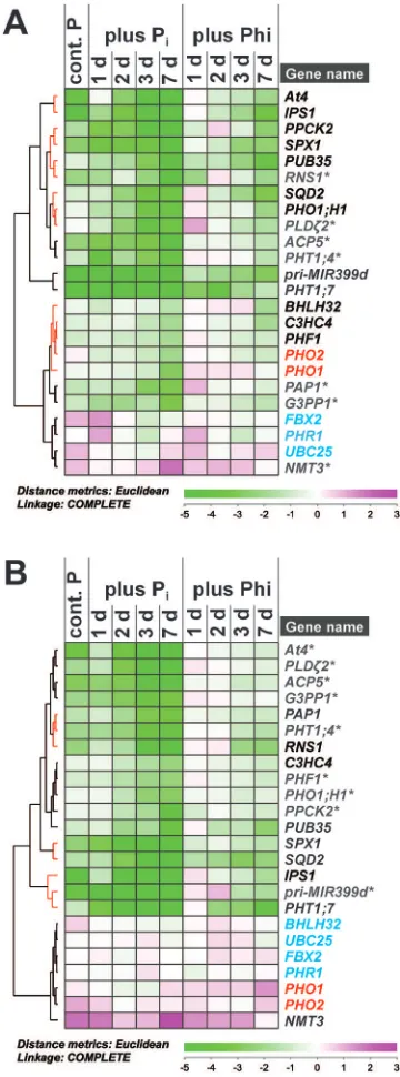
The selected PSR genes showed the previously docu-mented expression changes within 1 d of Pi resupply (Fig. 6). Surprisingly, 33% of the target genes showed no signiicant change in transcript abundance in response to Phi in shoots of P-limited plants over the 7-d treatment period (Fig. 6A, grey transcript names). Within this non-responsive group were the Pi transporter gene PHT1;4, as well as genes involved in Pi metabolism (ACP5, G3PP1, NMT3, PAP1, PLD ζ2, and
RNS1). In shoots, the majority of PSR genes tested showed an attenuated response to Phi treatment with a 1 d or 2 d delay compared with Pi resupply. This set included genes encoding regulatory components such as At4, IPS1, PHO1;H1, and SPX1, as well as genes encoding protein kinase PPCK2 and sulpholipid synthase SQD2 (Fig. 6A, red clusters). In con-trast, other genes responded strongly to Phi, as they did to Pi resupply. These responses included an 8-fold suppression within 24 h of Phi treatment for PHT1;7 transcript amounts, with a further 16-fold drop within 2 d of treatment. Similarly, transcripts encoding U-box-containing E3 ligase PUB35 were less abundant in shoots within 24 h of Phi treatment. A milder suppression compared with Pi was observed for the primary transcript of regulatory microRNA miR399d. Transcripts encoding transcription factor BHLH32, E3 ubiquitin ligase C3HC4, and transport facilitator PHF1 responded more slowly but similarly to both Pi resupply and Phi treatment, with a >4-fold lower abundance than in shoots of P-limited seedlings at the end of the experiment. PHO2
transcripts showed an unexpected proile in shoots, with 2- to 4-fold lower levels in Pi-resupplied over P-limited seedlings. Phi treatment triggered a similar 2-fold decline in PHO2 tran-scripts within 3 d of treatment.
Despite the fact that Phi accumulated as quickly as Pi in roots, 43% of the tested P-responsive transcripts did not respond to Phi in this organ (Fig. 6B, grey transcript names). Transcripts from ACP5, G3PP1, PLDζ2, and PHT1;4 were among those that were also identiied as being non-respon-sive to Phi in shoots. In roots, Phi-non-responnon-respon-sive transcripts
included those from At4, PHO1;H1, PHF1, and PPCK2, all of which responded to Phi to some extent in shoots. On the other hand, transcripts encoding phosphatase PAP1 and
Fig. 6. Effect of phosphate (Pi) and phosphite (Phi) on transcript
abundance in P-limited seedlings. Hierarchical cluster analysis of a time-course on relative transcript abundance in P-limited Arabidopsis thaliana
(A) shoots and (B) roots in response to Pi resupply or Phi treatment.
Mean log2 expression ratios (–ΔΔCt) relative to the normalized expression
in P-limited plants with three biological replicates for each sample are shown. Raw data were normalized against the transcript abundance of
PP2AA3 and UBC9 reference genes. Clusters that contain Phi-responsive transcripts are highlighted by red lines in the tree. Transcripts in black change abundance in response to both Pi and Phi treatment, while those in grey (*) are unresponsive to Phi treatment (P≤0.05). PHO1 and PHO2
transcripts are highlighted in red. Transcripts in blue show no signiicant change in abundance across treatments. Details on individual transcript expression patterns and statistical analysis can be found in Supplementary Table S2 at JXB online.
by guest on August 6, 2015
http://jxb.oxfordjournals.org/
2508 | Jost et al.
ribonuclease RNS1 were more responsive to Phi in roots compared with shoots. As in shoots, NMT3 transcript abun-dance in roots increased 2-fold in response to Phi within 24 h of treatment, but transcript levels did not continue to increase and were 11-fold lower compared with roots of Pi -resupplied plants on day 7 (Supplementaty Table S2 at JXB
online). PHT1;7, PUB35, SPX1, and SQD2 were highly Phi responsive in roots as well as in shoots. However, the response was relatively delayed in roots for PHT1;7 and PUB35, while
SPX1 and SQD2 transcripts were more quickly suppressed in roots than in shoots. C3HC4, IPS1, and pri-MIR399d tran-script abundance showed a weaker response to Phi in roots compared with shoots. In contrast to shoots, PHO1 transcript abundance did not respond to Pi resupply in roots. Curiously, within 48 h of Phi treatment, PHO1 transcript abundance was ~2-fold greater than that in roots of P-limited plants and continued to increase throughout the time-course. PHO2
transcript abundance in roots did not respond to either Pi or Phi treatment.
It has to be noted that seedlings were not severely P starved at the beginning of the experiment. Evidence for this was the small changes in transcript abundance in organs of P-limited control plants at day 1 of the experiment compared with transcript levels in plants continuously supplied with Pi (blue bar in Supplementary Table S2 at JXB online). As a conse-quence, transcript levels of the target Pi-starvation-induced genes continued to increase over the time-course in P-limited control plants. This was also the case for those transcripts that did not show a response to Phi in Phi-treated seedlings.
In contrast to the gradual response to Pi deprivation, Pi resupply led to the suppression of Pi-starvation-induced genes within 24 h (Supplementary Table S2 at JXB online). Thereafter, transcript abundance remained at the newly estab-lished lower levels for the rest of the time-course. Exceptions to this expression proile were those of microRNA antago-nists IPS1 and At4, which showed a more gradual response to Pi resupply in both roots and shoots. In shoots, PHT1;4 transcripts also showed this gradual decrease in abundance in response to Pi. Unlike all other target genes, transcripts from both IPS1 and PHT1;4 decreased in abundance to below the level observed in shoots of seedlings that were continuously supplied with Pi. In roots, PHO2 transcript levels tended to increase transiently within 24 h of Pi resupply, rather than showing a sustained increase over P-limited plants. PHO2
transcripts did not respond to Pi resupply in shoots.
Discussion
Phi has been demonstrated to suppress the induction of Pi -starvation responses. This conclusion was drawn from a series of experiments where P-suficient plants were transferred to Pi-containing or Pi-free media supplemented with increasing Phi concentrations, or where seeds were germinated on these media (Carswell et al., 1996; Ticconi et al., 2001; Varadarajan et al., 2002; Berkowitz et al., 2013; Eshraghi et al., 2014). Thus, these studies focused on the ability of Phi to interfere with the induction of PSR genes in response to Pi removal or the lack of Pi supply. The experimental set-up used in this
study allowed direct comparison of Phi and Pi effects on the suppression of Pi-starvation responses through monitoring plant growth, Pi anion and anthocyanin accumulation, as well as PSR gene expression. The experimental set-up has several advantages. (i) Withdrawal of Pi from the medium prior to Phi treatment avoids competition between the two anions for uptake. (ii) A direct comparison of Phi and Pi effects on the suppression of PSR genes can be conducted. (iii) Phi accu-mulation in the cytosol and organelles should be favoured over the vacuole under these conditions, so that more direct effects on metabolism and gene regulatory networks can be observed. (iv) The kinetic dependences of these effects on the accumulation of both P anions in roots and shoots can be determined.
Discrimination between Pi and Phi by PHT1 transporters
The differential movement of Phi and Pi into the shoots of plants suggests different afinities for these molecules within their transport routes. Measurements of transport kinet-ics in different systems have concluded that Pi transporters are able to transport Phi, albeit with a lower afinity than for Pi (d’arcy-Lameta and Bompeix, 1991; Pratt et al., 2004; Danova-Alt et al., 2008; Basheer et al., 2011). This means that Phi can bind to Pi transporter proteins without induc-ing the same conformational changes necessary for eficient transport (Basheer et al., 2011). It is unknown if all plant PHT transporters interact with Phi with the same afinity or whether some discriminate more strongly against Phi. In this study, the more pronounced delay in root-to-shoot trans-port of Phi in the pht1;8 and pht1;9-1 mutants than in wild-type seedlings, without a delay in Phi uptake, suggests that the encoded transporters discriminate more strongly against Phi than PHT1;1. The fact that discrimination is stronger in the absence of either PHT1;8 or PHT1;9 could mean that the two only partially complement each other (Lapis-Gaza et al., 2014) which would slow down transport even further. Alternatively, a third transport process, perhaps involving the Pi exporter PHO1 (Arpat et al., 2012), could be implicated in the stronger discrimination between Pi and Phi in both mutants. The alleviation of the Phi discrimination phenotype over time is most probably due to remobilization processes between sink and source organs involving other PHT trans-porters, such as PHT1;5 (Nagarajan et al., 2011).
Differential recognition of Phi by different PHT proteins may modulate not only transport activity, but also signalling events associated with this activity (Schothorst et al., 2013). It is unclear whether such a ‘transceptor’ function applies to the plant PHT family, but complex post-translational regu-lation has already been shown. Bayle et al. (2011) showed that some high-afinity PHT1 proteins undergo complex post-translational modiications, including protein phos-phorylation. PHT1 protein abundance is also controlled by ubiquitin-mediated protein degradation (Lin et al., 2013; Park et al., 2014). Both PHT1;8 and PHT1;9 proteins can be distinguished from other family members by the presence of a PEST [proline, glutamic acid (E), serine, threonine] domain
by guest on August 6, 2015
http://jxb.oxfordjournals.org/
Short-term phosphite effects on phosphate signalling networks | 2509
that mediates phosphorylation-dependent protein degrada-tion in many systems (Rechsteiner and Rogers, 1996), for example the high- and low-afinity Pi transporters in yeast (Lagerstedt et al., 2004; Estrella et al., 2008).
Differential expression of ‘PHO regulon’ genes in response to local Pi signalling in roots and shoots
There is mounting evidence that the local and systemic con-trol of PSR gene expression is governed by different signal-ling circuits in roots and shoots, and that different circuits within each organ respond either to the direct perception of Pi or to a more indirect process involving downstream metab-olites or other as yet unidentiied signals (Müller et al., 2004; Bari et al., 2006; Thibaud et al., 2010; Woo et al., 2012; Rojas-Triana et al., 2013). The discrimination between Pi and Phi by PHT1;8 and PHT1;9 shown in this study leads to a delayed accumulation of Phi in shoots. This delayed accumulation of Phi may hence be an elegant tool for dissecting direct sensing of Pi from other potential signals of P status in the shoot.
PHT1;7 and pri-MIR399d transcripts in the present study were suppressed earlier in shoots than in roots and responded before Phi accumulated to signiicant levels. This would place them into an early-response circuit more directly connected to a Pi-speciic sensor in the root-to-shoot transport route.
PHT1;7 and pri-MIR399d expression was deregulated in the pht1;9-1 (Lapis-Gaza et al., 2014) and the phr1 mutant, but not the pho2 mutant (Bari et al., 2006). Slower shoot accumulation of Phi correlated with an attenuated down-regulation of a select subset of PSR genes closely associated with the ‘PHO regulon’, such as At4, IPS1, SPX1, PHF1, and PHO1;H1. This would support their response to local Pi or, in this case, Phi availability in the shoot. Interestingly, these genes were also deregulated in both P-limited phr1 and Pi-resupplied pho2 mutants (Bari et al., 2006). These ind-ings may indicate that early Pi- and Phi-responsive genes are more directly connected to PHR1, possibly through a SIZ1-co-ordinated network in roots (Miura et al., 2005) and an unknown signalling component in shoots (Fig. 7) (Klecker et al., 2014). Only very few locally responsive PSR genes in the shoot seem to be PHO2 dependent (Pant et al., 2015). In a split-root system, genes that were systemically regulated in roots showed a strong enrichment of the P1BS element for PHR1 binding in their promoter regions (Thibaud et al., 2010). This may indicate differences in signal perception between roots and shoots. A clear distinction of regulatory groups of genes according to their responsiveness to Pi and Phi in space and time would therefore be useful to deine indi-vidual response circuits further.
PHO1 transcripts encoding a Golgi-localized Pi exporter (Arpat et al., 2012) showed a contrasting expression proile in roots to that of the other PSR genes tested in this study: instead of being suppressed by either Pi or Phi addition, they were more abundant in roots of Phi-treated compared with P-limited plants and did not respond to Pi resupply. In shoots, Pi resupply caused the down-regulation of PHO1, while Phi treatment caused a transient increase in PHO1 transcript lev-els similar to its effect in roots. PHO1 is therefore the only
PSR gene tested that responded to the more severe depletion of local cytosolic Pi pools that is expected in the presence of Phi (Pratt et al., 2009). Alternatively, PHO1 expression may be triggered by the strong inhibition of seedling growth in the presence of Phi. In this context, it is interesting to note that shoot growth in transgenic lines with reduced PHO1
expression is uncoupled from the actual P status of the shoot (Rouached et al., 2011). PHO1-associated signalling compo-nents could therefore integrate growth stimuli and P status.
PHO1;H1 transcript accumulation was suppressed by Pi in both roots and shoots, with a strong suppression by Phi in shoots. These results conirm the indings of Stefanovic et al. (2007) showing that PHR1-dependent PHO1;H1 expression is Phi responsive, while PHR1-independent PHO1 expression is not. PHO1 and PHO1;H1 are SPX (SYG1, Pho81, and XPR1) domain proteins (Secco et al., 2012). Transcripts encoding another SPX domain protein, SPX1, responded to Phi in both roots and shoots. SPX1 is a competitive inhibitor of PHR1 binding to the P1BS element in PSR gene promoters (Puga et al., 2014). Its interaction with PHR1 is also highly depend-ent on the presence of either Pi or Phi. In contrast to most PSR genes in the present study, it responded much more quickly to Phi in roots. Both PHO1;H1 and SPX1 are regulated in a PHR1- and PHO2-dependent manner (Bari et al., 2006), but
SPX1 is also controlled by SIZ1 (Duan et al., 2008). The lat-ter may explain its more direct response to local Phi concen-trations in the root (Miura et al., 2011). This would put SIZ1 into a position close to the local Pi- and Phi-sensing module in roots (Fig. 7). Surprisingly, SPX1 is also systemically regulated in P-limited roots in a split-root system (Thibaud et al., 2010).
PHO2 transcripts encoding an E2 ubiquitin conjugase (Aung et al., 2006; Bari et al., 2006) accumulated transiently in Pi-resupplied roots, but were largely unresponsive to Phi treatment. This suggests that PHO2 is connected to a signal-ling circuit that responds very sensitively to changes in over-all P status, perhaps through monitoring concentrations of a downstream P metabolite (Klecker et al., 2014; Pant et al., 2015). In support of this interpretation, P-suficient pht1;9
mutants showed a stronger accumulation of At4 and pri-MIR399d transcripts, and lower transcript accumulation of
PHO2 in shoots which did not correlate with Pi concentrations in pht1;9 roots or shoots (Lapis-Gaza et al., 2014). The decline in PHO2 transcripts over the treatment period could therefore be an early response to the Pi depletion of the media resulting in lower levels of a downstream P metabolite. This Pi deple-tion after 7 d of treatment would also explain the observed lower Pi concentration and the higher transcript abundance for PHO1 and SPX1 in roots as well as increasing transcript levels for PHT1;7 and pri-MIR399d in shoots of Pi-resupplied seedlings. In shoots of Pi-resupplied seedlings, PHO2 expres-sion was even lower than that in P-limited seedlings. Since At4
and IPS1 transcript levels were signiicantly lower in shoots in response to either Pi or Phi treatment, the late increase in
pri-MIR399d transcript abundance, which underlies PHO2
repression, might explain the further drop in PHO2 transcript amounts in the shoot. In contrast to roots, this response was mimicked by Phi to some extent, again highlighting the differ-ences in Pi perception between the two organs.
by guest on August 6, 2015
http://jxb.oxfordjournals.org/
2510 | Jost et al.
All the genes mentioned in this section respond very quickly to changes in P status. However, there is a clear distinction in the regulation of SPX1 that responds very early in roots,
PHO1, which seems to respond to signals associated with growth, PHO2 which responds to unknown downstream P sig-nals, and all other components of the ‘PHO regulon’ that do show strong responses to both Pi and Phi, especially in shoots. It is possible that the irst perception of Pi takes place during root-to-shoot transport or within the shoot itself. Conversely, PSR gene expression in the root largely responds to secondary, shoot-derived signals as previously demonstrated (Bari et al., 2006; Lin et al., 2008; Thibaud et al., 2010).
Phosphite-dependent expression changes in roots affect transcripts for local lipid-remodelling pathways
In roots, transcripts encoding sulpholipid synthase SQD2 that catalyses the last step in sulpholipid biosynthesis and phosphoethanolamine N-methyltransferase NMT3 that
synthesizes the head group of the phospholipid phosphatidyl-choline responded very quickly to both Pi and Phi, while their response was slower in shoots. By contrast, PLDζ2 transcripts encoding a phospholipase D isoform showed a response to Pi, but not to Phi. NMT3 is one of the few genes that respond to Pi independently of PHR1 and PHO2 in A. thaliana seedlings (Bari et al., 2006). In the study of Woo et al. (2012), many lipid-remodelling genes such as PLDζ2 and SQD2 were among the group of genes that speciically responded to Pi in both roots and shoots. Their response to Pi was PHR1-dependent, but undisturbed in pho2 seedlings (Bari et al., 2006). They were also systemically regulated in a split-root system, but their induction was attenuated compared with that in P-limited control roots (Thibaud et al., 2010). These genes were also highly responsive to Phi in an earlier acclimation study (Berkowitz et al., 2013). Kobayashi et al. (2006) demonstrated that the promoter of another lipid-remodelling gene, MGD2, responds very strongly to Phi in roots and shoots, and that Phi is able to cause the modiication of shoot lipid proiles in
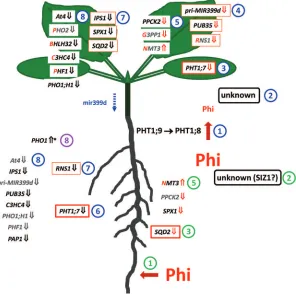
Fig. 7. A model for the sequence of changes in phosphate-starvation-responsive (PSR) gene expression observed in roots and shoots of phosphorus-limited Arabidopsis thaliana seedlings in response to phosphite (Phi) treatment. Blue pathway: the discrimination of Phi by PHT1;9 and subsequently by PHT1;8 during xylem loading (1) may indicate the recognition by a receptor that signals the availability of phosphate (Pi) and Phi to the shoot, possibly
involving SIZ1 (2). This sequence of events may primarily affect PHT1;7 expression in the shoot (3), followed by the consecutive suppression of other PSR genes within 24 h (4+5) or later after 3 d when Phi inally started to accumulate in the shoot (7+8). Green pathway: in roots, early local recognition of Phi is possibly restricted to the suppression of SQD2 within 24 h (3) and of the less responsive SPX1 and PPCK2 as well as to the induction of NMT3
(5). Compared with shoots, PHT1;7, RNS1 and a couple of transcripts in group 8 (in grey) responded more slowly in roots, most probably indicating that their expression in roots is regulated by PHO2 and relies on systemic signalling, perhaps through reduced levels of mir399d in the phloem (blue dotted arrow). Curiously, PHO1 expression in roots increased within 3 d of Phi treatment, which may indicate its connection to independent regulatory networks (purple 8) that directly respond to the overall P status of the plant or the growth inhibition triggered by Phi. Note that the number of genes responding equally well to either Pi or Phi (red irst letter in gene name) was greater in shoots than in roots. Gene names in black indicate a 2-fold expression change
in response to Phi over P-limited controls (Fig. 6). An orange border indicates a 4-fold expression change. A bold red border indicates an 8-fold change. Grey names indicate non-signiicant changes. Red arrows following gene names indicate suppression (↓) or induction (↑) within 24 h of Phi exposure, while black arrows indicate a response within 3 d of treatment. An asterisk indicates a Phi-speciic expression change that was not observed in Pi-resupplied
seedlings.
by guest on August 6, 2015
http://jxb.oxfordjournals.org/