Synthesis and stability test of
radiogadolinium(III)-DOTA-PAMAM G3.0-trastuzumab as SPECT-MRI
molecular imaging agent for diagnosis of HER-2
positive breast cancer
Hardiani Rahmania
a, Abdul Mutalib
b, Martalena Ramli
c, Jutti Levita
a,* aDepartment of Pharmaceutical Analysis and Medicinal Chemistry, Faculty of Pharmacy, Universitas Padjadjaran,Jl. Raya Bandung-Sumedang Km. 21, Jatinangor, Jawa Barat, Indonesia
bDepartment of Chemistry, Faculty of Mathematics and Natural Sciences, Universitas Padjadjaran,
Jl. Raya Bandung-Sumedang Km. 21, Jatinangor, Jawa Barat, Indonesia c
Radioisotopes and Radiopharmaceuticals Technology Centre, National Nuclear Energy Agency of Indonesia, Kawasan PUSPIPTEK, South Tangerang, Banten, Indonesia
a r t i c l e
i n f o
Article history:
Received 22 October 2014 Accepted 3 December 2014 Available online 15 December 2014
Keywords:
Gd-DOTA-PAMAM G3.0-trastuzumab
HER-2
Target-specific radiopharmaceutical Targeted contrast agent
a b s t r a c t
Nonivasive diagnosis of cancer can be provided by molecular imaging using hybrid mo-dality to obtain better sensitivity, specificity and depiction localization of the disease. In this study, we developed a new molecular imaging agent, radiogadolinium(III)-DOTA-PAMAM G3.0-trastuzumab in the form of 147Gd-DOTA-PAMAM G3.0-trastuzumab, that can be both target-specific radiopharmaceutical in SPECT as well as targeted contrast agent in MRI for the purpose of diagnosis of HER-2 positive breast cancer. 147Gd radionuclide emitsg-rays that can be used in SPECT modality, but because of technical constraint,147Gd radionuclide was simulated by its radioisotope, 153Gd. Gd-DOTA complex has also been known as good MRI contrast agent. PAMAM G3.0 is useful to concentrate Gd-DOTA com-pelexes in large quantities, thus minimizing the number of trastuzumab molecules used. Trastuzumab is human monoclonal antibody that can spesifically interact with HER-2. Synthesis of radiogadolinium(III)-DOTA-PAMAM G3.0-trastuzumab was initiated by conjugating DOTA NHS ester ligand with PAMAM G3.0 dendrimer. The DOTA-PAMAM G3.0 produced was conjugated to trastuzumab molecule and labeled with153Gd. Characteriza-tion DOTA-PAMAM G3.0-trastuzumab immunoconjugate was performed using HPLC sys-tem equipped with SEC. The formation of immunoconjugate was indicated by the shorter retention time (6.82 min) compared to that of trastuzumab (7.06 min). Radiochemical purity of radiogadolinium(III)-DOTA-PAMAM G3.0-trastuzumab was>99% after purification pro-cess by PD-10 desalting column. Radiogadolinium(III)-DOTA-PAMAM G3.0-trastuzumab compound was stable at room temperature and at 2e80Cas indicated by its
radiochem-ical purity 97.6±0.5%e99.1±0.5% after 144 h storage.
Copyright©2014, The Egyptian Society of Radiation Sciences and Applications. Production and hosting by Elsevier B.V. This is an open access article under the CC BY-NC-ND license (http://creativecommons.org/licenses/by-nc-nd/3.0/).
*Corresponding author.
E-mail addresses:[email protected],[email protected](J. Levita).
Peer review under responsibility of The Egyptian Society of Radiation Sciences and Applications.
ScienceDirect
Journal of Radiation Research and Applied
Sciences
j o u r n a l h o m e p a g e :h t t p : / / w w w . e l s e v i e r . c o m / l o c a t e / j r r a s
http://dx.doi.org/10.1016/j.jrras.2014.12.001
1.
Introduction
Recently, early detection and accurate diagnosis of cancer through noninvasive imaging at molecular level (molecular imaging) can be provided by magnetic resonace imaging (MRI), computed tomography (CT) and nuclear medicine imaging modalities, such as positron emission tomography (PET) and single photon emission computed tomography (SPECT), of course, using molecular imaging agents that are appropriate with the target molecules expressed by cancer cells. Nuclear medicine imaging modalities have high sensitivity and can distinguish between malignancy and necrosis. However, the spatial resolution of nuclear medicine imaging is low, so that is unable to provide the anatomical image required to pre-cisely localize lesions. On the other hand, MRI and CT mo-dalities can provide excellent anatomic details, but have low sensitivity, so do not provide functional details (Gambhir, 2007; Schillaci&Simonetti, 2004). This encourages the utiliz-ing of PET-CT and SPECT-CT hybrid modalities. The main advantages are better attenuation correction, increased specificity, and accurate depiction of the localization of dis-ease and possible involvement of adjacent tissues (Mariani et al., 2010). Because of spatial resolution of MRI is much higher compared to CT and because MRI only uses radio fre-quencies, instead of ionizing radiations, the utilizing PET-MRI and SPECT-MRI hybrid modalities in molecular imaging research have been popular and are potential in clinical use (Goetz, Breton, Choquet, Israel-Jost,&Constantinesco, 2008;
Pichler, Kolb, Nagele,&Schlemmer, 2010).
Function and performance of MRI and nuclear medicine imaging in molecular imaging are determined by the appro-priate biological interactions between molecular imaging agents administered to the patients and target molecules expressed by diseased cells. Molecular imaging agents for PET and SPECT are stated as target-spesific radiopharmaceuticals, whereas for MRI are called as targeted contrast agents. Ideally, molecular imaging agent fot PET-MRI and SPECT-MRI hybrid modalities is a single compound that acts both as target-spesific radiopharmaceutical for PET or SPECT, and as tar-geted contrast agent for MRI (Valliant, 2010).
Antigens and receptors are target molecules expressed by cancer cells that become main focus associated to the use of monoclonal antiody. Monoclonal antibodies are part of mo-lecular imaging agents that have a major role in process of biological interactions with the target molecules. Epidermal growth factor receptor (EGFR) or human epidermal growth factor receptor (HER) which is transmembrane tyrosine kinase protein is an important receptor (the target molecule) because it is recognized by monoclonal antibodies, and is also often found overexpressed in malignant cancers (Marmor, Skaria,&
Yarden, 2004). One family member of this receptor is HER-2, that is also known as ErbB2, overexpressed in 25e30% of
breast cancer cases (Carlsson, 2008; Leonard et al., 2002). One of the monoclonal antibodies that can spesifically interact with HER-2 is tastuzumab, an IgG monoclonal anti-body under the trade name Herceptin®
. Herceptin®
has been routinely used for the treatment of HER-2 positive breast cancer. The complement determinant region in variable domain (Fv) in the molecule structure of trastuzumab have a
role in recognizing epitope or binding site on extracellular domain of HER-2 when immunotherapy process desired takes place (Leonard et al., 2002).
Trastuzumab labeled with g-ray emitting radionuclides can spesifically interact with HER-2 that is overexpressed in HER-2 positive breast cancers. The g-ray radiation will be detected by SPECT detector placed in the patient's body sur-face, which is then displayed in the form of location of cancer cells image that subsequently used for diagnosis (Tamura et al., 2013). The same way is shown by trastuzumab conju-gated with MRI contrast agents that is able enhance water proton relaxation rate (1/T1) at surrounding area of interaction of trastuzumab with HER-2. Enhancement proton relaxation rate causes enhancement microwave intensity emitted and captured by MRI detector, which is then displayed in the form of brighter location image (Bellin, 2006).
Trastuzumab labeling with g-ray emitting radionuclides for diagnostic purposes has been carried out byPerik et al. (2006) and de Korte et al. (2007) by conjugating 111In-DTPA with trastuzumab.111In radionuclide decays by electron cap-ture followedg-ray emission that can be detected by SPECT. 111
In-DTPA-trastuzumab radiopharmaceutical wass used as molecular imaging agent for noninvasive SPECT imaging that was potential to HER-2 (Perik et al., 2006; de Korte et al., 2007), although SPECT has limitation in spatial resolution (Gambhir, 2007).
This study developed a new molecular imaging agent, radiogadolinium(III)-DOTA-PAMAM G3.0-trastuzumab in the form of147Gd-DOTA-PAMAM G3.0-trastuzumab that has dual roles in SPECT-MRI hybrid modality for diagnosis of HER-2 positive breast cancer. 147Gd radionuclide which decays by electron capture areg-ray emitting radionuclide (Eg¼229 keV,
t1/2¼38.1 h) that can be used in SPECT. But, due to technical constraints, 147Gd radionuclide was simulated by its radio-isotopes, 153Gd (E
g ¼ 102 keV; t1/2 ¼ 241.6 days). Gd-DOTA complex has also been known as good MRI contrast agent. Role of PAMAM G3.0 dendrimer is to concentrate Gd-DOTA complex in large quantitites and to minimize the use of expensive trastuzumab, so the molecular imaging agent dose administered to the patient can be minimized without reducing image quality as well as ensuring the safety related the dose of Gd-DOTA that can be reduced considerably lower than the LD50.
The aim of this study was to synthesize and test the sta-bility of molecular imaging agent147Gd-DOTA-PAMAM G3.0-trastuzumab through simulation of 153Gd-DOTA-PAMAM G3.0-trastuzumab which have dual roles as SPECT target-spesific radiopharmaceuticals and as MRI targeted contrast agents for diagnosis of HER-2 positive breast cancer.
2.
Methods
2.1. Materials and equipments
Materials used are trastuzumab (Herceptin®
1,4,7,10-tetraazacyclododecane-1,4,7,10-tetraacetic acid mono (N-hydroxysuccinimide ester) or DOTA NHS ester (Macrocyclics), NaCl, HCl, NaOH, ethylen diamine tetraacetic acid (EDTA), dipotassium hydrogen phosphate, potassium dihydrogen phosphate (Merck), sulfo-succinimidyl-4-(N-maleimidometh-yl) cyclohexane 1-carboxylate or sulfo-SMCC, Traut's reagent or 2-iminothiolane HCl (Thermo Scientific), sephadex G25 medium column, PD-10 desalting column (GE HealthCare), standard protein, protein dye, Chelex-100 ion exchange resin (Bio Rad), bovin serum albumin or BSA, 2 kD and 20 kD Mo-lecular Weight Cut Off (MWCO) dialysis cassette (Thermo Scientific), Instant Thin Layer Chromatography-Silica Gel or ITLC-SG (Pall), size exclusion column (Agilent Bio SEC-3; 7.8 300 mm), gadolinium(III) oxide (Strem Chemicals), 153Gd prepared by irradiating natural Gd in a multipurpose
G.A. Siwabessy Reactor, National Nuclear Energy Agency of Indonesia, Serpong.
Equipments used are thermomixer (Eppendorf), magnetic stirrer (ColeeParmer), orbital shaker, water bath (Fisher
Sci-entific), microplate spectrophotometer (Biotek), water purifier equipment (Sartorius), HPLC (Shimadzu) equipped with UV-Visible detector and size exclusion column (Agilent), gamma mini assay (DPC), dose calibrator (Biodex Medical Systems).
2.2. Synthesis of radiogadolinium(III)-DOTA-PAMAM
G3.0-trastuzumab
2.2.1. Purification of trastuzumab
Trastuzumab (5 mg mL 1) was purified from the excipients using 20 kD MWCO dialysis cassette with 0.1 M phosphate buffer saline (PBS) pH 7.4 (contains 5 mM EDTA and 1.2 g Chelex-100 ion exchange resin). Dialysis was carried out for 324 h at 2e8C. The concentration of purified trastuzumab
was measured using microplate spectrophotometer and was characterized using HPLC equipped with size exclusion col-umn, UV-Visible detector, and 0.01 M PBS pH 7.4 as mobile phase.
2.2.2. Synthesis of DOTA-PAMAM G3.0-trastuzumab immunoconjugate
2.2.2.1. Preparation and activation of DOTA-PAMAM G3.0 conjugate.DOTA NHS ester solution in 0.1 M phosphate buffer pH 7.2 was reacted with PAMAM G3.0 dendrimer (molar ratio 96: 1). The pH of the mixture was adjusted to 7.2. The mixture was then incubated for 24 h at 2e8C.
DOTA-PAMAM G3.0 conjugate produced was purified using 2 kD MWCO dialysis cassette with 0.05 M phosphate buffer pH 7.4 (contains 5 mM EDTA and 1.2 g Chelex-100 ion exchange resin). Dialysis was carried out for 324 h at 2e8C.
Purified DOTA-PAMAM G3.0 conjugate was then activated by adding Traut's reagent solution (1 mg mL 1) in phosphate buffer 0.05 M pH 7.4 (contains 5 mM EDTA) into DOTA-PAMAM G3.0 conjugate. The mixture was then incubated at room temperature under N2flow for 1 h.
Activated DOTA-PAMAM G3.0 conjugate was purified using PD-10 desalting column which has been conditioned by BSA and 0.1 M phosphate buffer pH 7.4 (contains 5 mM EDTA) as eluent. Eluates was retrieved in 250mL fraction with total of 40 fractions. Each fraction then was sampled and tested using
colorimetric protein dye (1:4 in H2O). Positive fractions to the colorimetric assay were collected.
2.2.2.2. Activation of trastuzumab.Sulfo-SMCC was dissolved in a small volume of DMF, and diluted by adding 0.1 M PBS pH 7.4 (contains 5 mM EDTA) to obtain 1 mg mL 1. The solution was then added into purified trastuzumab. The mixture was incubated for 1 h at room temperature followed by purifica-tion using PD-10 desalting column which has been condi-tioned by BSA and 0.1 M PBS pH 7.4 (contains 5 mM EDTA) as eluent. Eluates was retrieved in 250mL fraction with total of 40 fractions. Each fraction then was sampled and tested using colorimetric protein dye (1:4 in H2O). Positive fractions to the colorimetric assay were collected.
2.2.2.3. Preparation of DOTA-PAMAM G3.0-trastuzumab immunoconjugate. Activated trastuzumab was added into activated DOTA-PAMAM G3.0 conjugate. The mixture was incubated for 24 h at 2e8C. DOTA-PAMAM G3.0-trastuzumab
immunoconjugate produced was purified using 20 kD MWCO dialysis cassette with 0.1 M phosphate buffer pH 7.4 (contains 1.2 g Chelex-100 ion exchange resin). Dialysis was carried out for 412 h at 2e8C.
Purified DOTA-PAMAM G3.0-trastuzumab immunoconju-gate was then dispensed to the vials and stored at 2e8C. This
immunoconjugate was characterized using HPLC equipped with size exclusion column, UV-Visible detector, and 0.01 M PBS pH 7.4 as mobile phase.
2.2.3. Labeling of DOTA-PAMAM G3.0-trastuzumab immunoconjugate with153Gd
2.2.3.1. Preparation153GdCl
3.50 mg Gd2O3was irradiated in a
multipurpose G.A. Siwabessy Reactor (National Nuclear En-ergy Agency of Indonesia) for four days. Irradiated Gd2O3was dissolved in 5 mL of 2 N HCl.
2.2.3.2. Labeling process. Aliquots of DOTA-PAMAM G3.0-trastuzumab immunoconjugate in 0.1 M phosphate buffer pH 7.4 was reacted with GdCl3in the same buffer solution. The mixture was adjusted by adding 0.1 M NaOH to pH 7, then incubated at 37
C. An excess of 0.05 M EDTA solution (molar ratio EDTA:153Gd¼20: 1) was added at the end of the reaction, followed by incubating at 37C for 10 min. Optimization of reaction time was carried out at 2e8C.
Labeling percentage of 153Gd on DOTA-PAMAM G3.0-trastuzumab was determined using ITLC-SG strip (1 cm 10 cm) as stationary phase and saline solution as mobile phase. Labeling percentage was obtained by comparing the counts under peak of 153Gd-DOTA-PAMAM G3.0-trastuzumab to the total counts.
Purification of153Gd-DOTA-PAMAM G3.0-trastuzumab was carried out using PD-10 desalting column which has been conditioned by BSA and 0.1 M PBS pH 7.4 as eluent. Eluates was retrieved in 250mL fraction with total of 40 fractions. Each fraction was scanned using gamma mini assay. The fractions had high radioactivities was analyzed using the TLC system to determine the radiochemical purity.
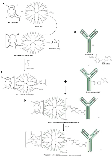
Fig. 1eSynthesis reaction scheme: A. DOTA-PAMAM sulfhydryl B. Trastuzumab-maleimide; C. DOTA-PAMAM
Gd. The mixture was adjusted by adding 0.1 M NaOH to pH 7, and incubated at the same condition with the labeling of DOTA-PAMAM G3.0-trastuzumab with153Gd.
An excess of 0.05 M EDTA solution (molar ratio EDTA: 153Gd¼20: 1) was added at the end of the reaction, followed by
incubating at 37C for 10 min. The mixture was loaded onto PD-10 desalting column which has been conditioned by BSA and 0.1 M PBS pH 7.4 as eluent. Eluates was retrieved in 250mL fraction with total of 40 fractions. Each fraction was scanned using gamma mini assay. The fractions had high radioactiv-ities was analyzed using the TLC system to determine the radiochemical purity.
2.2.3.4. Nonspesific binding test of trastuzumab.Trastuzumab solution in 0.1 M phosphate buffer pH 7.4 was reacted with 153Gd. The mixture was adjusted by adding 0.1 M NaOH to pH
7, was then incubated at the same condition with the labeling of DOTA-PAMAM G3.0-trastuzumab with153Gd.
An excess of 0.05 M EDTA solution (molar ratio EDTA: 153Gd¼20: 1) was added at the end of the reaction, followed by
incubating at 37C for 10 min. The mixture was loaded onto PD-10 desalting column which has been conditioned by BSA and 0.1 M PBS pH 7.4 as eluent. Eluates was retrieved in 250mL fraction with total of 40 fractions. Each fraction was scanned using gamma mini assay. The fractions had high radioactiv-ities was analyzed using the TLC system to determine the radiochemical purity.
2.3. Stability test of
radiogadolinium(III)-DOTA-PAMAM G3.0-trastuzumab
Stability test of radiogadolinium(III)DOTA-PAMAM G3.0-trastuzumab was carried out at room temperature and at 2e8 C. The radiochemical purity was the analyzed using
ITLC-SG as stationary phase and saline solution as mobile phase on 0, 48, 72, and 144 h.
3.
Results and discussion
Synthesis of radiogadolinium(III)-DOTA-PAMAM G3.0-trastuzumab in the form of 147Gd-DOTA-PAMAM G3.0-trastuzumab through simulation153Gd-DOTA-PAMAM G3.0-trastuzumab, as SPECT-MRI molecular imaging agent for diagnosis of HER-2 positive breast cancer, was carried out in several stages of the reaction (Fig. 1): a) the preparation and activation of DOTA-PAMAM G3.0 conjugate using Traut's re-agent; b) activation of trastuzumab using sulfo-SMCC; c) conjugation of activated DOTA-PAMAM G3.0 to activated trastuzumab; d) labeling of DOTA-PAMAM G3.0-trastuzumab immunoconjugate with153Gd.
The first reaction was conjugation of DOTA NHS ester ligand with PAMAM G3.0 dendrimer to form DOTA-PAMAM G3.0 conjugate (Fig. 1A). NHS ester-containing reagents react with nucleophiles by releasing the NHS leaving group to form an acylated product (Hermanson, 2008). The reaction of DOTA NHS ester ligand with PAMAM G3.0 dendrimer containing 32 primary amine groups yields DOTA-PAMAM G3.0 conjugate through stable amide bonds.
By adjusting the molar ratio of NHS ester ligand to target molecule, the level of modification and conjugation can be controlled to create an optimal product (Hermanson, 2008). In this study, molar ratio of DOTA NHS ester to PAMAM G3.0 used was 96: 1 or three folds of the number primary amines in PAMAM G3.0 to obtain an optimal DOTA-PAMAM G3.0 conju-gate, one molecule of PAMAM G3.0 could binds DOTA mole-cules in the maximum amounts.
DOTA-PAMAM G3.0 conjugate formed was then activated using Traut's reagent to create DOTA-PAMAM G3.0-sulfhydryl (Fig. 1A). This thiolation reaction introduce reactive sulfhydryl group at one end of target molecules. The sulfhydryl group is employed to direct the conjugation reaction to a particular part of a target macromolecule (Hermanson, 2008), in this study, the macromulecule is trastuzumab.
Sulfhydryl groups are susceptible to oxidation and tion of disulfide crosslinks. To prevent disulfide bond forma-tion, activation reaction of DOTA-PAMAM G3.0 conjugate using Traut's reagent was carried out under nitrogen gas flow to remove oxygen. In addition, EDTA (0.01e0.1 M) may be
added to buffers to chelate metal ions, preventing metal-catalyzed oxidation of sulfhydryls (Hermanson, 2008).
Traut's reagent reacts with primary amine in the range of pH 7e10, but its half-life in solution decreases as the pH
in-creases. Protein modification with Traut's reagent is very efficient and proceeds rapidly at slightly basic pH (Hermanson, 2008).
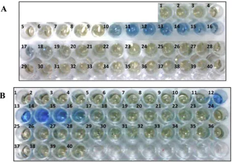
DOTA-PAMAM G3.0-sulfhydryl was purified using PD-10 desalting column with 0.1 M phosphate buffer containing 5 mM EDTA as eluent. The eluate fractions was sampled and tested by protein dye. The fractions containing DOTA-PAMAM G3.0-sulfhydryl was blue, while the fractions containing no DOTA-PAMAM G3.0-sulfhydryl was brown like origin color of protein dye. Colorimetric assay by protein dye showed the fractions 10e17 containing DOTA-PAMAM G3.0-sulfhydryl
(Fig. 2A). The fractions were then collected to be used in next step reaction.
The second reaction was activation of trastuzumab using sulfo-SMCC to form trastuzumab-maleimide (Fig. 1B). The NHS ester end of the reagent can react with primary amine
Fig. 2ePurification fractions of: A. Activated
groups on proteins to form stable amide bonds (Hermanson, 2008). Trastuzumab-maleimide was purified using PD-10 desalting column with 0.1 M PBS containing 5 mM EDTA as eluent. The eluate fractions was sampled and tested by
protein dye. Colorimetric assay by protein dye showed the fractions 12e16 containing trastuzumab-maleimide
(Fig. 2B). The fractions were then collected to be used in next step.
Fig. 3eHPLC chromatograms of: A. Trastuzumab; B. DOTA-PAMAM G3.0-trastuzumab; C. Water; using SEC (stationary
phase) and 0.01 M PBS pH 7.4 (mobile phase).
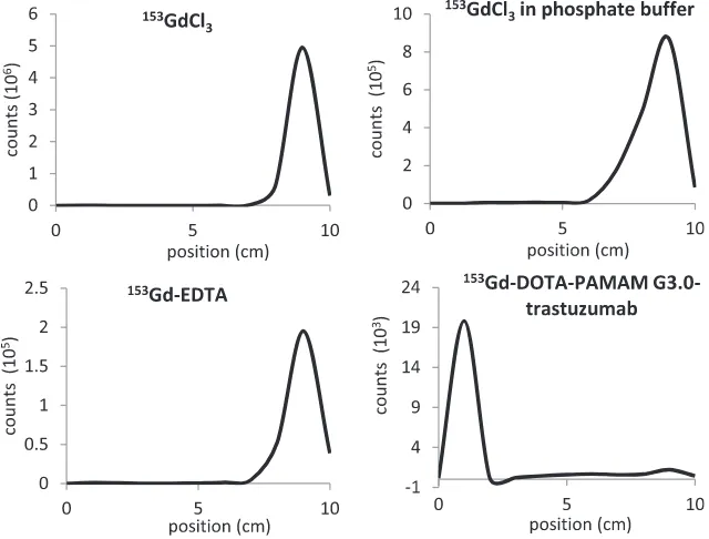
Fig. 4eRadiochromatogram of reagents involved in labeling DOTA-PAMAM G3.0-trastuzumab immunoconjugate with
The third reaction was conjugation of DOTA-PAMAM G3.0-sulfhydryl with trastuzumab-maleimide to generate DOTA-PAMAM G3.0-trastuzumab immunoconjugate (Fig. 1C). The double bond of maleimides that activated trastuzumab may undergo an alkylation reaction with sulfhydryl groups of DOTA-PAMAM G3.0-sulfhydryl to form stable thioether bonds. This reaction is spesific in the pH range 6.5e7.5 (Hermanson,
2008). DOTA-PAMAM G3.0-trastuzumab immunoconjugate formed was then purified using 20 kD MWCO dialysis cassette with 0.1 M phosphate buffer pH 7.4 containing 1.2 g Chelex-100 ion exchange resin.
DOTA-PAMAM G3.0-trastuzumab immunoconjugate was charactrized using HPLC equipped with SEC, UV detector at 280 nm. Eluent used was 0.01 M PBS pH 7.4 with flow rate of 0.8 mL min 1. The chromatogram obtained was compared to the trastuzumab which also characterized with the same HPLC system (Fig. 3).
The retention time of trastuzumab was 7.06 min, whereas DOTA-PAMAM G3.0-trastuzumab immunoconjugate's was 6.82 min. The difference of trastuzumab's retention time compared to DOTA-PAMAM G3.0-trastuzumab immuno-conjugate showed that there is an increasing of the product's molecular weight, thus smaller molecules of trastuzumab difused to column pores as they passed through the column, while larger molecules of the immunoconjugate remained in
interstitial space and flowed rapidly down the length of the column because they could not reside inside the pores. Larger molecules will be eluted first (Wu, 1995).
Labeling was carried out by adding 153GdCl3 to DOTA-PAMAM G3.0-trastuzumab immunoconjugate with molar ratio 30:1 to form 153Gd-DOTA-PAMAM G3.0-trastuzumab (Fig. 1D). Forming reaction of Gd-DOTA complex was carried out at pH 7 (Bryant et al., 1999; Hak et al., 2009). At the end of reaction, EDTA was added in excess to bind free153Gd which was not bound to DOTA-PAMAM G3.0-trastuzumab immunoconjugate.
Fig. 5eRadiochromatogram of153Gd-DOTA-PAMAM
G3.0-trastuzumab purification.
Fig. 6eRadiochromatogram of fraction 13 and 24 using TLC system.
Fig. 7eRadiochromatogram of nonspesific binding test of
Labeling percentage of 153Gd on DOTA-PAMAM G3.0-trastuzumab was determined using ITLC-SG strip and saline solution (Table 1). This method was adopted from previous studies concerning the labeling DOTA-trastuzumab immu-noconjugate with 177Lu by Humani, Ramli, Rustendi, and
Subur (2010)andRamli et al. (2011). The radiochromatogram of153Gd-DOTA-PAMAM G3.0-trastuzumab and the reagents involved shown inFig. 4.
Labeling DOTA-F(ab0)24.1 immunoconjugate with153GdCl3 was carried out byRamakrishnan et al. (2008)at 37
C for 16 h. Higher labeling percentage was obtained at 37C than at 2e8C. This condition was not time dependent. In this work
we minimized the heating time to prevent structural damage, and the labeling process was carried out at 37C for 3 h.
Purification of153Gd-DOTA-PAMAM G3.0-trastuzumab was carried out by PD-10 desalting column to obtain radiochemical purity of95% as requirement for good molecular imaging agent. Each fraction of eluate was counted its radioactivity and was plotted (Fig. 5). The first peak at chromatogram was predicted to be 153Gd-DOTA-PAMAM G3.0-trastuzumab molecule and the second peak was153Gd-EDTA. Both peaks (fraction 13 and 24) was analyzed using TLC system to deter-mine the radiochemical purity.
Rf of fraction 13 was zero (Fig. 6) indicated the presence of 153Gd-DOTA-PAMAM G3.0-trastuzumab molecule with the
radiochemical purity of 99.5%. Meanwhile, the Rf of fraction 24 was one indicated the presence of153Gd-EDTA molecule.
Peak area of 153Gd-DOTA-PAMAM G3.0-trastuzumab molecule included fraction 10e18 (Fig. 5). Radiochemical
pu-rity analysis of that fractions using TLC system showed that the fractions 10-17 had radiochemical purity of95%.
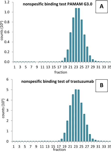
In this labeling process, we performed additional assays that were (1) nonspesific binding test of153Gd to PAMAM G3.0 and (2)153Gd to trastuzumab. Results were shown inFig. 7A and B that showed radioactivity peak was at fraction 24. This fraction was area peak of153Gd-EDTA molecule as described
previously in the purification of radioimmunoconjugate (Fig. 5). In the nonspesific binding test of153Gd to PAMAM G3.0, fractions 10e17 was positive to protein dye assay, but did not
have radioactivity peak as shown atFig. 7A. In the nonspesific binding test of 153Gd to trastuzumab, fractions 10
e15 was
positive to protein dye assay, but did not have radioactivity peak as shown atFig. 7B. This data showed that there were no nonspesific binding of153Gd to PAMAM G3.0 as well as153Gd to trastuzumab.
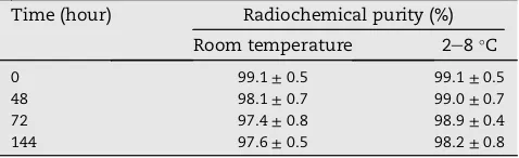
Molecular imaging agent should be stable, still intact, not degradated during storage. Stability test of 153 Gd-DOTA-PAMAM G3.0-trastuzumab was carried out in storage at room temperature and at 2e8C. The test was analyzed by TLC
system on 0, 48, 72, and 144 hTable 2showed153 Gd-DOTA-PAMAM G3.0-trastuzumab was stable, relatively intact, with radiochemical purity of respectively 97.6 ± 0.5% and 98.2±0.8% at room temperature and at 2e8C after 144 h of
storage. This result was similar to that reported by Nwe, Bernando, Regino, Williams, and Brechbiel (2010), which stated Gd-DOTA complex was very chemically stable.
4.
Conclusion
Synthesis radiogadolinium(III)-DOTA-PAMAM G3.0-trastuzumab as SPECT-MRI molecular imaging agent for diagnosis of HER-2 positive breast cancer had been success-fully carried out. DOTA-PAMAM G3.0-trastuzumab immuno-conjugate as precursor of radiogadolinium(III)-DOTA-PAMAM G3.0-trastuzumab had been successfully formed as shown by characterization using HPLC system equipped with SEC. Radiogadolinium(III)-DOTA-PAMAM G3.0-trastuzumab com-pound was stable at room temperature and at 2e8C.
Acknowledgment
This study was supported by the Radioisotopes and Radio-pharmaceuticals Technology Centre, National Nuclear Energy Agency of Indonesia.
r e f e r e n c e s
Bellin, M.-F. (2006). MR contrast agents, the old and the new.
European Journal of Radiology, 60(3), 314e323.
Bryant, L. H., Brechbiel, M. W., Wu, C., Bulte, J. W., Herynek, V., & Frank, J. A. (1999). Synthesis and relaxometry of high-generation (G¼5, 7, 9, and 10) PAMAM dendrimer-DOTA-gadolinium chelates.Journal of Magnetic Resonance Imaging, 9, 348e352.
Carlsson, J. (2008). EGFR-family expression and implications for targeted radionuclide therapy. In T. Stigbrand, J. Carlsson, & G. Adam (Eds.),Targeted radionuclide tumor therapy: Biological aspects(pp. 25e28). Dordrecht: The Netherlands: Springer.
Gambhir, S. S. (2007). Just what is molecular imaging.MI Gateway, The MICoE Newsletter, 1(1), 1e8.
Goetz, C., Breton, E., Choquet, P., Israel-Jost, V., &
Constantinesco, A. (2008). SPECT low-field MRI system for small-animal imaging.Journal of Nuclear Medicine, 49, 88e93.
Table 1eTemperature and time optimization of labeling
reaction of DOTA-PAMAM G3.0-trastuzumab
Table 2eStability test result of153Gd-DOTA-PAMAM
G3.0-trastuzumab.
Time (hour) Radiochemical purity (%)
Room temperature 2e8C
Hak, S., Sanders, H. M., Agrawal, P., Langereis, S., Gru¨ll, H., Keizer, H. M., et al. (2009). A high relaxivity Gd(III)DOTA-DSPE-based liposomal contrast agent for magnetic resonance imaging.European Journal of Pharmaceutics and Biopharmaceutics, 72, 397e404.
Hermanson, G. T. (2008).Bioconjugate technique(2nd ed.). USA: Elsevier, 67e68, 171e172, 183e184, 283e284.
Humani, T. S., Ramli, M., Rustendi, C. T., & Subur, M. (2010).
Preparasi and uji stableitas177Lu-DOTA-nimotuzumab sebagai
radiofarmaka terapi kanker. Seminar Nasional VI: SDM Teknologi Nuklir (pp. 663e669).
de Korte, M. A., de Vries, E. G., Lub-de Hooge, M. N., Jager, P. L., Gietema, J. A., van der Graaf, W. T., et al. (2007).111
Indium-trastuzumab visualises myocardial human epidermal growth factor receptor 2 expression shortly after anthracycline treatment but not during heart failure: a clue to uncover the mechanisms of trastuzumab-related cardiotoxicity.European Journal of Cancer, 43, 2046e2051.
Leonard, D. S., Hill, A. D., Kelly, L., Dijkstra, B., McDemott, E., & O'Higgins, N. J. (2002). Anti-human epidermal growth factor receptor 2 monoclonal antibody therapy for breast cancer.
British Journal of Surgery, 89, 261e271.
Mariani, G., Bruselli, L., Kuwert, T., Kim, E. E., Flotats, A., Israel, O., et al. (2010). A review on the clinical uses of SPECT/CT.
European Journal of Nuclear Medicine and Molecular Imaging, 37, 1959e1985.
Marmor, M. D., Skaria, K. B., & Yarden, Y. (2004). Signal transduction and oncogenesis by ErbB/HER receptors.
International Journal of Radiation Oncology, Biology, Physics, 58, 903e913.
Nwe, K., Bernando, M., Regino, C. A., Williams, M., & Brechbiel, M. W. (2010). Comparison of MRI properties between derivatized DTPA and DOTA gadoliniumedendrimer
conjugates.Bioorganic&Medicinal Chemistry, 18, 5925e5931.
Perik, P. J., Lub-De Hooge, M. N., Gietema, J. A., van der
Graaf, W. T., de Korte, M. A., Jonkman, S., et al. (2006). Indium-111-labeled trastuzumab scintigraphy in patients with human epidermal growth factor receptor 2-positive metastatic breast cancer.Journal of Clinical Oncology, 24, 2276e2282.
Pichler, B. J., Kolb, A., Nagele, T., & Schlemmer, H.-P. (2010). PET/ MRI: paving the way for the next generation of clinical multimodality imaging applications.Journal of Nuclear Medicine, 51(3), 333e336.
Ramakrishnan, M., Wengenack, T. M., Kandimalla, K. K., Curran, G. L., Gilles, E. J., Ramirez-Alvarado, M., et al. (2008). Selective contrast enhancement of individual alzheimer's disease amyloid plaques using a polyamine and Gd-DOTA konjugated antibody fragment against fibrillar Ab42 for magnetic resonance molecular imaging.Pharmaceutical Research, 25(8), 1861e1872.
Ramli, M., Hidayat, B., Ardiyanto, C. N., Aguswarini, S., Karyadi, Rustendi, C. T., et al. (2011). Uji preklinis177
Lu-DOTA-trastuzumab radiofarmaka potensial untuk terapi kanker payudara positif HER-2. InProsidisng Pertemuan Ilmiah Radioisotop, Radiofarmaka and Siklotron(pp. 117e125).
Schillaci, O., & Simonetti, G. (2004). Fusion imaging in nuclear medicinedapplications of dual-modality systems in oncology. Cancer Biotherapy&Radiopharmaceutical, 19(1), 1e10.
Tamura, K., Kurihara, H., Yonemori, K., Tsuda, H., Suzuki, J., Kono, Y., et al. (2013, November). 64Cu-DOTA-Trastuzumab PET imaging in patients with HER2-positive breast cancer.
Journal of Nuclear Medicine, 54(11), 1e7.
Valliant, J. F. (2010). A bridge not too far: linking disciplines through molecular imaging probes.Journal of Nuclear Medicine, 51, 1258e1268.