*Corresponding author: E-mail: [email protected]
Available online at http://medpet.journal.ipb.ac.id/
Phytochemical Screening and
in Vitro
Ovicidal, Larvacidal, and Nematicidal Effects
of
Murraya paniculata
(L.) Jack Extract on Gastrointestinal Parasites of Goats
G. E. Tresiaa, D. Evvyernieb*, & R. Tiuriac
a Study Program of Nutrition and Feed Science, Faculty of Animal Science, Postgraduate School,
Bogor Agricultural University
b Department of Nutrition and Feed Science, Faculty of Animal Science, Bogor Agricultural University c Department of Parasitology and Helminthology, Faculty of Veterinary, Bogor Agricultural University
Jalan Agatis, Kampus IPB Darmaga Bogor 16680, Indonesia
(Received 10-06-2016; Reviewed 01-08-2016; Accepted 23-11-2016)
ABSTRACT
In our previous research, kemuning leaves (Murraya paniculata L. Jack) was shown to have the capability as an anthelmintic candidate for PE (Ettawa crossbred) lactating dairy goats by reduc-ing 43.67% of the egg per gram (EPG) of Strongylida compared to 0.0005% in orally treated with Oxfendazole as a control. To confirm it, the aim of this in vitro study was to determine the effective dosage of kemuning leaves from two extraction methods (infuse and maceration) to reduce the Trichostrongylidae and to evaluate the bioactive compounds of the leaves. The research was conducted using a randomized complete design with 11 treatments and 5 replications. The treatments consisted of control (0.0005% and 0.005% Oxfendazole), kemuning leaves infuse extract (KIE) and maceration extract (KME) each with level of 1%, 3%, 5%, and 7% (w/v). The results showed that the LT50 gradu-ally decreased (shortening the lethal time) and the mortality of Trichostrongylidae gradugradu-ally increased associated with the increased concentration of treatment (P<0.01). The infusion of 7% kemuning ex-tract demonstrated the highest efficiency in reducing the larval development, infective larvae, and the adult Trichostrongylidae by 93.16%, 94.39%, and 90%, respectively. This treatment could be developed as the most prospective herbal anthelmintic drug in controlling the infection by Trichostrongylidae.
Key words: infusion, kemuning (Murraya paniculata L. Jack), maceration, Trichostrongylidae
ABSTRAK
Penelitian kami sebelumnya menunjukkan bahwa herbal daun kemuning (Murraya panicu-lata L. Jack) memiliki kemampuan sebagai kandidat obat cacing pada ternak kambing PE (Peranakan Ettawa) laktasi yang dapat mengurangi telur tiap gram tinja (TTGT) dari Strongylida sebesar 43,67% dibandingkan dengan pemberian oxfendazole 0,0005% secara oral sebagai kontrol. Untuk mengkon-firmasi hasil tersebut, penelitian secara in vitro ini bertujuan untuk menentukan dosis efektif daun kemuning dengan dua metode ekstraksi (infusa dan maserasi) dalam mereduksi Trichostrongylidae dan mengidentifikasi senyawa bioaktif yang terkandung dalam daun kemuning. Penelitian ini menggunakan rancangan acak lengkap (RAL) dengan 11 perlakuan dan 5 ulangan. Perlakuan terdiri atas kontrol (0,0005% dan 0,005% Oxfendazole), ekstrak infusa daun kemuning (KIE) dan ekstrak maserasi (KME) dengan tingkatan 1%, 3%, 5%, dan 7% (b/v). Hasil penelitian menunjukkan bahwa LT50 secara bertahap menurun (mempersingkat) dan kematian Trichostrongylidae secara bertahap meningkat seiring meningkatnya konsentrasi perlakuan (P<0,01). Ekstrak infusa (KIE) 7% memiliki efektivitas paling tinggi dalam menurunkan perkembangan larva, larva infektif, dan cacing dewasa Trichostrongylidae (masing-masing 93,14%; 94,39%; dan 90%). Dapat disimpulkan bahwa secara in vitro 7% ekstrak infusa daun kemuning (KIE) dapat dikembangkan sebagai kandidat terbaik herbal obat cacing dalam mengendalikan infeksi Trichostrongylidae.
INTRODUCTION
Nematodes are often found in the gastrointestinal tract of goats and mostly from Ordo Strongylida, Family
Trichostrongylidae which produce strongylid-type eggs.
A parasitic infection caused by the gastrointestinal nematodes is one of the common diseases in goats. This
infection accounts for significant economic loss due to
weight loss, delayed growth, and reduced milk
produc-tion by 6.25%-21.5% (Alberti et al. 2009). The parasites are controlled with anthelmintic drugs. Repeated ad-ministration of the anthelmintic, especially the synthetic
ones, is a common method (Molento et al., 2011). This
method, however, has some adverse effects, such as the
development of drug-resistant strains of worms and the presence of drug residues in the derived products
(Kinsella et al., 2009). This condition encourages the
search for new active compounds which are safer and
more efficient. Medicinal plants could be an alternative
for the problem.
As a tropical country, Indonesia has high biodi-versity, including the medicinal/herbal plants. The
efficacy of herbs is determined from the absence of
clinical symptoms and improvement of performance of the patients. The knowledge on this recovery process is usually obtained from experience, being passed down
through generations but this emphirical efficacy needs scientifical and pharmacological testing. Plants offer
advantages over synthetic drugs for being more vastly
available, environmentally friendly, and more effective
to control parasitic worms.
Kemuning belongs to Class Magnoliopside, Ordo
Geraniales, Family Rutaceae, Genus Murraya, and
Species Murraya paniculata. The bioactive compounds in its leaves vary according to the solvent used during
extraction and the growing media (Gautam et al., 2012;
Vaghasiya et al., 2012; Syahadat & Aziz, 2012). Generally,
they include flavonoids, saponins, and tannins, posing a
high potential for antioxidant, anticancer, antimicrobial,
and antidiabetic agent (Ng et al., 2012).
Our previous study showed that the administration
of 0.7% kemuning leave meal (KLM) into the feed re
-duced the egg number per gram feces (EPG) by 21.28% at the fourth week in lactating Ettawa crossbred goats (average milk production 940 mL/head/d) which were
infected by natural parasite of Ordo Strongylida. The
EPG reduction increased to 43.6% at the fifth week when then the concentration of KLM being increased to 1%.
Lactating Ettawa crossbred goats (average milk produc
-tion of 861 mL/head/d) which have a similar infec-tion
and were treated with Oxfendazole, were used as a
con-trol. The same positive effects were observed with both
treatments, including a detected improvement on blood
profiles (close to normal) and a reduction of somatic cell
count in the milk. This condition implied that although
the administration of KLM could not increase the milk
production, it did improve the health status of the
lactat-ing Ettawa grade goats. Apparently, the right dose to significantly reduce the effect of parasitic infection was
yet to be searched. This study tested the in vitro effective dose of kemuning leave extract as anthelmintic agents
that can both inhibit the larval development and reduce the number of the adults.
MATERIALS AND METHODS
Research Design
Control, kemuning infuse extract (KIE), kemuning maceration extract (KME), and Oxfendazole (OXF) were
compared in this research. The experiment consisted 11
treatments (P0= Control; P1= KIE 1%; P2= KIE 3%; P3= KIE 5%; P4= KIE 7%; P5= KME 1%; P6= KME 3%; P7= KME 5%; P8= KME 7%; P9= OXF 0.0005%; dan P10= OXF 0.005%) with 5 replications.
Extraction Procedure (Depkes, 2000)
Infuse method. Kemuning leaves meal (KLM) was diluted with distilled water (1:10 w/v), and the solution was placed into an infused pan, stirred, heated for 15
min at 90oC, and filtered with filter cloth. The process
was repeated several times for the same KLM. The fil
-trate was concen-trated by spray drying (mini spray dry
Buchi 190). This extract was used in in vitro test.
Maceration method. KLM was diluted in 96% ethanol
(1:10 w/v), left for 24 h, and filtered. The process was repeated several times for the same KLM. The solvent was removed with vacuum rotary evaporator (Buchi
Rotavapor R-205) at 55oC. This extract was used in in
vitro test. The yield of the extract was calculated as:
Yield (%)= [Extract mass (g) / KLM mass (g)] x 100%
Phytochemical Analysis (Harbone, 1987)
Alkaloid test. KLM sample (1-2 g) was mixed with chloroform-ammonia solution (10 mL) and filtered.
Some drops of H2SO42N were added to the filtrate and the solution was shaken until 2 distinct layers were
formed. The acid layer (no color) was pipetted into
another reaction tube and divided into 3. Some drops of
Dragendrorf, Mayer, and Wagner reagents were added
into each of them. Positive result is indicated by the for-mation of orange, white-yellowish, and brown
precipita-tion following the addiprecipita-tion of Dragendrorf, Mayer, and Wagner reagent, respectively.
Flavonoid test. The sample was added with few mL
of ethanol or distilled water until completely soaked,
followed by boiling and filtration. Few drops of 10%
NaOH were added which would result in the formation of yellow color. This color should fade out when being added with concentrated H2SO4, indicating the presence
of flavonoid.
Saponin and tannin test. The sample was diluted with
hot distilled water, boiled, and filtered. The filtrate was
divided into 2 reaction tubes, each for saponin and tan-nin test. The one for sapotan-nin test was cooled down and shaken manually. A positive result would be indicated
into the filtrate. A positive result would be indicated by
the formation of green, blue, and black colors.
Triterpenoid and steroid test. Sample was diluted with
25 mL of ethanol (50oC), filtered into a porcelain dish,
and let to evaporate. Ether was added to the residue. The ether extract was transferred to drop plate and was added with 3 drops of (CH₃CO)₂O and one drop of con-centrated H2SO4 (Lieberman-Burchard test). Formation of red-violet and green color would indicate the pres-ence of triterpenoid and steroid, respectively.
Quantitative Phytochemical Analysis (Depkes, 2009)
The color intensity from the positive qualitative tests was quantitatively assessed. The analyses were as follow:
Analysis of flavonoids (as quercetin). Sample (1 g) was added distilled water and refluxed for 3 h. The volume was then adjusted to 500 mL with distilled water. After filtration, 1 mL of the filtrate was added 2 mL of
Folin-Denis and 5 mL of Na2CO3 Denis anhydrous and adjust
-ed to 500 mL volume with 100 mL distill-ed water. From tannic acid solution (100 ppm), the standard was made
with concentrations of 0 ppm, 1 ppm, 2 ppm, 3 ppm, 4
ppm, 5 ppm. Samples and standards were measured for their absorbance with a spectrophotometer at 725 nm (Hitachi U-2000 250 V).
Analysis of saponins. Sample (±1 g) in 25 mL volumet
-ric flasks was added with distilled water up to a quarter of the flask’s volume and was refluxed for 2 h. The volume was then adjusted to 25 mL with distilled water followed by 24 h storage. After filtration, some filtrate (μL) was applied on aluminum plate silica gel 60 GF 254
with 5 μL of water-diluted saponin (190 ppm) as a com
-parison. The plate was eluted in CHCl3:ethanol (49:1) added with some drops of ethyl acetate until it reached
the limit of the eluent (±15 cm). The plate was let to dry and detected with Thin Layer Chromatography (TLC)
scanner Camag 3 at 292 nm.
Analysis of tannins. From tannic acid solution (0.1 mg/ mL) as the standard, 0.02 mL, 0.04 mL, 0.06 mL, 0.08 mL, and 0.10 mL were taken and added with 0.5 mL distilled water, 0.25 mL Folin-Ciocalteu reagent, and
1.25 mL NaCO3 solution. The solution was vortexed
and measured for their absorbance with a
spectropho-tometer at 725 nm. The sample solution was diluted 6x with distilled water. Then, 0.05 mL of it was taken,
diluted 10x with distilled water, and treated similarly to standard solution. The tannin/saponin concentration was determined as:
[(Absorbance of sample / Absorbance of standard) x tannin or saponin concentration (g x 10-2 g) x dilution
factor]
Larval Development Assay (Nery et al., 2010)
Infective larvae were developed from the eggs col-lected from the feces of naturally infected goats. Feces
(4 g) were ground, mixed with vermiculite with ratio of 1:3 (v/v), and added with treatments. The mixtures were placed in a petri dish (diameter= 7 cm) lined with Whatman paper. The surface was leveled, covered, and stored for 7 d at 25-29 ᵒC. As much as 20 mL of the
larvae-containing liquid was collected. The solution
was applied to 10 objective glasses (0.01 mL each) and observed under a microscope (Nikon YS 100) with 10 x 10 magnification. The number of infective larvae in 20 mL was calculated as follow:
N1= [V1 x N2] / V2
with N1= the number of infective larvae in 20 mL, N2= number of infective larvae in 0.1 mL, V1= volume of solution 20 mL, V2= volume of solution 0.1 mL.
Larvicidal Assay (Akhter et al., 2015)
Feces (300 g) were fertilized according to Akhter
et al. (2015) and the larvae were harvested following
modified Baermann larval collection technique (1917). The larvae-containing liquid (0.01 mL) was transferred into counting plate (diameter= 2 cm) and added with
treatments. Every hour for 4 h, the number of larvae was
observed under a microscope at 4 x 10 magnification
within a range of 60-130 infective larvae.
Identification of Infective Larvae of Trichostrongylidae
(van Wyk & Mayhew, 2013)
Larvae-containing liquid (0.01 mL) was pipetted into the objective glass, added with lugol solution, and observed under microscope (Nikon Eclipse E600) at 10 x 10 magnification and with a total of 100 larvae
being observed. The larvae were distinguished based on the presence of sheath, head shape, the presence of refractile, and the size of tail sheath. For every type of larvae, three larvae were measured for their length and diameter with microscope connected to a visual scaler
(SVS 3000) and monitor.
Nematicidal Assay (Akhter et al., 2015)
The adult worms obtained from necropsied goats’
abomasum were put in petri dish (diameter= 9 cm) con
-taining 10 mL of the treatment solution. The mortality,
indicated by no response upon touching, was observed every 30 min for 6 h.
Data Analysis
The experimental design used was completely
randomized design (CRD) with 11 treatments and 5
replications. The data were analyzed with ANOVA and compared with Duncan Test in SPSS 16.0. The lethal
concentration was analyzed using probit Minitab 14.0.
RESULTS
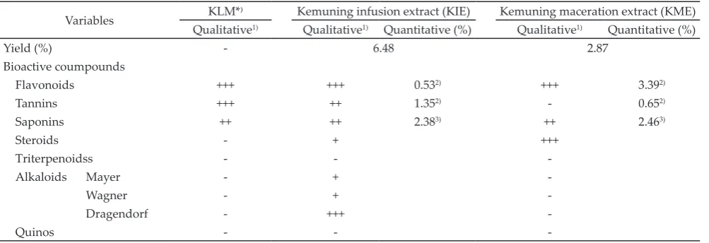
Table 1 showed that the bioactive compounds
in KIE were more diverse than in KLM and KME.
identified in the three of them. KIE was 2.73% lower in flavonoids as quercetin, 0.08% lower in saponins, and 0.7% higher in tannins compared to KME. The values indicated a high level of flavonoids other than quercetin
in KIE.
Larvae of Trichostrongylus sp., Haemonchus
sp., and Cooperia sp. which belong to the family of
Trichostrongylidae were identified in coprocultures con
-trol (P0) (Table 2). The success rate of the larval devel
-opment in the control group was 40%. The average
lethal time and percentage of mortality during larval development phase, infective larvae, and the adults of
Trichostrongylidae were presented in Table 3. The
analy-sis of variance (ANOVA) showed a gradual decrease in LT50 (shortening the lethal time) and an increase in mor -tality of Trichostrongylidae with increasing concentration
of treatment (P<0.01). The 7% KIE (P4) which had higher
concentration of tannins, was more effective than KME
(P8) and Oxfendazole (P10) in inhibiting larval develop
-ment, infective larvae (L3), and adult Trichostrongylidae
(by 93.14%, 94.39%, and 90% respectively). It was
expected that this concentration of KIE contained
0.04% quercetin, 0.09% tannins, and 0.17% saponins. Compared to the 7% KIE, the 5% KIE showed the same inhibition effect toward larval development but differ
-ent effect toward infective larvae (L3) and the adults. This ovicidal effect was also comparable with 7% KME (P8) and Oxfendazole (P9 and P10). Infusion method was more effective than the maceration as a potential
ovicidal, larvicidal, and nematicidal agent as the former
took lower concentration than the latter to generate the same efficacy.
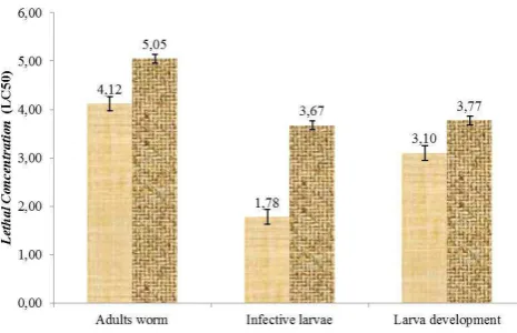
Figure 1 showed that higher concentration was needed to kill the adult helmints, followed by that for
Variables KLM*⁾ Kemuning infusion extract (KIE) Kemuning maceration extract (KME)
Qualitative1) Qualitative1) Quantitative (%) Qualitative1) Quantitative (%)
Yield (%) - 6.48 2.87
Bioactive coumpounds
Flavonoids +++ +++ 0.532) +++ 3.392)
Tannins +++ ++ 1.352) - 0.652)
Saponins ++ ++ 2.383) ++ 2.463)
Steroids - + +++
Triterpenoidss - -
-Alkaloids Mayer - +
-Wagner - +
-Dragendorf - +++
-Quinos - -
-Note: *)Kemuning Leave Meal; 1)Analysis in Biofarmaka Laboratory (2016); 2)Analysis in Balittro (2016); 3)Analysis in Balitnak (2016). (-) Negative, (+)
Positive weak, (++) Positive, (+++) Strong positive, (++++) Very strong positive. Table 1. Bioactive compounds and yield of kemuning leave extract
Note: *)Lenght of larva infective based on Van Wyk & Mayhew (2013).
Variables Genus infective larvae
Trichostrongylus sp. Haemonchus sp. Cooperia sp.
Total (%) 83.10 8.92 7.98
Classification factor
Sheat Present Present Present
Head shape Square Round Square
Refractile bodies Absent Absent Present
Diameter(μm) 18.87±0.21 22.49±1.36 33.84±3.55
Lenght (μm) 653.40±39.74 803.30±30.63 919.07±5.37
Literature* (μm) 730±50 882.13±102.63 1005±123
Figure
larval development phase and infective larvae, respec-tively. The concentration of KIE needed to kill the
nema-todes at each phase was always lower than that of KME.
DISCUSSION
Bioactive Compounds and Yield of Kemuning Leave Extract
Kemuning, especially its bioactive compounds with their various biochemical characteristics, was already known for its potential to control gastrointesti-nal nematodes in ruminants. To further investigate its
effectiveness, two types of extracts obtained with two different solvents (ethanol and distilled water) were
used. Between the two, KIE had a higher yield and
bioactive compounds, especially flavonoids and tannins.
This suggested that kemuning leave extract as a polar one. Possibly, the bioactive compounds were easier to be extracted in the solvent by infusion than by
macera-tion, explaining the high bioactive compounds and yield
in KIE. In addition to the effect of different polarities, a
heating process in infusion method could increase their solubilities, although it might also reduce the tannin
concentration. Khattab & Arntfield (2009) reported that
heating at 121oC for 20 min reduced tannin
concentra-tion by 1.42%-4.75%.
The anthelmintic activity of kemuning leave extract is accounted by their high saponin and tannin content which are known as antinutrition. High consumption of
tannins (4% dry matter (DM)) and saponins (3g/day or 0.4% DM) will cause detrimental effects for ruminants,
including a decrease in appetite, growth inhibition, as well as interferences in the morphology and proteolytic
activity of ruminal microbes (Mao et al., 2010; Hu et al., 2006; Barman & Rai, 2008). Based on the calculation on the usage of the concentration of the extract with total
tannins or saponin level, it was estimated that 7% KIE
contained 0.09% and 0.17% while the same concentra
-tion of KME contained 0.04% and 0.17% of tannins and
saponins respectively. It suggested that the saponins
and tannins in both extracts at 7% concentration were
at tolerable levels and were expected to be potential anthelmintic agents with no negative impacts on the livestock.
Ovicidal, Larvicidal, and Nematicidal Activity of Kemuning Leave Extract
In vitro test showed that the kemuning leave
extracts with both infusion and maceration method
were effective against Trichostrongylidae. However,
KIE showed a significant anthelmintic activity against larval development, infective larvae (L3), and the adults.
Similar results as in treatment P3, P4, and P8 were re-ported by Akhter et al. (2015) which studied the effect
of infuse (10%) and maceration (5%) extracts of ofneem, tamak, korola flower, chatim, and sharma on infective
larvae and the adults of Haemonchus contortus. However,
Treatments Mortality (%) Lethal time 50% (h)
Adult worms Infective larvae Larval development Adult worms Infective larvae
P0 4.0± 5.48ᵍ 8.50± 2.09f 0.00±0.00ᵉ * **
P1 16.0± 5.48f 75.39± 6.46c 33.14±6.11ᵈ * 2.05±0.08cd
P2 40.0± 7.07ᵈ 87.16± 4.45ab 36.48±5.78cd * 1.72±0.20ᵉ
P3 70.0± 7.07ᵇ 91.85± 4.64ab 88.23±3.49ᵃ 5.34±0.35ᵃ 1.09±0.47f
P4 90.0± 7.07ᵃ 94.39± 3.78ᵃ 93.16±5.93ᵃ 4.00±0.39ᵇ 1.09±0.45f
P5 28.0± 8.37ᵉ 42.06± 4.62ᵉ 54.92±2.26ᵇ * 3.38±0.11ᵃ
P6 42.0±10.95ᵈ 61.75± 6.11ᵈ 44.91±2.76c * 2.39±0.13ᵇ
P7 48.0±13.04cd 75.58±15.56c 40.15±6.44cd * 1.83±0.36de
P8 72.0±13.04ᵇ 84.34± 7.53ᵇ 86.64±6.44ᵃ 4.09±1.02ᵇ 1.72±0.22ᵉ
P9 28.0± 4.47ᵈ 63.07± 2.11ᵈ 90.24±8.46ᵃ * 2.24±0.05bc
P10 54.0± 5.48c 73.49± 2.55c 93.32±2.89ᵃ 5.12±0.50ᵃ 2.04±0.23cd
Table 3. The average lethal time and percentage of mortality of Trichostrongylidae
Note: Means in the same column with different superscript differ significantly(P<0.01). * = percentage of mortality was less than 50%; ** the time of death was more than 4 h. P0= Control; P1= KIE 1%; P2= KIE 3%; P3= KIE 5%; P4= KIE 7%; P5= KME 1%; P6= KME 3%; P7= KME 5%; P8= KME 7%; P9= OXF 0.0005%; and P10= OXF 0.005%. KIE= kemuning infusion extract; KME= kemuning maceration extract.
Figure 1. Lethal Concentration (LC50) of kemuning infuse ex
-tract (KIE, ME) ( K) and kemuning maceration extract
different results were obtained by Nery et al. (2010) with
infuse (5%) and maceration (4%) extract of Anarcadium
humile (5%) on larval development.
According to the classification of efficacy index
by Parasitology World Association for the Advancement of
Veterinary Parasitology, >90% anthelmintic activity char
-acterizes an effective product, 80%-90% for moderately effective, 60%-80% for less effective, and <60% for not effective product (Nery et al., 2010). Therefore, 7% KIE
(P4) was considered effective in inhibiting larval devel
-opment, infective larvae, and adult Trichostrongylidae. The toxicity or anthelmintic activity level of a plant
extract could be seen from its LC50, in which the lower
the LC50, the more toxic and the higher the anthelmintic
effect. It always took lower concentration for infuse
extract than the maceration to generate the same
mor-tality effect. It indicated that the former had a better anthelmintic function than the latter. Additionally, the
extract concentration increased in line with the level of development phase of the nematodes. It was due to the
difference in morphological development such as the difference in the structure between larval cuticle and
nematodes (Page et al., 2014), metabolism, and the
im-mune system of each phase of Trichostrongylidae.
In our previous study, the administration of KLM (equal to 1% extract) in the feed additives reduced EPG
of Strongylid by 43.67% in Ettawa crossbred goats. This
response was higher than the in vitro mortality level of adult worms in the current study. It suggested that the
herbal mechanism of kemuning leave is more effective
in inhibiting the reproduction than in causing mortal-ity of the worms. This hypothesis was supported by
Martínez-Ortíz-de-Montellano et al. (2010) who reported
that the administration of tannins (1200 μg/mL) from tzalam reduced EPG, the fecundity (damaging the
vulva), and the length of the worms.
The anthelmintic activity of kemuning leave
extract is attributed to its level of flavonoids, sapo
-nins, and tannins. Sapo-nins, tan-nins, and flavonoids
are expected to work synergistically as anthelmintic agents. Saponins and tannins damage the cuticle of
Trichostrongylidae. Saponin’s main action could alter
the pore shape and the cuticle permeability of the parasites while tannins could bind their cuticle protein
(Kerboeuf et al., 2008; Wang et al., 2010). These
mecha-nisms are expected to ease the diffusion of flavonoids
and increase the exposure to the compounds. Tannins
and flavonoids inhibit enzyme secretion (tyrosin kinase, nonspcific colinesterase, and esterase) that may cause
fatal intracellular instability, such as disorganization of
neuromuscular (neuron and muscle) cells and energy
metabolism. These conditions would lead to impaired
flexibility, paralysis, energy depletion, and eventu
-ally the death of Trichostrongylidae (Kerboeuf et al., 2008; Hoste et al., 2006). Hence, these three bioactive com-pounds have a potential to be alternative anthelmintic
drugs. Oxfendazole or benzimidazole (BZ) derivatives
have been widely used as anthelmintic drugs and are known to have a broad spectrum of actions against
nematode and cestode. BZ inhibits microtubule polym
-erization pathway by selectively binding the β-tubulin
subunit, hampering the absorption of glucose as an
energy source that leads to energy depletion and death
(Munguia et al., 2015).
In addition, the lethal time and mortality level of
Trichostrongylidae are also affected by in vitro condition
which is not 100% similar to its natural habitat (in vivo). The natural habitat of Trichostrongylidae, the goat’s ab
-omasum, produces abomasum juice containing nutrition
and enzyme that support their life and NaCl and help in
the regulation of ionic and fluidic balance.
In the previous study, kemuning leaves were used at such low amount that the reduction achieved was
still <50% even after 5 weeks of administration. The ef -fectiveness of kemuning leaves was lower compared to
Oxfendazole, in which 0.0005% of the latter (once, no replicate) could reduce EPG by 100%. Additionally, it
was possible that the amount of bioactive compounds being absorbed was still far from that for an optimal
reduction of EPG. Hence, to confirm the potential of
kemuning as an alternative to Oxfendazole, the
con-centration of 7% needs to escape rumen, implying that it can 90% act as anthelmintic agent. Further study was
expected to modify kemuning leaves for in vivo test that allows the bioactive compounds to escape rumen and gives the same response as in this in vitro test. It is important that the administration of kemuning leaves
must not negatively affect the ruminal microorganisms
for they are sensitive to antinutrition.
CONCLUSION
The 7% kemuning infusion extract demonstrated the highest efficiency in reducing the larval develop -ment, infective larvae, and the adult Trichostrongylidae
by 93.16%, 94.39%, and 90%, respectively. This
treatment can be developed as the most prospective herbal anthelmintic drug in controlling the infection by Trichostrongylidae.
REFERENCES
Akhter, S., A. R. Dey, S. Hossain, T. R. Dey, & N. Begum.
2015. In vitro anthelmintic effect of some medicinal
plants against Haemonchus contortus. J. Anim. Sci. Adv. 5:
1162-1170.
Alberti, E. J., S. A. Zanzani, N. Ferrari, G. Bruni, & M. T. Manfredi. 2012. Effects ofgastrointestinal nema -todes on milk productivity in three dairy goat breeds. Small Rumin. Res. 106: 12–17. https://doi.org/10.1016/j. smallrumres.2012.04.027
Barman, K. & S. N. Rai. 2008. In vitro nutrient digestibility, gas production and tannin metabolites of Acacia nilotica pods in goats. Asian-Australas. J. Anim. Sci. 21: 59-65. https:// doi.org/10.5713/ajas.2008.60161
Departemen Kesehatan RI. 2000. Acuan Sediaan Herbal.
Direktorat Jendral POM –Depkes RI, Jakarta.
Departemen Kesehatan RI. 2009. Farmakope Herbal Indonesia.
Direktorat Jendral POM –Depkes R, Jakarta.
Gautam, M. K., M. Gangwar, G. Nath, C. V. Rao, & R. K. Goel.
2012. In–vitro antibacterial activity on human pathogens
and total phenolic, flavonoid contents ofMurraya panicula-taLinn. Leaves. Asian. Pac. J. Trop. Biomed. 2:S1660-S1663. https://doi.org/10.1016/S2221-1691(12)60472-9
Harborne, J. B. 1987. Metode Fitokimia: Penuntun Cara Modern
Menganalisa Tumbuhan. 2nd ed. Institut Teknologi
Hoste, H., F. Jackson, S. Athanasiadou, S. M. Thamsborg, & S. O. Hoskin. 2006. The effects of tannin-rich plants on
parasitic nematodes in ruminants. Trends Parasitol. 22:
253-261. https://doi.org/10.1016/j.pt.2006.04.004
Hu, W. L., J. X. Liu, Y. M. Wu, Y. Q. Guo, & J. A. Ye. 2006. Effects
of tea saponins on in vitro ruminal fermentation and growth performance in growing Boer goat. Arch. Anim. Nutr. 60: 89–97. https://doi.org/10.1080/17450390500353119
Kerboeuf, D., M. Riou, & F. Guegnard. 2008. Flavonoid and Related Compounds in Parasitic Disease Control.
Mini. Rev. Med. Chem. 8: 116-128. https://doi. org/10.2174/138955708783498168
Khattab, R. Y. & S. D. Arntfield. 2009.Nutritional quality of
legume seeds as affected by some physical treatments 2.
Antinutritional factors. JFST 42:1113–1118. https://doi.
org/10.1016/j.lwt.2009.02.004
Kinsella, B., S. J. Lehotay, K. Mastovska, A. R. Lightfield, A. Furey, & M. Danaher. 2009. New method for the
analy-sis of flukicide and other anthelmintic residues in bovine
milk and liver using liquid chromatography–tandem mass spectrometry. Anal. Chim. Acta. 637: 196–207. https://doi.
org/10.1016/j.aca.2008.10.072
Mao, H. L., J. K. Wang, Y. Y. Zhou, & J. X. Liu. 2010. Effects
of addition of tea saponins and soybean oil on methane production, fermentation and microbial population in the
rumen of growing lambs. Livest. Sci.129:56–62. https://doi. org/10.1016/j.livsci.2009.12.011
Munguia, B., M. Michelena, E. Melian, J. Saldana, X. Ures, E. Manta, & L. Domínguez. 2015. Development of novel
valerolactam-benzimidazole hybrids anthelmintic
de-rivatives: Diffusion and biotransformation studies in hel
-minth parasites. Exp. Parasitol. 153: 75–80. https://doi. org/10.1016/j.exppara.2015.03.013
Molento, M. B., F. Fortes, D. Pondelek, F. A. Borges, A. C. S. Chagas, J. F. J. Torres-Costa, & F. Geldhof. 2011. Challenges of nematode control inruminants: focus on
Latin America. Vet. Parasitol. 180: 126–132. https://doi. org/10.1016/j.vetpar.2011.05.033
Martínez-Ortíz-de-Montellano, C., C. Arroyo-López, I. Fourquaux, J. F. J. Torres-Acosta, C. Sandoval-Castro, & H. Hoste. 2013. Scanning electron microscopy of
Haemonchus contortus exposed to tannin-rich plants under
in vivo and in vitro conditions. Exp. Parasitol. 133:281-286.
https://doi.org/10.1016/j.exppara.2012.11.024
Nery, P. S., F. A. Nogueira, E. R. Martins, & E. R. Duarte.
2010. Effect of Anacardium humile on the larval develop-ment of gastrointestinal nematodes of sheep. Vet Parasitol. 171:361–364. https://doi.org/10.1016/j.vetpar.2010.03.043
Ng, M. K., N. Y. Abdulhadi, Y. K. Cheah, S. K. Yeap, & N. B. Alitheen. 2012. Bioactive studies and chemical constitu-ents of Murraya paniculata (Linn) Jack. IFRJ 19:1307-1312.
Page, P. A., G. Stepek, A. D. Winter, & D. Pertab. 2014. Enzymology of the nematode cuticle: A potential drug tar-get?. Int. J. Parasitol. 4: 133-141. https://doi.org/10.1016/j.
ijpddr.2014.05.003
Syahadat, R. M. & S. A. Aziz. 2012. Pengaruh komposisi media dan fertigasi pupuk organik terhadap kandungan bioaktif
daun tanaman kemuning (Murraya paniculata (L.) Jack) di pembibitan.Bul. Littro. 23: 142-147.
Vaghasiya, Y., R. Dave, & S. Chanda. 2011. Phytochemical analysis of some medical plants from western region of
India. Res. J. Med. Plant 5: 567-576. https://doi.org/10.3923/ rjmp.2011.567.576
Van Wyk, J. A. & E. Mayhew. 2013. Morphological identifica -tion of parasitic nematode infective larvae of small
rumi-nants and cattle: A practical lab guide. Onderstepoort J.
Vet. Res. 80:1-14. https://doi.org/10.4102/ojvr.v80i1.539
Wang, Y., Y. Zhang, Z. Zhu, S. Zhu, Y. Li, M. Li, & B. Yu. 2007. Exploration of the correlation between the structure, hemo-lytic activity, and cytotoxicity of steroid saponins. Bioorg.
Med. Chem. 15: 2528–2532. https://doi.org/10.1016/j. bmc.2007.01.058