RESULTS OF CLAVICLE FRACTURE TREATMENT IN
CHILDREN
Anko Antabak1, Nikša Matković2, Lana Stanić2, Judith Adrianne Deutsch3, Dino Papeš1, Robert Karlo4, Ivan Romić1, Nino Fuchs1 and Tomislav Luetić1
1Clinical Department of Surgery, Zagreb University Hospital Center; 2School of Medicine, University of Zagreb; 3Department of Anesthesiology, Zagreb University Hospital Center, Zagreb; 4Department of Pediatric Surgery,
Zadar General Hospital, Zadar, Croatia
SUMMARY – Treatment of clavicle fracture is principally outpatient. Operative treatment is accompanied by the need for more x-rays and possible complications. Fractures with absolute indica-tions for operative treatment occur only sporadically and these indicaindica-tions are relatively clear, but children often undergo surgery because of relative indications (shortening, fragment displacement, multifragmentary fractures), which are open to debate. In a retrospective study on 256 children, of 44 (17%) patients that received operative treatment only one 17-year-old boy had an absolute indi-cation for surgical intervention. Other indiindi-cations were fragment distraction (22 mm on average), age, associated injuries, and multifragmentary fracture. The placement of K-wire of appropriate thickness is often difficult, since the wire tends to bend and break, and patients have to undergo two additional operations of plate and screw fixation and later removal. In this retrospective study, we considered the advantages of using titanium or an elastic steel pin. All patients had favorable outcome, although some experienced numbness around the operation scar (4.5%), skin infections around the wire (15%), and/or the implanted K-wire damage (7%).
Key words: Clavicle – injuries; Fractures, bone – surgery; Fractures, bone – therapy; Child
Correspondence to: Assist. Prof. Anko Antabak, MD, PhD, Clini-cal Department of Surgery, Zagreb University Hospital Center, Šalata 2, HR-10000 Zagreb, Croatia
E-mail: [email protected]
Received February 27, 2014, accepted August 21, 2015
Introduction
The clavicle holds the shoulder away from the ster-num and is constantly affected by bending forces. Due to the child’s gentle skeletal system and more frequent injury exposure, clavicle fractures are twice as com-mon as in adults, accounting for 10%-15% of all child age fractures1. These fractures heal quickly,
complica-tions are rare, and operative treatment is occasionally needed.
Clavicle growth length develops mostly from its medial epiphysis (until 25 years of age), and its width from the periost. Fracture shortenings are
compensat-ed for by acceleratcompensat-ed growth length and angular de-formities due to enhanced periost activity. The periost fibrous layer, which is rich in collagen and elastin fi-bers, is firmer than the cortical layer and is responsible for clavicle bending tolerance. In preschool children, the diaphysis bows without cortical layer breaking, but with stronger bending forces; this leads to partial breaking of the periost and cortical layers with an-gular deformity (greenstick fracture). The germina-tive layer gives enormous regeneration potential to the periost, which is expressed by huge periosteal callus around the fracture site. Fractures are most com-mon in the mid-third, which account for more than 80% of all clavicle fractures2. Clavicle biomechanical
features change during the growth period. Children older than 6 years usually sustain displaced fractures because of bending forces that cause complete frac-tures of the cortex and periost. The older the child,
the more likely they are to sustain complete fracture, larger fragment dislocation and shortening. Multi-fragmentary clavicle fractures occur exclusively in older adolescents. The treatment purpose is to restore complete shoulder movement within as a short time as possible using the simplest procedure. Clavicle frac-ture treatment is principally ambulatory and nonop-erative. However, there are fractures and cases where the choice of treatment is not that simple3. Experts in
pediatric traumatology agree that absolute indications for operative treatment are open fractures, fractures with neurovascular bundle injury, skin penetration, and skin tenting by sharp fragments4. Luckily, the
prevalence of these fracture types is low. Open reposi-tions and mid-shaft dislocated fracture stabilization are still disputable topics5. The decision on a relative
indication for operative treatment is principally based on patient age, but also some other factors6. Patient
expectations, parent demands, fracture layout, general patient condition and associated injuries have to be taken into consideration. The decision must be indi-vidualized and made after comprehensive discussion among the child, parents and attending physician, with their complete understanding the risks and ben-efits of every treatment plan. Some authors rarely do operative fracture stabilizations in children7, whereas
others use this treatment in pubertal age children ac-tive in sports and following the indications as in adults (fragment distraction by two or more centimeters)8,9.
The operative procedure itself has evolved over time. Now, surgeons insert a wire or needle through the clavicle (intramedullary), maintaining the shaft and preventing further fragment displacement. How-ever, the risk of spontaneous implant migration may arise; therefore, some surgeons use plate and screw fixation. More recently, elastic steel pins or titanium pins are used for stabilization, which can be placed intramedullary percutaneously backwards through the back side of the lateral third of the clavicle. Using this method, resuming full sports activity is recom-mended in 4 to 6 weeks after full recovery. With the previous method, return to sports activity is usually 3 months after surgical fixation. Regardless of the method of treatment for clavicle fracture, results are good in children. The most severe complications are pseudoarthrosis and prolonged healing. They occur rarely and almost never in children younger than 11
years. After treatment, some patients experience pain, reduced shoulder movement, weakness and paresthe-sia, but also hypertrophic callus and aesthetic flaws. Infections, bleeding, neurovascular damage, pneu-mothorax, osteosynthetic material migration, pro-longed healing and pseudoarthrosis can accompany operative treatment10.
In this article, pediatric patients with clavicle frac-tures, treatment procedures and results are presented and analyzed.
Patients and Methods
This retrospective study included 284 children aged up to and including 18 years, treated for clavicle fractures from June 1, 2008 until June 1, 2013 at Za-greb University Hospital Center. Data were collected from archived medical documentation, medical files, and outpatient/inpatient treatment protocols. A table file was assembled and data filled in for each study patient, including age at fracture occurrence, gender, side and site of fracture, ad latus fragment displace-ment, shortening, angulations, fragment impaction, location in the city where the injury was inflicted, mechanism of injury, number of x-rays, outpatient/ inpatient treatment, and treatment outcome. For op-eratively treated children, the method used, osteo-synthesis device, plate or Kirschner wire were noted. Fracture side, the fractured third, ad latus displace-ment, shortening, angulations and impaction were read from x-ray files taken on initial examination in the emergency room. In this article, angulation ex-cludes the presence of shortening and/or ad latus dis-placement, but does not exclude fragment impaction. Children with multiple injuries were considered only by the number of clavicle x-rays taken. Intraoperative diascopy used in operatively treated children was not added to the x-ray count, and computed tomography scans were not included. In cases where data on the location and mechanism of injury were incomplete, parents were contacted by phone to collect this infor-mation. Associated injuries were also coded using the ICD-10 classification and classified as superficial or complicated injuries, open wounds and fractures. The injuries treated at the hospital were described as severe injuries, as opposed to less severe ones that were treat-ed outpatiently. Of 284 children, 28 were excludtreat-ed
from the statistics due to incomplete documentation. All procedures were performed in accordance with the institutional ethical standards, regional Commit-tee on Human Experimentation and Helsinki Decla-ration from 1975. Patient names, initials and hospital numbers were not disclosed.
Results
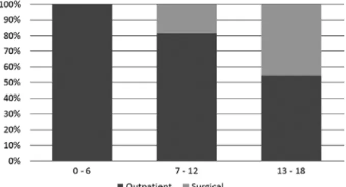
In this study, 256 children were analyzed, includ-ing 160 (62.5%) boys and 96 (37.5%) girls, mean age 8.3 years (9.7 years in boys and 5.9 years in girls). Of all study children, 142 (55%) had right sided fractures and 114 (45%) left sided fractures. Boys broke equally the left and the right clavicle, while girls had a ten-dency to fracture the left side twice as often as the right side. There were 226 children with mid-third fractures, 26 with lateral third, and four with medial third fractures. Multifragmentary fractures were seen in 20 (7.8%) children, of which 16 received operative treatment. Hospital (operative) treatment was carried out in 44 (17.2%) and ambulatory management in 212 (82.8%) children. The mean age was 7.3 years in children treated as outpatients and 14.4 years in op-eratively treated patients. Age distribution of children treated as outpatients and inpatients (operatively) is shown in Figure 1. Only three of operatively treated children were younger than 10 years, the youngest one was aged seven years. According to initial radiologi-cal findings, there were 135 (53%) fractures without significant displacement and 121 (47%) fractures with clavicle shortening (Fig. 2). As for deformation types, the most common were angulations (20%), and the most infrequent ones were impactions (3%). The rates of ambulatory and operative treatment
accord-ing to types of displacement are shown in Figure 3. Superficial injuries (excoriations, skin abrasions and hematomas) were recorded in 75 (44 inpatient and 31 outpatient) children, localized on the head (34.2%), shoulders (26.3%), and knees and lower legs (13.1%). Wounds that required primary treatment were seen in ten children, localized on the head in six, two on the forearms and two on lower legs, all of which re-ceived hospital treatment. There were 15 associated fractures and all of them received hospital treatment. All the 4 skull bone fractures were caused by a car hit-ting the pedestrian, and the children were of school age. Among male patients, one had concussion, one had nasal bone fracture accompanied by subarachnoid hemorrhage, and one had pelvis fracture. As a result of falling from a tractor, one boy sustained upper leg fracture, brachial plexus injury, and open head and forearm wounds. Two boys had associated rib frac-tures. Fist bone fractures, foot bone fractures and open head wounds with numerous superficial wounds were sustained by two boys that also broke their clavicles by falling from a bicycle driving down a slope through
Fig. 1. Type of treatment by age.
Fig. 2. Occurrence of clavicle fragment displacement types in 256 children.
Fig. 3. Nonoperatively and operatively treated children according to clavicle displacement type.
the woods. Concussion, radius and clavicle fractures occurred together in a passenger in the overturned car. Another four children had broken forearm bones. Ambulatory treatment was performed with the appli-cation of a figure-eight bandage in 173, sling in 31, Dessault’s bandage in three, and hard reinforced Des-sault’s bandage in five children. Immobilization was maintained for one to four weeks. Outpatient physical therapy was conducted in 54 children. The patients had a mean of 1.45 radiological follow up visits over a period of 20 days. Upon finishing physiotherapy, 22 children experienced moderate pain, 19 children had enhanced callus formation, and three children experi-enced brachial plexus irritation. Terminal movement difficulty was seen only in one 15-year-old girl. Be-cause of prolonged healing, professional sports activ-ity for her was not allowed for about eight months. The end result of her treatment was completely fa-vorable. The operation was performed in general an-esthesia, including open reposition and adaptation of fragments under visual control and fixation with intramedullary placed Kirschner wire. An absolute indication for surgical intervention was seen in only one 17-year-old boy (having sharp bone fragments threatening skin penetration) (Fig. 4). Other indica-tions were fragment distraction (22 mm on average), age, and associated injuries. Three children (aged 16, 17 and 18 years) received fixation with a rigid osteo-synthetic plate and screws. Children that received operative treatment were followed-up by conventional radiological films 3.2 times on average, but there were
also intraoperative diascopies to be added for each child. K-wires were removed outpatiently after 3 to 5 weeks, while plates were operatively removed at the one-day surgery outpatient clinic. At follow up exam-inations, four weeks after K-wire removal, all of the children had full range of motion. There were 5 local skin infections around the K-wire and one boy had a large pyogenic granuloma. Three children had signifi-cant bowing of the K-wire (Fig. 5), and in an athlete the K-wire broke at the fracture site. Long term par-esthesia was noted in two cases at the scar site. In one 17-year-old handball player, fracture union failed to occur even seven months after operative intervention and with severe K-wire bowing. A repeat operation was conducted to remove the old wire and fixation was achieved with a plate and screws. The outcome of this patient was excellent. In another patient, frac-ture union did not occur after nine months, even af-ter K-wire placement. The patient developed painful atrophic pseudoarthrosis. Stabilization was achieved again using a plate and screws, and spongious bone graft. Four months later, the clavicle healed properly. Plate fixation was initially done once, while in two patients it was a second therapeutic choice.
Discussion
Clavicle fractures are frequent in all ages, especial-ly in school and preschool children. The average pa-tient age in our study was eight years, and there were slightly more boys (average age 10) than girls (average age 6). The most common clavicle fractures were mid-third fractures; they comprised 88.3% of all cases. Distribution by age, gender and side was similar to other series. The surgeon’s decision on treatment was
Fig. 4. Clavicle fragment under the skin, an absolute in-dication for open treatment.
Fig. 5. A 16-year-old boy injured in traffic accident as a motorcycle driver. First follow up x-ray on day 7 after open reposition and intramedullary placed K-wire with 1.5 mm radius.
made on the basis of fracture appearance and child’s age. Treatment of fractures without or with minimal offset fragments (seen in 53% cases) was ambulatory (immobilization). Healing and recovery was quick and complete. This group had no complications. Chil-dren up to 12 years of age with clavicle fractures with minimal offset fragments almost exclusively received ambulatory treatment (176 children). Operative treat-ment was performed in polytraumatized patients and those with the fragments offset of one or more centi-meters (14 children). At this age, the complication rate was low and pseudoarthrosis almost never appeared. There were no severe complications in this group (190 children). At the age of 16, the occurrence of compli-cations increased. In a retrospective study of operative treatment in 24 children (mean age 12.6, range 7-16 years), Mehlman et al. have reported only mild com-plications. No infection or pseudoarthrosis occurred. Complications included scars, local sensitivity in two patients, and transient neurapraxia of the ulnar nerve. The authors accentuate that the growth and remod-eling in adolescents and teenagers (near skeletal ma-turity) is not that much foreseeable and can appear as that in adult bone11. It is disputable how to treat
children aged from 13 to 18 years with displaced frac-tures. There are almost no studies analyzing clavicle fracture treatments in this age group. Vanderhave et
al. treated a group of children aged 12-18 years
op-eratively in 27% of cases, with a mean shortening of 2.7 cm. For stabilization they used the plate and screw method. Four nonoperatively treated children had angulation and excessive pain after the fragments had grown together, so corrective osteotomy was nec-essary12. Apart from operative treatment frequency
differences among similar age groups, differences be-tween operative methods were also significant. Some authors used only one procedure. Vander et al. report treatment results in 43 adolescents (mean age 15.4 years), 17 (39.6%) of them treated operatively. The au-thors recommend fixation with a plate and screws13.
Other authors used several methods in adolescents. Prinz et al. describe treatment results in 60 children, ten (20%) of which underwent surgery, all of them older than 10 years. Fragments were stabilized by K-wire (in 2 cases) or by elastic stable intramedullary osteosynthesis (in 8 cases). Pseudoarthrosis was de-scribed in one 8-year-old girl with ambulatory
treat-ment, but after surgical resection and TEN fixation the end result was good. In their study, all the chil-dren aged up to 10 years had excellent outcome. Older children, regardless of treatment method, had good functional outcome, but operatively treated children had a less favorable aesthetic result. They recommend the ESIN method14. Some authors used five
differ-ent methods for fragmdiffer-ent stabilization. Kubiak and Slongo report on a retrospective study that included 15 children (14 boys and one girl, mean age 13.1, age range 9-15 years) having undergone operative treat-ment in a period from 1989 to 2000. They used in-tramedullary stabilization (n=5), outer fixation (n=2), Dexon bone suture (n=3), K-wires (n=4), and/or a screw (n=2). They report no serious complications four months after the procedure15. Nearly all studies report
good early operative results, and there was no need for further investigations. Nevertheless, Namdari et
al. analyzed functional outcomes of operative
treat-ment in 14 adolescents (mean age 12.9, age range 10-15 years). Using demographic and radiologic indices, radiologic and functional outcomes, arm and shoulder disability (by DASH questionnaire and SST shoulder test) were evaluated. There was no lack in function, only pain that occurred with physical activity, and long term numbness of the incision area, operation or fracture site16.
In our study, operative treatment was used in 44 (17%) children. Only one boy had an absolute indica-tion for operative treatment. Operative treatment was most commonly used in children older than 14 years, with shortening by two or more centimeters, and children with associated serious injuries. All children younger than 6 years, without offset, with impacted fractures and fractures with angulation were treated on outpatient basis. Only two 17-year-olds, with shortening of 1 cm, lower leg fractures and commo-tional syndrome were treated operatively (motorcycle fall and a passenger in traffic). Analysis of ambulatory treatment results revealed that there were no compli-cations (only one prolonged union). In the operatively treated children, numbness at the operation scar site persisting for several months was recorded in two (4.5%), skin infection around the wire in six (15%), and implanted K-wire damage in three (7%) patients. An insufficiently thick K-wire was the cause of its de-formation and rupture. The clavicle has two arches,
the medullary canal is narrow, and because of this an adequately thick K-wire placement is often not pos-sible, whereas a rigid plate osteosynthesis has a major downside of additional hospitalization, anesthesia and operative removal. Postoperative scar as an aesthetic defect cannot be ignored in girls. There are also anxi-ety and discomfort during hospital treatment, which must be considered as well.
Operative treatment of clavicle fractures with intramedullary positioned Kirschner wire in adoles-cents is accompanied by frequent complications. The authors of this study are considering a new method to be adopted for closed reposition and intramedullary stabilization, this being achieved by elastic stable os-teosynthesis with titanium or an elastic steel pin.
References
1. England S, Sundberg S. Management of common pediatric fractures. Pediatr Clin North Am. 1996;43:991-1012. 2. Staheli LT. Pediatric orthopedic secrets. Philadelphia:
Han-ley & Belfus, 1998, pp. 189-202.
3. Hill JM, McGuire MH, Crosby LA. Closed treatment of displaced middle-third fractures of the clavicle gives poor re-sults. J Bone Joint Surg Br. 1997;79:537-9.
4. Curtis RJ Jr. Operative management of children’s fractures of the shoulder region. Orthop Clin North Am. 1990;21:315-24. 5. Hosalkar HS, Parikh G, Bittersohl B. Surgical fixation of
displaced clavicle fracture in adolescents: a review of litera-ture. Orthop Rev (Pavia). 2013;5:e29.
6. Seif El Nasr M, Essen H, Teichmann K. Clavicular fractures in pediatric traumatology. Unfallchirurg. 2011;114:300-10. 7. Weber BG, Bruner C, Freuler F. Treatment of fractures in
chil-dren and adolescents. New York: Springer, 1980,pp. 58-64. 8. Kocher MS, Waters PM, Micheli LJ.Upper extremity
inju-ries in the paediatric athlete. Sports Med. 2000;30:117-35. 9. Wiesel BB, Getz CL.Current concepts in clavicle fractures,
malunions and non-unions. Curr Opin Orthop. 2006;17: 325-30.
10. Manske D, Szabo R.The operative treatment of mid-shaft clavicular non-unions. J Bone Joint Surg Am. 1985;67:1367-71. 11. Mehlman CT, Yihua G, Bochang C, Zhigang W. Operative
treatment of completely displaced clavicle shaft fractures in children. J Pediatr Orthop. 2009;29:851-5.
12. Silva SR, Fox J, Speers M, Seeley M, Bovid K, Farley FA, Vander Have KL, Caird MS. Reliability of measurements of clavicle shaft fracture shortening in adolescents. J Pediatr Or-thop. 2013;33:e19-22.
13. Vander Have KL, Perdue AM, Caird MS, Farley FA. Opera-tive versus nonoperaOpera-tive treatment of midshaft clavicle frac-tures in adolescents. J Pediatr Orthop. 2010;30:307-12. 14. Prinz KS, Rapp M, Kraus R, Wessel LM, Kaiser MM.
Dis-located midclavicular fractures in children and adolescents: who benefits from operative treatment?. Z Orthop Unfall. 2010;148:60-5.
15. Kubiak R, Slongo T. Operative treatment of clavicle frac-tures in children: a review of 21 years. J Pediatr Orthop. 2002;22:736-9.
16. Namdari S, Ganley TJ, Baldwin K, Rendon Sampson N, Hosalkar H, Nikci V, Wells L. Fixation of displaced mid-shaft clavicle fractures in skeletally immature patients. J Pe-diatr Orthop. 2011;31:507-11.
Sažetak
REZULTATI LIJEČENJA PRIJELOMA KLAVIKULA U DJECE
A. Antabak, N. Matković, L. Stanić, J. A. Deutsch, D. Papeš, R. Karlo, I. Romić, N. Fuchs i T. Luetić
Liječenje fraktura klavikule je prevenstveno ambulantno. Operativno liječenje zahtijeva dodatne rentgenske snimke i praćeno je mogućim komplikacijama. Frakture s apsolutnom indikacijom za operativno liječenje se pojavljuju sporadično i te indikacije su jasne, ali često djeca budu operirana zbog relativnih indikacija koje nisu sasvim jasne. U retrospektivnoj studiji na 256 djece, od kojih je 44 (17%) operativno liječeno, samo jedan dječak od 17 godina je imao apsolutnu indikaciju za operaciju. Ostale indikacije su bile distrakcija (prosječno 22 mm), dob, udružene ozljede i multifragmentarne frakture. Postavljanje Kirschnerove žice odgovarajuće debljine je često nemoguće zbog savijanja i pucanja žice pa bolesnici moraju ići na dva dodatna zahvata, fiksaciju pločicom i vijcima i vađenje osteosintetskog materijala. U sklopu ove studije razmotrili smo prednosti korištenja titanskog čavla. Svi bolesnici su imali dobar ishod, iako se u određenom broju operiranih pojavila utrnutost oko ožiljka (4,5%), infekcija rane (15%) i oštećenje Kirschnerove žice (7%).