University of Zagreb
FACULTY OF PHARMACY AND BIOCHEMISTRY
Iva Krtalić
DEVELOPMENT OF INNOVATIVE IN SITU
FORMING GELS FOR TOPICAL
OPHTHALMIC DELIVERY
DOCTORAL DISSERTATION
UNIVERSITY OF ZAGREB
FACULTY OF PHARMACY AND BIOCHEMISTRY
Iva Krtalić
DEVELOPMENT OF INNOVATIVE
IN SITU
FORMING GELS FOR TOPICAL
OPHTHALMIC DELIVERY
DOCTORAL DISSERTATION
Supervisor:
Jasmina Lovrić, Ph.D.
SVEUČILIŠTE U ZAGREBU
FARMACEUTSKO-BIOKEMIJSKI FAKULTET
IV
A KRTALIĆ
RAZVOJ INOVATIVNIH
IN SITU
GELIRAJUĆIH PRIPRAVAKA ZA
TOPIKALNU PRIMJENU OFTALMIČKIH
LIJEKOVA
DOKTORSKI RAD
Mentorica:
i
zv. prof. dr. sc. Jasmina Lovrić
The doctoral thesis was submitted to the Faculty Council of the Faculty of
Pharmacy and Biochemistry, University of Zagreb in order to acquire a PhD degree
in the area of Biomedicine and Health, the field of Pharmacy, the branch of
Pharmacy.
The work presented in this doctoral thesis was performed at the Faculty of
Pharmacy and Biochemistry and PLIVA CROATIA Ltd. under supervision of
Associate Prof.
Jasmina Lovrić
, PhD. The research conducted was financed by the
project „Development of
in vitro and ex vivo models for the permeability testing of
new topical ophthalmic formulations“ (04.01/56, „Partnership in research“,
ZAHVALE / ACKNOWLEDGEMENTS
Na kraju ovoga izazovnoga, ali i ispunjujućega te poprilično dugoga putovanja koje je snažno
obilježilo moj život, željela bih zahvaliti dragim ljudima, bez kojih ono ne bi bilo isto, a do čijega bi kraja bilo značajnoteže stići.
Prvenstveno, od srca veliko hvala mojoj mentorici izv. prof. dr. sc. Jasmini Lovrić na iznimnoj
pomoći u nastajanju ove disertacije, na jasnoj viziji i vodstvu tijekom našega istraživanja, savjetima, potpori i strpljenju. Hvala na svemu što si me naučila, ostavilo je veliki trag u meni.
Hvala kolegicama i kolegama iz I&R, mojim Fizikalkama (Liljani, Dunji i Ivani) kojima nije bilo
teško montirati haubicu na reometar po milijunti put. Dragoj Ani i Nikolini velika zahvala na
pomoći pri izradi uzoraka - vjerojatno će još dugo i naširoko zaobilaziti ledene kupelji.
Hvala Senki na nemjerljivoj pomoći u svijetu statistike. Tvrtku zahvala na lijepim grafičkim
idejama i rješenjima. Tanja, hvala puno na logističkoj, a često i na emocionalnoj potpori kada mi je bila potrebna.
Mojim Pliva-curkama - Ivani, Jasmini, Ivi, Mihaeli, Maji, Marini te cimeru - veliko hvala za
potporu sve ove godine, lako je raditi kada si okružen prijateljima. Hvala dugogodišnjim
prijateljima (mojim Metkovcima, curama iz Gorjanske i trešnjevačkoj ekipici) – dugo su slušali o
nekim gelovima i davali mi potporu da ustrajem u ovom cilju.
Neizmjerno sam zahvalna mojoj obitelji, najdražim Ujdurima – mami, tati, Kati i Tomi što su, bez iznimke, uvijek i u svakom smislu, pravo značenje izraza ''biti tu'', lijepo je biti dio ove obitelji.
Hvala i mojoj ''novoj'' obitelji, Krtalićima, na velikoj podršci tijekom nastajanja ovoga rada.
I na kraju, hvala mojim dečkima Viti i Vedranu, dvojcu najvrjednijem u mom životu, mojoj radosti, veselju, miru i snazi… hvala na podršci, razumijevanju, razgovorima, zagrljajima,
TABLE OF CONTENTS
SUMMARY ... 1
SAŽETAK ... 3
1. INTRODUCTION ... 8
2. THEORETICAL PART ... 11
2.1. Ophthalmic drug delivery ... 12
2.1.1. Topical ophthalmic drug delivery ... 13
2.2. Topical ophthalmic dosage forms ... 20
2.2.1. Innovative ophthalmic drug delivery systems... 22
2.3. Hydrogels in ophthalmic drug delivery... 25
2.3.1. In situ forming ophthalmic gels ... 26
2.3.2. pH-responsive in situ forming gels ... 30
2.3.3. Ion-responsive in situ forming gels ... 31
2.3.4. Temperature-responsive in situ forming gels ... 33
2.4. Poloxamer-based in situ forming ophthalmic gels ... 36
2.5. Considerations for formulation development of poloxamer-based in situ forming ophthalmic gels ... 40
2.6. Chitosan in ophthalmic drug delivery ... 42
2.7. Rheological characterization of in situ forming gels ... 44
2.8. Quality by Design and Design of Experiments in pharmaceutical development ... 51
2.8.1. DoE workflow ... 53
2.9. Ocular tolerability assessment... 55
3. AIMS OF THESIS ... 57
4. MATERIALS AND METHODS ... 59
4.1. Materials ... 60
4.2. Methods ... 61
4.2.1. Preparation of in situ forming ophthalmic gels ... 61
4.2.2. Osmolality, pH and transparency determination ... 61
4.2.3. Measurement of temperature of gelation and complex viscosity ... 62
4.2.4. Design of experiments... 63
4.2.5. Oscillatory rheological characterization – strain sweep, frequency sweep and gelation time testing ... 65
4.2.6. Rotational rheological characterization... 66
4.2.7. Sterilization ... 66
4.2.8. Physical stability of samples at room temperature ... 67
4.2.9. In vitro release testing ... 67
4.2.9.1. Solubility testing and choice of IVR medium ... 69
4.2.9.2. Membrane selection study... 69
4.2.9.3. Data presentation and statistical analysis ... 70
4.2.10. UPLC analyses ... 70
4.2.10.1. Validation of UPLC method ... 71
4.2.11. In vitro biocompatibility study ... 72
4.2.11.1. Cell culture conditions ... 72
4.2.11.2. Cultivation of the HCE-T cell-based model... 73
4.2.11.3. MTT Assay ... 73
5. RESULTS AND DISCUSSION ... 74
5.1. In situ forming ophthalmic gel development and optimization using a Design of Experiments ... 75
5.2. The model of temperature of gelation ... 79
5.3. Models of rheological properties: complex viscosity and storage modulus ... 83
5.3.1. The model of complex viscosity ... 83
5.3.2. The model of storage modulus ... 86
5.4. Selection of rheologically improved in situ forming ophthalmic gel ... 89
5.4.1. In depth rheological characterization of in situ forming ophthalmic gel – oscillatory tests ... 91
5.4.2. Dynamic viscosity evaluation by rotational rheology ... 97
5.5. Effect of active pharmaceutical ingredients on temperature of gelation and complex viscosity ... 100
5.5.1. Evaluation of dynamic viscosity of systems containing APIs ... 103
5.5.2. Gelation time ... 104
5.6. Drug release from in situ forming gels ... 105
5.6.1. Validation of UPLC method for quantification of TIMO ... 106
5.6.2. Development of in vitro release method ... 110
5.6.2.1. Selection of receptor medium – solubility testing... 110
5.6.2.2. Membrane selection ... 110
5.6.3. Evaluation of IVR from the optimal in situ forming gel ... 116
5.7. Sterilization ... 117
5.8. Stability study ... 121
5.9. Biocompatibility assessment of in situ forming ophthalmic gel ... 123
6. CONCLUSIONS ... 125
REFERENCES ... 128
1
2 The anatomical barriers and the tear fluid dynamics in the precorneal area have a huge effect on the eye-related drug bioavailability. In situ forming gels stand out as the formulation candidates for the improvement of bioavailability and efficacy of topical ophthalmic drugs. These are prepared as liquid dosage forms that undergo phase transition on eye surface to form viscoelastic gel in response to an environmental stimulus following topical administration. During formulation development, in situ forming ophthalmic gels need to be fine-tuned considering all the biopharmaceutical challenges of the front of the eye. The aim of this thesis was to develop temperature-responsive in situ forming poloxamer P407/ poloxamer P188/chitosan gel fine-tuned in terms of polymer content, temperature of gelation, viscosity and storage modulus by applying Quality by Design principles. Minimizing the total polymer content, while retaining the advantageous rheological properties, has been achieved by means of D-optimal statistical design.
In situ forming gels were prepared by cold method and analyzed by rheological temperature
sweep test. Statistical software was used to set experimental design, establish the mathematical model and analyze the collected data. Validated mathematical models enabled the selection of 4 candidates of poloxamer 407/poloxamer 188/chitosan systems, which viscoelastic behavior was studied in depth by oscillatory rheological tests under biorelevant conditions (after dilution with simulated tear fluid in the ratio of 40:7). The optimal in situ forming gel was selected based on minimal polymer content (P407, P188 and chitosan concentration of 14.2, 1.7 and 0.25%, w/w, respectively), favorable rheological characteristics and in vitro resistance to tear dilution. The formulation robustness against entrapment of active pharmaceutical ingredients (API) with different physicochemical characteristics was demonstrated using four APIs in the concentrations and combinations relevant for topical ophthalmic use. Through discriminative in vitro release test, adequate release profile of model drug was proven. Biocompatibility of the optimal in situ
forming gel was confirmed employing 3D corneal epithelial cell models. In conclusion, a systematic approach to in situ forming gel development implemented in this thesis resulted in a stable and robust formulation characterized by ease of formulation preparation, ability for sterile filtration and autoclaving, ease of administration, accuracy of dosing, avoidance of eye-related discomfort, biocompatibility and prolonged residence at the eye surface.
KEYWORDS: ophthalmic drug delivery, quality by design (QbD), poloxamers, chitosan, rheology, temperature-responsive in situ forming gel, temperature of gelation.
3
SAŽETAK
Uvod: Topikalna je primjena glavni put primjene oftalmičkih lijekova, a kapi za oko najčešće
korišten farmaceutski oblik. Međutim, pri primjeni kapi za oko, zbog dinamičnih barijera prekornealonog područja, dolazi do brze eliminacije lijeka s površine oka. Stoga je lijek dostupan
za apsorpciju kroz samo nekoliko minuta što rezultira vrlo niskom bioraspoloživošću, uglavnom
manjom od 5%. Klasični tehnološki pristupi, s ciljem produljenja vremena zadržavanja lijeka na
površini oka, uključuju pripravu viskoznih otopina, gelova i masti. Međutim, ti su sustavi
povezani sazamućenjem vida i otporom gibanju vjeđa pri treptanju te poteškoćama pri doziranju
što umanjuje suradljivost bolesnika. Iz navedenih je razloga velika potreba za inovativnim
oftalmičkim pripravcima koji bi omogućili poboljšanje suradljivosti bolesnika i terapijske
učinkovitosti u odnosu na klasične farmaceutske oblike. In situ gelovi predstavljaju jedno od
inovativnih rješenja suvremene farmaceutske tehnologije. Izrađuju se od polimera koji pokazuju reverzibilne fazne prijelaze, a zbog povoljnih reoloških svojstava nastaloga gela, minimiziraju
smetnje s treptanjem, osjećaj stranoga tijela u oku, kao i zamućenje vida. Takav je sustav
oblikovan kao otopina lijeka pogodna za primjenu u oko jer gelira pri fiziološkim uvjetima. In situ oftalmički gelovi trebaju biti pomno dizajnirani s ciljem produljenja vremena zadržavanja
lijeka na mjestu primjene, povećanja bioraspoloživosti i učinkovitosti. Sustavan pristup razvoju farmaceutskih proizvoda provodi se po načelima kvalitete ugrađene u dizajn proizvoda (eng.
Quality by design, QbD). Implementacija QbD principa rezultira uspješnim razvojem
farmaceutskoga proizvoda, smanjenjem troškova te skraćenjem vremena do pojave lijeka na tržištu. Cilj je ovoga doktorskoga rada razviti in situ gel za oftalmičku primjenu korištenjem
smjese polimera (poloksamera P407, poloksamera P188 i kitozana) specifično dizajnirane u
smislu ukupnoga sadržaja polimera te reoloških parametara temperature geliranja (Tgel),
kompleksne viskoznosti (η*) i modula pohrane (G').
Metode: In situ oftalmički gelovi poloksamera P407 i P188 i kitozana (CS) pripravljeni su
''hladnom'' metodom dok je izotoničnost postignuta dodatkom NaCl. Osmolalnost je određena metodom snižavanja ledišta, a transparentnost mjerenjem indeksa refrakcije. S obzirom da su reološka svojstva formulacije pri sobnoj temperaturi i pri temperaturi površine oka ključna za točnost doziranja, vrijeme zadržavanja lijeka na površini oka, kao i njegovo oslobađanje iz in situ
4 Temeljem eksperimentalne situacije, odabran je D-optimalni dizajn. Koncentracije P407, P188 i kitozana odabrane su kao nezavisne varijable, dok su kao odgovori od interesa odabrane Tgel, η* i
G' pri temperaturi površine oka (γ5°C) mjerene reološkim testom promjene temperature (engl.
temperature sweep) pri konstantnoj kutnoj frekvenciji i smičnoj deformaciji. Statistički je softver
korišten za postavljanje eksperimentalnoga dizajna, utvrđivanje modela, generiranje sekvenci i za statističku obradu prikupljenih podataka. Kao optimalni raspon za Tgel odabran je 28 – γβ°C; za
G' vrijednost minimuma je postavljena na 5 Pa, dok se optimalni raspon za vrijednosti viskoznosti odredio na osnovu karakterizacije komercijalno dostupnih in situ oftalmičkih gelova.
Sva su reološka mjerenja provedena na reometru povezanim s posebnom jedinicom koja
osigurava točnu temperaturu mjerenja i minimalizira isparavanje uzorka.
Viskoelastična svojstva in situ gelova ispitivana su testom promjene sile smičnog naprezanja pri
konstantnoj frekvenciji (eng. strain sweep). Testom promjene frekvencije (eng. frequency sweep)
procijenila se ovisnost viskoznosti i viskoelastičnih modula o promjeni frekvencije. Evaluacija in
situ gelova provedena je i pri biorelevantnim uvjetima (nakon razrjeđenja umjetnom suznom
tekućinom u omjeru 40:7) u svrhu procjene otpornosti formulacije prema razrjeđenju suzama. Za procjenu dinamičke viskoznosti razvijenih sustava, u usporedbi s komercijalno dostupnim oftalmičkim lijekovima, korišten je rotacijski reološki test u bioreleavantnim uvjetima.
Robusnost razvijene formulacije prema uklapanju djelatnih tvari ispitana je evaluacijom utjecaja
djelatnih tvari različitih fizičko-kemijskih svojstava na reološke karakteristike gela. Korištene su
četiri oftalmičke djelatne tvari (timolol, deksametazon, dorzolamid i tobramicin) u
koncentracijama i kombinacijama relevantnim za topikalnu oftalmičku primjenu.
In vitro oslobađanje lijeka ispitano je korištenjem vertikalne difuzijske ćelije. Za određivanje
djelatne tvari u prikupljenim uzorcima korištena je tekućinska kromatografija ultravisoke
djelotvornosti (UPLC). Sterilizacija odabranih formulacija provedena je filtracijom te filtracijom
i autoklaviranjem. Stabilnost sterilizirane formulacije pohranjene u dobro zatvorenim bočicama
pri sobnoj temperaturi tijekom šest mjeseci evaluirana je mjerenjem ključnih reoloških svojstava. Biokompatibilnost razvijenoga optimalnoga in situ gela ispitana je korištenjem MTT testa
vijabilnosti stanica na 3D HCE-T staničnim modelima rožnice (kultiviranima na filtrima veličina
5
Rezultati: Temeljem eksperimentalnoga plana i mjerenjem Tgel, η* i G' uspješno su dobiveni i
validirani matematički modeli koji opisuju ovisnost mjerenih zavisnih varijabli o
koncentracijama P407, P188 i kitozana. Matematičkim modelom za Tgel pokazan je redoslijed
učinaka koncentracija na odabranu varijablu: P407 (koeficijent = -13,84) > P188 (koeficijent =
6,70) > CS (koeficijent = β,74). Negativna vrijednost koeficijenta upućuje na obrnut odnos
između faktora (koncentracija) i odgovora (Tgel), što za posljedicu ima da porast koncentracije
P407 rezultira smanjenjem Tgel. Pozitivna vrijednost uočena za koeficijente vezane za
koncentracije C188 i CS sugerira da povećavanjem njihovih koncentracija dolazi do pomicanja Tgel prema višim temperaturama. U svrhu dobivanja matematičkih modela za η* i G', podatci
dobiveni mjerenjima su logaritmirani. Povećanje koncentracije P188 rezultiralo je smanjenjem
log(η*) (negativan koeficijent = -1,60), dok povećanje koncentracije P407 za posljedicu ima
porast log(η *) (koeficijent = 1,5γ). Učinak koncentracije CS na log(η *) statistički se pokazao manje značajnim od učinka koncentracija poloksamera. Slično modelu za log(η*), povećanjem koncentracije P188 smanjivao se log(G') (koeficijent = -0,76), dok je porast koncentracije P407 rezultirao porastom odabrane zavisne varijable (koeficijent = 2,16).
Temeljem dobivenih modela, odabrana su četiri vodeća sustava (PL1-4) koja su u detaljnoj
reološkoj karakterizaciji pokazala postojanje strukture gela na temperaturi oka, ali i nakon
razrjeđenja suznom tekućinom. Reološki profil razvijenih sustava sugerirao je na strukture tzv. mekanih gelova koji nastaju pri srednjim koncentracijama poloksamera. Uloga kitozana, promotora mehaničke čvrstoće gela, dokazana je reološkim testom promjene frekvencije. Usporedbom dinamičkih viskoznosti razvijenih sustava s komercijalno dostupnim oftalmičkim in situ gelirajućim pripravcima u biorelevantnim uvjetima, utvrđeno je njihovo odgovarajuće
pseudoplastično ponašanje.
S obzirom na najmanji udio polimera (P407, P188 i CS od 14,2, 1,7 i 0, 25%, m/m), poželjnim
reološkim svojstvima (Tgel= γ1,1°C, η* = β,1 Pa s; niskoj dinamičkoj viskoznosti pri visokim
brzinama smicanja) te in vitro rezistentnosti na razrjeđenje suznom tekućinom (G’ = β1β,4 Pa
prije razrjeđenja i 175,γ Pa nakon razrjeđenja), sustav PL1 odabran kao optimalni in situ gel.
Procijenjen je učinak djelatnih tvari na temeljna reološka svojstva (Tgel, η*, dinamičku viskoznost
6 snizile Tgel. Najizraženiji je učinak imao deksametazon, a taj se učinak može pripisati promociji
samoorganizacije P407 u sustavima koji sadrže hidrofobne djelatne tvari. Otopljene djelatne tvari
(dorzolamid i timolol) pokazale su nešto slabiji učinak od deksametazona na snižavanje Tgel.
Takav se utjecaj djelatnih tvari na Tgel može pripisati efektu isoljavanja. Djelatne tvari nisu
značajnije utjecale na promjenu η*, dok su produljile vrijeme geliranja optimalne formulacije
PL1. Međutim, raspon vremena geliranja od 41,1 do 87,4 sekunde prihvatljiv je za oftalmičku
primjenu.
U svrhu praćenja oslobađanja modelnoga lijeka iz optimalnoga in situ gela, razvijena je
odgovarajuća in vitro metoda za ispitivanje oslobađanja. Prikladnost razvijene metode dokazana je kroz testove topljivosti, odabir najmanje rezistentne membrane i kroz evaluaciju diskriminatornosti metode. Kvantifikacijska (UPLC) je metoda validirana u skladu s ICH smjernicama. Dobiveni profil oslobađanja timolola iz in situ gela ukazao je na produljeno
oslobađanje, značajno sporije od kontrolne otopine timolola, slično oslobađanju lijeka iz gela usporedive viskoznosti. Time je potvrđena viskoznost kao najkritičniji parametar što kontrolira oslobađanje lijeka iz te vrste pripravaka.
Ključna reološka svojstva PL1 optimalnoga in situ gela ispitana su prije i nakon filtracije kroz
filtar pora 0,β µm te nakon filtracije i autokalviranja (β0 minuta na 1β1°C). Filtracija je rezultirala smanjenjem Tgel za oko 2°C, dok su vrijednosti η* dobivene u testu promjene
frekvencije povećane, što se može pripisati već poznatom utjecaju čistoće poloksamera na
viskoznost njihovih gelova.
Stabilnost sterilizirane formulacije, pohranjene u dobro zatvorenim bočicama pri sobnoj
temperaturi tijekom šest mjeseci, procijenjena je ispitivanjem kritičnih reoloških parametara koji
mogu utjecati na performanse razvijene platforme in vivo. Analize Tgel, η* i G’ ukazale su na
zadržavanje bitnih reoloških svojstava razvijenoga sustava tijekom ispitanoga razdoblja.
U svrhu preliminarnoga ispitivanja biokompatibilnosti PL1 optimalnoga in situ gela korišteni su
γD epitelni modeli rožnice. Izloženost modela in situ gelu u vremenu od 30 minuta, rezultirala je smanjenjem vijabilnosti stanica manjoj od β0%, ukazujući na prikladnost razvijenoga sustava za
7
Zaključak: Sustavan pristup razvoju in situ gelirajućeg pripravka, proveden u sklopu
doktorskoga rada, kroz uspješnu primjenu D-optimalnoga statističkoga dizajna, rezultirao je sustavom smanjene ukupne koncentracije polimera, temperature geliranja u fiziološki relevantnom rasponu, pseudoplastičnog ponašanja te otpornim na razrjeđenje suznom tekućinom.
Posljedično, zadržana su pozitivna reološka svojstva koja osiguravaju jednostavnost primjene,
točnost doziranja te izbjegavanje nelagode nakon primjene. Razvijeni se sustav pokazao robusnim na uklapanje djelatnih tvari, što ga čini prikladnom platformom za oftalmičku primjenu djelatnih tvari različitih fizičko-kemijskih svojstava. Nadalje, razvijeni se sustav pokazao stabilan
i biokompatibilan te je omogućio kontrolirano oslobađanje modelne djelatne tvari.
KLJUČNE RIJEČI: oftalmička primjena, kvaliteta uključena u dizajn (eng. QbD), poloksameri, kitozan, reologija, geliranje uzrokovano povećanjem temperature, temperatura geliranja.
8
9 The anatomical barriers and the tear fluid dynamics in the precorneal area of the eye have a huge effect on the eye-related drug bioavailability (1,2). This problem can be overcome by using in situ forming ophthalmic gels prepared from polymers that exhibit reversible phase transitions and pseudoplastic behavior to minimize interference with blinking, avoid foreign body sensation and blurring of vision (3–5). In situ forming ophthalmic gels need to be fine-tuned considering all the biopharmaceutical challenges of the front of the eye in order to increase drug residence time at the application site resulting in its improved bioavailability and efficacy (6). Such a system should be formulated as drug containing liquid suitable for instillation into the eye that shifts to the gel phase upon exposure to physiological conditions (7). The optimal in situ forming gel should be characterized by: (i) temperature of gelation in physiologically relevant range; (ii) pseudoplastic behavior that allows gel to thin during blinking, making it more comfortable and easier to spread across the eye surface (8); (iii) suitable gel strength to endure the dilution with tears. Moreover, in vivo performance of in situ forming ophthalmic gels is governed also by drug release and biocompatibility issues.
Poloxamers (trade name Pluronic®), a series of closely related block copolymers of polyethylene
oxide and polypropylene oxide, have been investigated as in situ forming gels (9). At high concentrations of poloxamers, the aqueous solutions exhibit a dramatic change of the viscoelastic moduli and become soft solids or gels. A phase transition from liquid to gel upon reaching physiological temperatures is presently one of the most important phenomenon for their applications (10). Poloxamer 407 (P407) was mainly studied for ophthalmic delivery, however, concentrated P407 aqueous solutions (>18%, w/w) gel already at the ambient temperature (11). In that case, in situ forming ophthalmic gel has to be stored in refrigerator and applied cold (7), and therefore, its instillation to the front of the eye can possibly be irritating and painful.
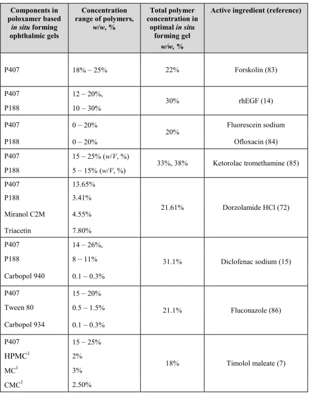
Use of P407 in mixture with other poloxamers is considered as a strategy to decrease the P407 concentration, modulate the temperature of gelation and rheological properties of in situ forming gel. Among others, poloxamer 188 (P188) was shown to be a good modulator of P407 in situ
forming gels (11–13). In previous studies, both poloxamers were used in in situ forming ophthalmic gels at very high total concentration, e.g. P407 and P188 in concentration of 21% and 10% (11), 16% and 14% (14), 20% and 11% (15) (w/w), respectively.
The additional improvements in the biopharmaceutical properties of poloxamer ophthalmic gels can be achieved by the presence of different additives in the formulation (16–20). Chitosan, a
10 biocompatible and biodegradable polycationic polymer, has been demonstrated to increase the mechanical strength and mucoadhesivness of in situ forming ophthalmic gels (20). In addition, there are other benefits of including chitosan in ophthalmic formulations such as improvement of eye-related permeability due to its paracellular permeation enhancing effects (21), antimicrobial activity (22) and corneal wound healing effect (23).
The aim of this doctoral thesis was to develop stabile and robust in situ forming ophthalmic poloxamers/chitosan gel fine-tuned in terms of polymer content and rheological properties, i.e. temperature of gelation, storage modulus and viscosity. Minimizing the total polymer content while retaining the advantageous rheological properties has been achieved by means of D-optimal statistical design. The selection of the leading candidate for drug formulation development has been based on the ability to keep its rheological properties upon dilution under biorelevant conditions. The robustness of selected mixed system has been evaluated upon entrapment of four ophthalmic active pharmaceutical ingredients (1).
11
12
2.1.
Ophthalmic drug delivery
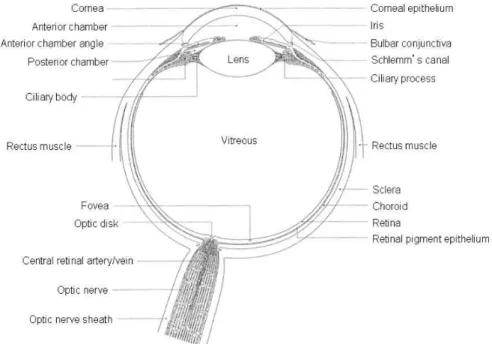
Although evidently exposed organ, in the terms of drug delivery, eye is a specific and extremely complex from multiple perspectives. Drugs must be transported across several protective barriers regardless of which administration route is utilized (i.e. topical, subconjunctival, sub-tenon’s intravitreal, peribulbar and retrobulbar administration). These barriers for effective drug absorption exist on physical, chemical and biochemical levels and each of them are active depending on the delivery route. Most of these obstacles are anatomical and physiological barriers that normally protect the eye from exogenous particles and toxicants (24). Anatomically, the eye is divided in anterior and posterior segment. The anterior segment includes approximately one-third of the eye consisting of cornea, conjunctiva, pupil, aqueous humor, iris, lens, ciliary body and the anterior portion of sclera. The posterior segment consists of vitreous humor, retina, choroid, sclera and optic nerve making up the remaining two-thirds in the back of the eye, Figure 1 (25).
Figure 1. The anatomy of the eye, picture reprinted with permission from (26).
The anterior and posterior segment of the eye can be affected by various vision impairing conditions. Diseases affecting anterior segment include glaucoma, allergic conjunctivitis, anterior
13 uveitis and cataract while age-related macular degeneration (AMD) and diabetic retinopathy are the most prevalent diseases affecting posterior segment of the eye (27).
2.1.1. Topical ophthalmic drug delivery
Topical administration is the most common drug administration route for the treatment of the anterior segment of the eye. This is due to the fact that it is the simplest, most convenient, self-administrable and non-invasive drug administration route for the management of anterior segment diseases/disorders. Drugs are delivered locally by this administration route, avoiding the blood-aqueous barrier, and the side effects and first-pass metabolism that may occur in some systemically administrated drugs. Depending on the formulation and drug physiochemical properties, drugs can reach different external (cornea, conjunctiva, sclera) and internal (aqueous humor, iris, ciliary body, vitreous humor, retina, etc.) sites in the eye after topical administration (28). However, the number of protective mechanisms play the decisive role in the fate of topically administrated drugs (Figure 2).
Ocular absorption of topically applied ophthalmic drugs is limited by: (i) rapid precorneal drug elimination
(ii)corneal/conjunctival epithelial barrier.
Consequently, eye-related bioavailability of topically administered products is low, typically less than 5%. The major fraction of the instilled dose is usually systemically absorbed via the naso-lacrimal duct or through the conjunctiva. For the treatment of diseases affecting anterior segment, the therapeutic concentrations can be achieved by frequent drug administration, however, for diseases affecting posterior segment, the intraocular levels achieved are often below minimal effective concentrations. Frequent administration is often associated with undesirable side effects caused by systemic drug absorption (2,24,29). Anterior segment diseases are mostly treated using eye drops (90% of marketed ophthalmic products) and ointments (27,28).
14
Figure 2. Ocular absorption and elimination pathways after topical administration, picture reprinted with permission from (28).
Precorneal factors
Precorneal factors, which include tear fluid dynamics, solution drainage and nonproductive absorption, determine the residence time of the drugs on the eye surface and therefore are of great importance for eye-related drug bioavailability.
Tear fluid dynamics
The tear film is a liquid layer covering the eye surface and acting as a barrier between the eye and environment (30). It is created by tears which are a complex aqueous mixture of peptides, proteins, electrolytes, lipids and small molecule metabolites (2,28). Immediately after the administration, drug/ophthalmic product is mixed with the tear film, which then presents a dynamic barrier to drug absorption. It is undergoes a constant renewal and consequently limits drug residence time on the eye surface.
15
Figure 3. Schematic representation of tear film, picture reprinted from (31).
It is composed of three layers (Figure 3): the superficial lipid layer, the middle aqueous layer and the deep mucous layer. It plays several important roles:
(i) it is forming and maintaining a smooth refracting surface over the cornea, as it moisturizes the environment for the epithelial cells of the cornea and conjunctiva,
(ii)it helps with transportation of metabolic products (primarily oxygen and carbon dioxide) to and from the corneal epithelial cells,
(iii) it provides a pathway for white blood cells in case of injury, and
(iv) it is diluting and washing away noxious stimuli and exhibits bactericidal properties (32). Lipids in the lipid layer of the tear film are secreted by the Meibomian glands, and they include triacylglycerols, free sterols, sterol esters and free fatty acids. The main function of lipid layer is to lower the surface tension of the tear fluid and prevent its evaporation. The aqueous portion of tear film, containing inorganic salts, glucose and urea, as well as biopolymers, proteins and glycoproteins, is secreted by the main and accessory lacrimal glands. The deepest layer of the tear film is the mucous layer. The mucous layer contains mucins, which are produced by the goblet cells of the conjunctiva. The mucins form a gel-like structure providing an easily wettable surface and thus assisting in water re-spreading after blinks (30). The mucus layer of a tear film is
16 restored slowly (~15 to 20 h) while the aqueous component is replaced approximately every 10 min (33).
Solution drainage
Administration of eye drops induces responsive protective mechanisms such as lacrimation and blinking, which results in more pronounced drug elimination. Commercially available eye-droppers provide average drop volume of 40 µl (range 25–56 µl). Available tear volume is about
7 µl, while conjunctival cul-de-sac can contain around γ0 µl of applied eye drop. After administration, drug has a short residence time of approximately 1–2 min at the eye surface, because of the permanent production of lacrimal fluid (0.5–β.β µl/min). Even though the tear
turnover rate is only approximately 1 µl/min, the excess volume of the instilled fluid flows to the
nasolacrimal duct in a couple of minutes (4).
Nonproductive absorption
In addition to tear fluid dynamics and solution drainage, systemic absorption can cause nonproductive drug removal instead of ocular absorption. Systemic absorption can occur either directly from the conjunctival sac via local blood capillaries or after the solution has arrived into the nasal cavity. Tear fluid containing the drug is transported from the lacrimal sac into the nasolacrimal duct, which empties into the nasal cavity, where the drug is then absorbed into the bloodstream. This absorption leads to drug wastage and, more importantly, possible side effects (4,24,34).
Anatomical barriers
After the topical administration, there are two main pathways of drug entry into the anterior chamber: via the cornea and via the conjunctiva. Small and lipophilic molecules (i.e. most clinically used drugs) are absorbed via the cornea, whereas large and hydrophilic molecules (e.g. new potential biotech-drugs such as protein and peptide drugs, gene medicines) are preferably absorbed through the conjunctiva and sclera. However, as mentioned previously, an important route of drug loss from the lacrimal fluid is systemic absorption through the conjunctiva.
17
Cornea
Cornea (Figure 4) is an optically transparent and avascular tissue which acts as the principal refractive element of the eye (34). Its surface and thickness are 1.04 cm2 and 0.5 mm, respectively. It comprises five layers: epithelium, Bowman’s membrane, stroma, Descemet’s membrane and the endothelium layer. The corneal upper layer, the epithelium, is endowed with microvilli, and covered by a charged glycocalyx, which maximizes surface area with the tear film (33). The epithelium and stroma are significant barriers to drug absorption, while endothelium participates only marginally in barrier function (35).
The permeation of drugs across corneal epithelium is a complex and dynamic process that includes passive diffusion through either a cell (transcellular) or intercellular space between the cells (paracellular), influx- and/or efflux-mediated transport by membrane proteins, endocytosis and transcytosis. Permeation is determined by both the physicochemical properties of the drug and barrier properties. Lipophilic drugs permeate via the transcellular route, while hydrophilic drugs permeate primarily via the paracellular pathway. The main physicochemical properties that determine the passive transport of drugs across cornea are:
(i) the degree of ionization at the pH of tears;
(ii) the drug lipophilicity (e.g. the optimal log P for transcorneal absorption is 2–3); and (iii) the charge, size and shape of the drug molecule (2).
The corneal epithelium consists of a basal layer of columnar cells, two to three layers of wing cells and one or two outermost layers of squamous polygonal shaped superficial cells (34). Epithelium is lipophilic in nature with superficial cells tightly connected by tight junctions. Tight junctions serve as a selective barrier for small hydrophilic molecules and they completely prevent the diffusion of macromolecules via the paracellular route. They are composed of a belt-like bands of intimate strands and the number of tight junction strands seems to be the most important determinant of paracellular resistance (34).
18
Figure 4. Schematic representation of corneal layers, picture reprinted with permission from (29).
From these reasons, epithelium is major obstacle for permeation of hydrophilic molecules. Compared to many other epithelial tissues (intestinal, nasal, bronchial, tracheal) corneal epithelium is relatively impermeable, but it is more permeable compared to the stratum corneum of the skin (34).
90% of corneal thickness is stroma, the hydrophilic layer containing collagen fibrils, which, due to its structure, blocks the transport of lipophilic molecules (24).
Monolayer of hexagonal cells forms the corneal innermost layer, the endothelium, separates the stroma from aqueous humor, and it is responsible for maintaining normal corneal hydration (24). Presence of endothelial leaky tight junctions allows free movements of molecules between aqueous compartments (stroma and aqueous humor) (36). From these reasons, corneal endothelium does not act as a significant barrier to the transcorneal diffusion of most drugs.
Conjunctiva
The conjunctiva of the eyelids and globe is a thin, vascularized mucus membrane which forms a continuous surface area of 17.65 cm2 (2), which is around 17 times bigger than the surface of cornea. Functionality of conjunctiva is related to the formation and maintenance of the tear film and in protection of the eye (34). However, in comparison to corneal, conjunctival drug absorption is considered to be nonproductive due to the presence of conjunctival blood capillaries and lymphatics, which can cause significant drug loss into the systemic circulation and, therefore, lower eye-related bioavailability. Tight junctions of the superficial conjunctival epithelium are the major barrier for drug penetration across conjunctiva. Intercellular spaces in the conjunctival epithelium are, however, wider than those in the cornea1 epithelium and therefore, the
19 conjunctival permeabilities of hydrophilic drugs are higher for an order of magnitude than their corneal permeabilities (24,34).
Sclera
The sclera constitutes the posterior five-sixths of the globe and provides the structural integrity that defines the shape and length of the eye (34). The sclera mainly consists of collagen fibers and proteoglycans embedded in an extracellular matrix (24). Permeability through the sclera is considered to be comparable to that of the corneal stroma and is inversely proportional to the molecular radius of the drug. Also, charge of a drug is important because positively charged molecules permeate poorly possibly due to their binding to the negatively charged proteoglycan matrix. Drugs penetrate across the sclera through perivascular spaces, through the aqueous media of gel-like mucopolysaccharides or through empty spaces within the collagen network (34) to reach the anterior segment (trans-scleral pathway).
20
2.2.
Topical ophthalmic dosage forms
The clinical performance of topical ophthalmic drug products is dependent on the interplay of the physicochemical factors of drug and delivery system along with physiological and anatomical environment of the complex eye surface. Conventional ophthalmic drug products include eye drops in form of solution or suspension and ointments mainly of the oleaginous type, and are still the mainstream treatment of anterior segment eye diseases.
Having in mind the complexity of eye surface environment and obstacles to eye-related bioavailability, formulation approaches for its improvement are to:
(i) improve drug concentration within the formulation; (ii)extend retention time on eye surface;
(iii) provide sustained drug release; and/or (iv) enhance corneal permeability.
To improve the drug concentration within the eye drops, the diverse solubility enhancers can be used (37). This could also allow a smaller volume of eye drop to be administered, which would then be less suspectible to removal from eye surface by solution drainage. The contact time of topical ophthalmic solutions with eye increases with the increase of formulation viscosity. Several synthetic polymers like polyvinylalcohol, polyvinylpyrrolidone, polyethylene glycol, polyacrylic acid, and many cellulose derivatives such as hypromellose and hydroxyethylcellulose, are commonly used as viscosity enhancers due to their physiologic compatibility and satisfactory physicochemical properties (38). However, increase in viscosity is followed by increase in bioavailability only to a certain extent (5).
Permeation enhancers can also be included into ophthalmic liquid formulations. They include preservatives (frequently added to multiple use ophthalmic systems and preventing microbial contamination) and surfactants (acting as drug solubilizers, and/or as spreading agents), and they modify transcorneal permeability by inducing ultrastructural changes in the corneal epithelium (39). Such molecules are, for example, ethylendiaminetetraacetic acid (EDTA), benzalkonium chloride (BAK), polyoxyethylene glycol ethers (lauryl, stearyl and oleyl), sodium taurocholate,
21 saponins and cremophor EL (40). However, there are safety concerns related to frequent use of these molecules (27).
Other formulation approaches to improve drug concentration within formulation, extend retention time on eye surface and provide sustained drug release include ophthalmic suspensions and emulsions. Ophthalmic suspensions are biphasic systems consisting of solid particles dispersed throughout a liquid phase (buffer), often with a help od suspending/dispersing agent (41). Suspensions are employed to deliver poorly water soluble drugs so frequently, corticosteroids are marketed in the form of suspension. Suspension particles retained in conjunctival sac subsequently improve drug retention time on eye surface and therefore the duration of action. The main challenge in their formulation, stability, efficacy and patient acceptance is the size and size distribution of particles. If not adequate, particle size can create several undesirable consequences like aggregation and resuspendability related issues. Particles in ophthalmic suspensions need to be micronized (less than 10 µm, preferably less than 5 µm) to avoid irritation/foreign body sensation (42). Smaller size particle restores the drug absorbed into ocular tissues from conjunctival sac, while larger particle size helps retain particles for longer time and provide slow drug dissolution (27). Among marketed ophthalmic, suspension of dexamethasone combined with tobramycin (TobraDex®) is one of the widely used for treatment of ocular bacterial infections. Drawbacks of ophthalmic suspensions are relatively high costs and complexity of manufacturing process since sterilization can cause physical or chemical instability. From this reasons, in some cases, aseptic processing is only viable manufacturing option, which increases costs even more (43).
Ophthalmic emulsions are colloidal pharmaceutical dosage forms consisting of one phase (water/oil) dispersed in other (oil/water) with an aid of surfactants (emulsifiers) (27). An example of such system a topical corticosteroid ophthalmic emulsion of difluprednate (DUREZOL®), indicated for the treatment of inflammation and pain associated with ocular surgery, currently available in the US market (44). Positive side of ophthalmic emulsions is their lubricating nature, while drawbacks associated to these systems are related to the use of surfactants (local toxicity issues) and to physical instability (flocculation).
Eye ointments are semisolid products usually intended for application to the conjunctiva, cornea, or eyelid. Ointments offer advantages over eye drops due to the significantly prolonged retention
22 time on the eye surface, minimization of tear dilution, reduced nasolacrimal drainage which all results in the improvement of bioavailability (45). Ophthalmic ointments account for approximately 10% of all of the ophthalmic dosage forms in the market (46). They are associated to poor patient compliance due to the greasy feeling, discomfort reflex tearing and blurring of vision (because of the difference in refractive index between nonaqueous ointment base and tears). Therefore, they are mostly applied at night. Furthermore, drawbacks of using ophthalmic ointments are related with potential content uniformity and dosing issues (45,46).
2.2.1. Innovative ophthalmic drug delivery systems
To improve eye-related drug bioavailability, there is a significant effort directed towards new drug delivery systems for ophthalmic administration (3,47–49). Innovative delivery platforms should lead to reduced dosing frequency, better patient compliance, reduced systemic adverse effects and finally improved clinical outcome. These delivery platforms include variety of drug delivery nanosystems and hydrogel-based systems.
Drug delivery nanosystems comprise nanosuspensions, nanoemulsions, polymeric micelles, nanoparticles, liposomes, niosomes, cubosomes and dendrimers (25,28,36,50–52). Using the nanosystems all the strategies to increase eye- related bioavailability can be tackeled (solubility, retention time, sustained release, increased permeability).
Nanosuspensions consist of nanosized drug particles suspended in an appropriate dispersion
medium and stabilized by polymer(s) or surfactant(s) (50). In comparison to conventional suspensions, nanosuspensions offer advantages such as faster dissolution and improved solubility of the drug, enhanced bioavailability, and reduced irritation to the eye. Nanosuspension approach provides opportunity to address many of the deficiencies associated with water insoluble drugs.
Nanoemulsions are biphasic dispersion of two immiscible liquids (most commonly oil-in-water),
stabilized using an appropriate surfactant, suitable for for the entrapment of hydrophobic drugs (51). The direct consequence of their small droplet size (20–200 nm) is their clear or hazy appearance and long-term physical stability, which differs them from milky white color and less stabile coarse emulsion (51,53). Moreover, nanoemulsions possess higher solubilization capacity than simple micellar dispersions. Therefore, nanoemulsion were shown to to improve
23 permeability, retention time and overall eye-related bioavailability (51). The most known ophthalmic nanoemulsion is RESTASIS®, a cyclosporine nanoemulsion for treatment of dry eye.
Polymeric micelles (10–200 nm) are based on amphiphilic molecules or block copolymers which
can generally self-assemble into organized core-corona/supramolecular structures in aqueous media at concentrations exceeding their critical micellar concentrations (CMC) (52). Polymeric micelles have shown potential for enhancing the solubility and therapeutic efficacy of poorly water soluble drugs and for protection of the drugs from degradation. Due to their small size and hydrophilic nature, micelles can efficiently permeate the corneal barriers and produce therapeutic concentrations both in the anterior as well as the posterior segment of the eye (52). To prolong the residence time at the ocular surface, polymeric micelles should intimately contact the mucus layer. Positively charged corona of polymeric micelles can enhance the interactions with negatively charged mucins at the corneal surface, facilitating precorneal retention (54).
Nanoparticles are particles consisting of various biodegradable materials which frequently have
multifunctional surface groups, with a diameter from 10 to less than 1000 nm providing a large surface area (28). Drugs can be either encapsulated in the matrix or attached to the surface of the particle. They offer a great opportunity for efficient local delivery of drugs to the anterior segment such as permeability enhancement across the blood-aqueous barrier and cornea, longer drug contact time with ocular tissues, specific/controlled delivery of drugs to the target site and protection of drugs from degradation and metabolism. Due to their size, low irritation and sustained release properties are expected, as well as less frequent administration (55).
Liposomes are lipid vesicles with one or more phospholipid bilayers enclosing an aqueous core.
Liposomes are adequate ophthalmic delivery systems due to the suitable biocompatibility, cell membrane-like structure and ability to encapsulate both hydrophilic and hydrophobic drugs (36). They have been investigated in ophthalmic drug delivery since 1980's. The earliest applications of liposomes were for topical drug delivery in the form of the eye drops and intravitreal injections for prolonged action (25). Although liposomes could entrap a wide range of molecules, their use is limited in topical ocular delivery because of relatively poor stability (36). Various modifications were evaluated to circumvent this drawback and one of the interesting approaches appears to be light activated liposomes which enable drug release at specified time and site with the projected laser light (25).
24
Niosomes are non-ionic surfactant vesicles structured in bilayers, ranging from 10-1000 nm,
which can entrap both hydrophilic and lipophilic drugs (36). Niosomes are suitable for topical ocular delivery due to the following benefits: chemical stability, ease of surface formation and modification because of the functional groups on their hydrophilic heads; improvement of bioavailability and effectiveness in the modulation of drug release properties (32,47,55). Furthermore, they are biodegradable, biocompatible, and non-immunogenic.
Cubosomes are defined as nanoparticles of a liquid crystalline phase with cubic crystallographic
symmetry formed by the self-assembly of amphiphilic or surfactant-like molecules (36). Glycerol monoolein is one of the most common surfactants used to make cubosomes. Cubic phases have useful properties, such as their ability to solubilize hydrophobic, hydrophilic, and amphiphilic molecules, their biodegradability by simple enzyme action and their suitability as sustained release delivery systems. Furthermore, the cubic phase is strongly bioadhesive and is thought to be a permeation enhancer (57). Several preclinical studies evaluated and showed promise for use of cubosomes as ophthalmic drug delivery systems (36,57).
Dendrimers are nanoscale, highly branched and reactive three-dimensional macromolecules with
a high degree of molecular uniformity, narrow molecular weight distribution, specific size and interesting structural properties like internal voids and cavities and a highly functional terminal surface formed by amino, carboxyl and/or hydroxyl groups (28). In addition to the outstanding structural control, their unique property is mimicry of globular proteins. They are referred to as
“artificial proteins”, based on their systematic, electrophoretic, dimensional length scaling and their biomimetic properties (58). Several preclinical studies showed their potential for ophthalmic use (28). Reports describe multiple use of dendrimers in ophthalmology for delivery of various drugs (genes, antioxidants, peptides) (36).
25
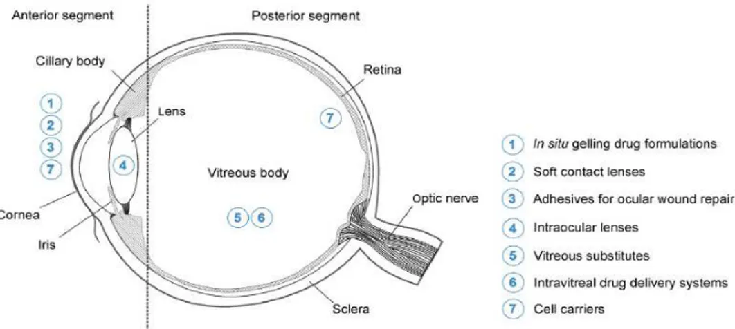
2.3.
Hydrogels in ophthalmic drug delivery
Aqueous gels or hydrogels formulated using hydrophilic polymers along those based on stimuli responsive polymers (in situ forming gels) are in the focus for the ophthalmic drug delivery via different routes of administration (Figure 5). Hydrogels allow the incorporation of various ophthalmic drugs (hydrophilic, lipophilic and biologicals). An interesting delivery systems based on hydrogels are hydrogel contact lenses. Due to their high water content and favourable properties they are highly compatible with human tissues (47). They extend the retention time of the drug, control and extend its release to several days or even months and enhance its eye-related bioavailability to more than 50% in comparison to conventional eye drops (47). Mostly used polymers in these lenses are hydroxyethyl methacrylate, methacrylic acid, polyvinyl alcohol and silicones.
In situ forming gels stand out as the main candidates for topical ophthalmic drug delivery and due
to their unique properties they have great market potential. They are prepared as liquid dosage forms (provides dose accuracy) which undergo phase transition on eye surface or conjunctival sac to form viscoelastic gel in response to an environmental stimulus following topical administration. The environmental stimulus can be of physical (temperature, light), chemical (pH, redox potential, ionic strength) and biological origin (inflammation, enzymes) (3,47).
Figure 5. Application sites of hydrogels in ophthalmology, picture reprinted with permission from (3).
26
2.3.1. In situ forming ophthalmic gels
One of the solutions of innovative pharmaceutical science for efficient ophthalmic drug delivery
are in situ forming ophthalmic gels. They are prepared from polymers that exhibit reversible
phase transitions and, due to their favorable rheological properties, minimize interference with blinking, avoid foreign body sensation and blurring of vision (3–5). Such systems are formulated as drug containing liquid suitable for instillation into the eye that shifts to the gel phase upon exposure to physiological conditions (7). Till now, for the purpose of ophthalmic delivery physical and chemical stimuli such as temperature, pH and ions were investigated (4,59). These triggers can be modified to control gel formation, retention time on the eye surface and drug release (6). Since they are not performed gels, but (viscous) solutions at administration, dosing is easier, more accurate and reproducible in comparison to the application and dosing of preformed gels. Additionally, in situ forming gels systems have gained major interest since no organic solvents or copolymerization agents are needed to trigger gelation (47).
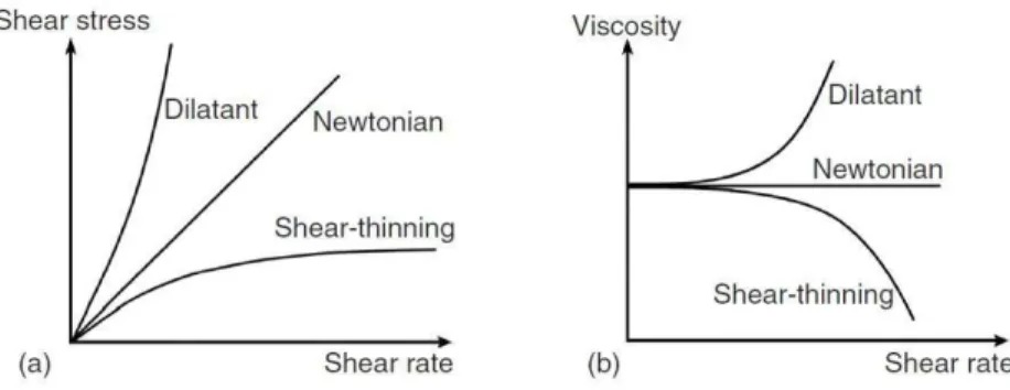
Since gelling can be affected by various factors such as salt concentration, ionic strength, pH, temperature, presence of a drug, adequate characterization of in situ forming gels is prequisite for their ophthalmic application. Pseudoplastic flow characteristics should be demonstrated with thixotropic properties which would assure easy spreadability across the ocular surface upon blinking and transformation to a more viscous material under low shear rate conditions to prolong the ocular residence time. Very important are safety issues which can be addressed with appropriate in vitro biocompatibility and in vivo ocular tolerability tests. Desirable properties of ideal ophthalmic gels and analytical methodology for their determination are summarized in the Table 1.
27
Table 1. Characteristics and mode of investigation of topical ophthalmic gel formulations, reprinted with permission and modified from (47).
Characteristic Analytical methods
Clarity and optical transmittance Visual inspection, refractive index or spectrophotometric analysis
Safety and good ocular tolerance Observation for possible adverse effects Visual acuity
Ocular tolerance (Draize, HETCAM, BCOP and histology tests) Cell-based cytotoxicity assays
Isotonicity test (osmommeter)
Suitable pH Measurement of pH at biorelevant temperature Pseudoplastic and thixotropic flow
Viscoelasticity
Cone and plate in rotational or oscillation rheometer
Gelling capacity Qualitative observation of gel formation in simulated tear fluid Mucoadhesive properties Polymeric mucoadhesion tests (rheometer)
Bioadhesive force of the gel
Extended residence time on eye surface Precorneal residence assessed using gamma scintigraphy
Ease and reproducibility of application Gel consistency (rheometer), firmness and cohesiveness assessed by texture analyzer
Compatible excipients Fourier transform infrared spectroscopy used to analyse potential drug-polymer interactions
Physical properties Solid-state properties (X-ray powder diffraction and particle size analyzed by dynamic light scattering)
Sustained/modified drug release In vitro release studies
Enhanced transocular permeation Ex vivo transcorneal and transscleral permeation studies Sterility Direct inoculation method
Stability Storage stability investigated
Polymers used in these systems are of natural, semisynthetic or synthetic origin, providing the unique property of significant structure conversion from viscous solution to gel upon reception of physical, chemical or biological stimulus (47). Advantages associated to hydrogels consisting of
28 the polymers of natural origin usually are non-toxicity, biodegradation, interaction with proteins and cells non-specific or specific binding (3). However, their potential drawbacks are weak mechanical strength, batch-to-batch inconsistency and immunogenicity. On the contrary, synthetic polymers offer well defined hydrogels in terms of network architecture, mechanical properties and prolonged stability, while they do not inherently interact with proteins or cells and their biocompatibility and biodegradability must be thoroughly assessed (3).
Polymers used in formation of in situ forming gels can be classified, according to the environmental stimuli that triggers sol-gel conversion, as temperature-responsive (thermoresponsive), pH or ion-responsive system (47).
Various studies have shown that these drug delivery platforms could improve the treatment of diseases affecting the surface and anterior segment of the eye, such as glaucoma, cataracts, dry eye disease, uveitis and ocular microbial infections, including conjunctivitis or keratitis. Some of these delivery platforms which are already either commercially available, or in clinical trials are summarized in the Table 2. Marketed ophthalmic products are indicated mainly for glaucoma, dry eye and ocular infections (6).
29
Table 2. In situ forming ophthalmic gels; either commercially available or currently in (pre)clinical trials.
Trade name Indication Manufacturer
Polymer(s) (mechansim of gelation)
Active
ingredient market/status
Betoptic S® Glaucoma Alcon®
Carbomer (pH-responsive) Amberlite® IRP-69 (ion exchange resin)
Betaxolol On the US and EU market
Carteol® LP Glaucoma Bausch & Lomb, Inc. Alginic acid (ion-responsive) Carteolol hydrochlorate On the EU market Timogel® Glaucoma Théa PHARMA
S.A.
Gellan gum
(ion-responsive) Timolol maleate
On the EU market
Timolol GFS Glaucoma Sandoz, Inc.
Gellan gum and xanthan gum (ion-responsive)
Timolol maleate On the US market
Timoptic XE Glaucoma Merck & CO, Inc. Gellan gum
(ion-responsive) Timolol maleate
On the US market Timoptol®-LA Glaucoma Santen UK Ltd. Gellan gum
(ion-responsive) Timolol maleate
On the UK market Rysmon® TG Glaucoma Wakamoto Pharmaceutical CO., LTD. Methylcellulose (temperature-responsive) Timolol maleate On the Japanese market Refresh
liquigel® Dry eye Allergan, Inc.
Carboxymethylcellul ose sodium (temperature-responsive) / On the US market REFRESH OPTIVE® Gel Drop
Dry eye Allergan, Inc.
Carboxymethylcellul ose sodium (temperature-responsive) Glycerin On the US market Systane gel
drops® Dry eye Alcon
® Hydroxypropyl-guar (pH-responsive and ion-responsive) Polyethylene Glycol 400 Propylene Glycol On the US and EU market Systane®
Balance Dry eye Alcon
® Hydroxypropyl-guar
(pH-responsive) /
On the US and EU market TobraDex ST Blepharitis Alcon® Xanthan gum
(ion-responsive)
Tobramycine Dexamethasone
On the US market
30
Trade name Indication Manufacturer
Polymer(s) (mechansim of gelation) Active ingredient market/status AzaSiteTM Bacterial conjunctivitis Inspire Pharmaceuticals, Inc. Polycarbophil (pH-responsive) Azithromycin Dexamethasone On the US market BesivanceTM Bacterial conjunctivitis
Bausch & Lomb, Inc. Polycarbophil (pH-responsive) Besifloxacin On the US, Argentina and Canadian market AzaSiteTM Plus Ocular infections and inflammation
InSite vision, Inc. Polycarbophil
(pH-responsive)7 Azithromycin
Phase III trials (US)
AzaSite Xtra™ Ocular
infections InSite vision, Inc.
Polycarbophil (pH-responsive) Azithromycin GLP tox studies finished (US) BromSiteTM Ocular inflammation Sun Pharmaceutical Industries Ltd. Polycarbophil/
(pH-responsive) Bromfenac FDA approved
DexaSite TM
Ocular pain and
inflammation
InSite vision, Inc. Polycarbophil
(pH-responsive) Dexamethasone
Phase III trials (US)
2.3.2. pH-responsive in situ forming gels
Ophthalmic formulations containing pH-responsive polymers (polyelectrolytes), which exhibit sol-gel phase transition in response to environmental pH changes (47) are promising tool for improvement of eye-related bioavailability. These systems exploit the difference of pH of tear fluid (7.4) and the pH of formulation. Such gels are composed of polymers with backbones containing large number of charged pendant groups (60,61). The sol-gel phase transition results from changes in the ionisation state of the weakly acidic (carboxylic or phosphoric) or weakly basic (ammonium) groups present in the polyelectrolyte (47). Common characteristic of these polyelectrolytes is pKa value between 3 and 10 which renders them suitable for pH-responsive systems (62). Most widely used pH-responsive polymers for ophthalmic formulations are polyacrylic acid and its derivatives (polymethacrylic acid, polycarbophils), chitosan and cellulose acetate phthalate (4).
Polyacrylic acid (also known as Carbomer, Carbopol) and its derivatives are anionic high molecular weight polymers whose acidic aqueous solutions are less viscous, while they are transformed into gels upon increasing the pH (Figure 6).
31
Figure 6. pH-dependent ionization poly(acrylic acid), picture reprinted with permission from (61).
Sol to gel transition is related to their polyanionic nature. Upon pH increase, as a result of the electrostatic repulsion of the negatively charged groups, polymers swell and gel is formed. Gel structure further entraps the drug molecules and delivers them to the environment in controllable manner. Sol-gel transitions of polyacrylic acid is dependent on the polymer molecular weight. Another beneficial feature of these polymers is mucadhesion, which is a result of physical entanglements or secondary bonds (63). Due to these favorable characteristics with positive impact on the eye related bioavailability, polyacrylic acid and its derivatives are frequently used in ophthalmic formulations (6).
Cellulose acetate phthalate is another polymer whose solutions show pH dependent gelation. It has a potential for sustained drug delivery to the eye because it is a free flowing solution at a pH of 4.4 and undergoes gelation when the pH is increased by the tear fluid to pH 7.4. This shift in pH results in almost instantaneous conversion of the fluid to a viscous gel. Gelation is triggered by neutralization of the acid groups of the polymer chain (4).
2.3.3. Ion-responsive in situ forming gels
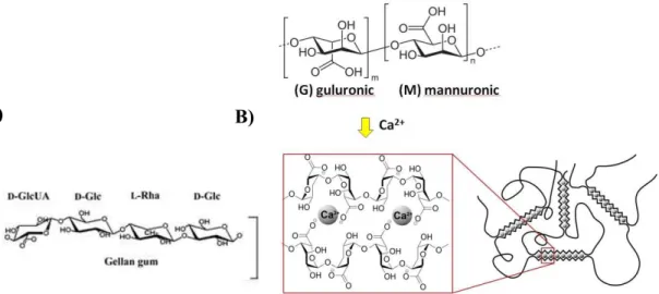
Polymers which solutions undergo phase transition to gel in the presence of different ions are known as ion-activated polymers. The ones with the potential for ophthalmic administration are those which interact with the ions present in the tear fluid (e.g. sodium, potassium and calcium). The most widely used ion-activated polymers in ophthalmic formulations are (deacetylated) gellan gum, xanthan gum, carrageenan and alginate (4,64).
Deacetylated gellan gum (Gelrite®) is anionic polysaccharide and it composed of tetrasaccharide repeating-units (Figure 7A). Cross-linking of the negatively charged polysaccharide helices by
32 monovalent and divalent cations (Na+, K+, Ca2+) is driving the change of structure form sol to gel. Na+ was found to be the most gel-promoting ion in vivo and gel formed is liquid crystalline comprising three-fold helical chains organized in parallel fashion in an intertwined duplex stabilized by hydrogen bonds (65).
Deacetylated gellan gum and alginate are approved as a pharmaceutical excipients for ophthalmic use. Gellan gum is marketed in two controlled-release glaucoma formulation called Timoptic-XE® (Merck) and Timolol GFS (Sandoz), while alginate, also for glaucoma therapy is marketed in France as Carteol LP by Laboratoire Chauvin, a Bausch and Lomb company.
Figure 7. Structural formula of gellan gum (A) and alginate with mechanism of gelation in presence of Ca2+ (B), reprinted from(66).
Similarly to gellan gum, aqueous solutions of alginate (Figure 7B) (a natural polysaccharide extracted from brown sea algae) also form gels when instilled into the eye. Alginates, due to their excellent biodegradable, biocompatible and mucoadhesive properties and are often used as constituents of numerous drug delivery systems (67). Alginic acid is a linear and anionic block copolymer polysaccharide comprising b-D-mannuronic acid and a-L-glucuronic acid residues joined by 1,4-glycosidic linkages. Dilute aqueous solutions of alginates form strong gels on addition of di- and trivalent cations by a cooperative process involving consecutive glucuronic residues in the a-L-glucuronic acid blocks of the alginate chain.
33 Rupenthal et al. characterized several ion-activated polymeric systems in vitro and in vivo (64). Among number of anionic polysaccharides (gellan gum, xanthan gum, carrageenan and alginate), formulations based on gellan gum and carrageenan exhibited the most favorable characteristics in terms of phase transition, rheological and textural properties, as their viscosity significantly increased upon contact with cations of the tear fluid, thus prolonging precorneal retention time and reducing nasolacrimal drainage (64). All tested polymeric formulations were found to be non-irritant and classified as safe for in vivo use. Systems based on gellan gum, xanthan gum and carrageenan exhibited the most favorable characteristics in terms of precorneal retention as well as delayed release of the model hydrophilic drug (pilocarpine hydrochloride) in vivo (8).
2.3.4. Temperature-responsive in situ forming gels
The sol-gel transition of this class of in situ forming gels is triggered by a temperature change from storage to physiological temperature i.e. temperature of the eye surface. A literature overview of the temperature-responsive in situ forming ophthalmic gels is summarized in the Table 3. Summarized data show that the major driving force for sol-gel transition for temperature-responsive in situ forming gels is predominance of hydrophobic interactions of amphiphilic (co)polymer molecules with the temperature increase.