Research Journal of Pharmaceutical, Biological and Chemical
Sciences
Docking and Cytotoxicity Test on Human Breast Cancer Cell Line (T47d) of
N
-(Allylcarbamothioyl)-3-chlorobenzamide and
N
-(Allylcarbamothioyl)-3,
4-dichlorobenzamide.
Siswandono, Tri Widiandani*, and Suko Hardjono.
Department of Pharmaceutical Chemistry, Faculty of Pharmacy, Universitas Airlangga Surabaya, Indonesia.
ABSTRACT
The specific objective of this research is to investigate the biological activity of thiourea derivativesby in silico study and the cytotoxicity test on human breast cancer cell lines. In this present study, the molecular docking of the new compound N-(allylcarbamothioyl)-3-chlorobenzamide (BATU-02) and N -(allylcarbamothioyl)-3,4-dichlorobenzamide (BATU-04) were evaluated on EGFR (1M17.pdb) using MVD v5.5 and showed that the re-rank scores of BATU-02 and BATU-04 are smaller than5-fluorouracil (5-FU). From the docking result, we can predict that the compounds have a higher biological activity. The cytotoxicity test were evaluated on human breast cancer cell lines (T47D) using MTT assay. Relevant result showed that these compounds(BATU-02 and BATU-04) demonstrated are more potent compared to 5-FU as the commercial anticancer drug, with respective IC50were 128μg/mL (BATU-02); 86 μg/mL (BATU-04); and 213 μg/mL (5-FU).
It can be concluded that the modification compounds of thiourea can be further developed as a potential anticancer drug.
Keywords: Docking, thiourea, cytotoxicity, T47D, 1M17
INTRODUCTION
Cancer is one of the leading causes of death in the world and it is becoming a serve problem in the world of wellness. The success of cancer treatment is a challenge in the 21st century, so it underlines the most
urgent to develop novel and safe chemotherapeutic agents with greater anticancer [1].
In the development of anticancer drugs, especially to obtain a better activity, it is performed modification of the structure of thiourea derivatives [2-4]. Thiourea derivatives constitute one class of anticancer promising for further development. This derivative work as inhibitors of EGFR to inhibit receptor tyrosine kinases (RTKs) in the intracellular region [5]. In addition, thiourea can also serve as a conjugate of the anti-EGFR monoclonal antibody[6,7].
In this study, the compoundshave been made by reacting allylthiourea with benzoyl chloride derivatives (3-chloro and 3,4-dichloro) to form the compound N-(allylcarbamothioyl)-3-chlorobenzamide (BATU-02) and N-(allylcarbamothioyl)-3,4-dichlorobenzamide[9].To predict the anticancer activity of the compounds, it is conducted in silico (docking) test using Molegro Virtual Docker version 5.5 that uses epidermal growth factor receptor (EGFR) kinase with pdb code 1M17, i.e. a receptor model of erlotinib constituting GFR a receptor inhibitor [5,10].
The BATU-02 and BATU-04 are investigated for cytotoxicity activity in human breast cancer cell line (T47D) [12, 15].The cytotoxicity test is determined through MTT method or 3-(4,5-dimethylthiazole-2-yl)-2-5-diphenyltetrazolium bromide. IC50is determined based on 50% T47D cells alive. For comparability, it is used
5-fluorouracyl, an anticancer compound that has been used clinically for the treatment of cancer. MATERIALS AND METHODS
In Silico
Materials: A computer (HP, core i7), Epidermal Growth Factor Receptor (1M17.pdb).
Method: The docking method is using Molegro Virtual Docker version 5.5. The docking began with a preparation on EGFR receptor with 1M17 code taken from the Protein Data Bank. The test ligand was prepared by making the 2-D and the 3-D structure of the compound using ChemBioOffice program Ultra 11.0 and its energy was minimized using MMF94. The resulted in Rerank Score describing the minimal energy required by the compound in interaction with the receptor [9].
In Vitro
Materials: (N-(allylcarbamothioyl)-3-chlorobenzamide (BATU-02), N -(allylcarbamothioyl)-3,4-dichlorobenzamide (BATU-04), 5-fluorouracyl(Sigma, USA); DMSO (Sigma, USA); Rosewell Park Memorial Institute (RPMI) 1640; 3-[4,5-dimetiltiazol-2-il(-2,5-difenil tetrazolium bromida)]= MTT (Sigma, USA); Fetal Bovine Serum (FBS) 10% (Gibco, USA).
Method: The Cytotoxicity assay was determined in vitro using cell lines T47D usingthe MTT method. This method required four test groups, namely the compound (A), a positive control (B), a control cell (C), and a control medium (D). It was prepared seven concentrations obtained from stratified dilution with RPMI medium
(1000; 500; 250; 125; 62.5; 31.25; 15.625) μg/mL. T47D cell culture was prepared in the microplate of 96 wells,
with a density of 5 x 10³ cells/well.
MTT assay
The assay was carried out following Mossman method [16] with modification in stopper reagent. T47D cells were distributed into 96 well plates, then were incubated for 24 hours under CO2. Test solutions in
followed by overnight incubation at room temperature. Absorbance was read with an ELISA reader at 595 nm. The absorbance was converted to percentage of living cells (cell viability) [17].
RESULTS AND DISCUSSION
Docking
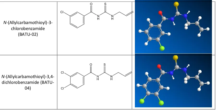
The result of preparing the structure of 2-Dimensional and 3-Dimensional of the N -(allylcarbamothioyl)-3-chlorobenzamide (BATU-02), N-(allylcarbamothioyl)-3,4-dichlorobenzamide (BATU-04)using ChemBio3D Ultra 11.0 program is shown in Figure 1 [12].
N -(Allylcarbamothioyl)-3-chlorobenzamide
(BATU-02)
N -(Allylcarbamothioyl)-3,4-dichlorobenzamide
(BATU-04)
Figure 1: The structure of 2-Dimensional and 3-Dimensional of the N
-(allylcarbamothioyl)-3-chlorobenzamide (BATU-02), N-(allylcarbamothioyl)-3,4-dichlorobenzamide (BATU-04) of which the energy has been minimized using MMFF94.
Docking results of 5-FU and BATU-02 and BATU-04on EGFR kinase (1M17.pdb) can be seen in Table 1 [13].
Table 1: Docking Results of 5-FU and BATU Derivatives
Compound Code RerankScore BATU-02 -94.4187 BATU-04 -93.1137 5-FU -48.9354
From the docking results, the Rerank Score of all BATU derivatives is smaller than the Rerank Score of 5-FU. This suggests that the modified compound has smaller bond energy and so thatthe binding is more stable [14]. Thus, the modified compound can be expected to have greater biological activity by in silico test.
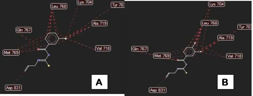
The overview of the amino acids involved in the interaction process of compounds between BATU-02 and BATU-04 versus EGFR is presented in Figure 2 [13].
Cytotoxicity Assay
Figure 2: The amino acids involved in the interaction process of compounds between BATU-02 (A) and
BATU-04 (B) and EGFR kinase(1M17)
Figure 3: The cells viability overlay profile of 5-FU and BATU-02 and BATU-04
To provide clearer pictures on the influence of different concentrations of 5-FU and BATU-02 and 04 on cell viability, the data is plotted as profiles of cell viability as can be seen in Figure 3. It is obvious that cell viability is getting decreased as the concentration of 5-FU and BATU-02and BATU-04 increased.
Table 2: T47D cells viability (%) after treatment at various concentrations
Compound The percentage of living cells T47D (%) at various concentrations 15.625
μg/mL μg/mL31.25 μg/mL62.5 μg/mL125 μg/mL250 μg/mL500 μg/mL1000
BATU-02 96.86±1.26 88.23±2.67 82.86±13.89 50.29±31.49 30.94±13.02 12.10±1.98 14.19±2.61 BATU-04 103.97±9.3
5
90.73±1.96 88.22±4.97 10.84±0.48 13.42±1.43 13.17±0.11 16.82±1.92 FU 61.70±7.98 54.23±4.07 52.91±2.66 52.85±4.58 46.58±4.96 39.41±2.19 27.28±1.05
Table 3: IC50 of the test compounds on T47D
Compound IC50(μg/mL)
BATU-02 128±11.0 BATU-04 86±3.6
FU 213±2.2
The results of probitanalysis to obtain the score of IC50 BATU-02; 04 and 5-FU as a comparison can be
seen in Table 3.
Based on the cytotoxicity results on IC50, it has been known that N
-(allylcarbamothioyl)-3-chlorobenzamide (BATU-02)and N-(allylcarbamothioyl)-3,4-dichlorobenzamide (BATU-04) havehigher cytotoxicity activity comparedwith 5-fluorouracyl. This result is linear with in silico approach. Therefore, these compounds are feasible to be developed as a potential anticancer drug.
CONCLUSION
Based on the results of this study can be concluded that the new compounds of thiourea derivatives (BATU-02 and BATU-04) can be further developed as a potential anticancer drug.
ACKNOWLEDGEMENTS
This experiment was supported in part bya grant fromthe Ministry of Research, Technology, and Higher Education, Republic of Indonesia and the Faculty of Pharmacy Universitas Airlangga.
REFERENCES
[1]. Liu, W., Zhou, J., Zhang, T., Zhu, H., Qian, H., Zhang, H., Huang, W., Gust, H., 2012, Design and synthesis of thiourea derivatives containing a benzo[5,6]cycloheptal[1,2-b]pyridine moiety as potential antitumor and anti-inflammatory agents, Bioorganic & Medicinal Chemistry Letter, Volume 22: 3701-2704.
[2]. Li, H.Q.; Lv, P.C.; Yan, T.; Zhu, H.L., 2009, Urea derivatives as anticancer agents.Anticancer Agents Med Chem., Volume 9: 471-480
[3]. Li, H.Q, Yan, T., Ying, Y., Shi, L., Zhou, C. F., Zu, H.L., 2010, Synthesis and structure-activity relationship of N-benzyl-N-(X-2-hydroxybenzyl)-N’-phenylureas and thioureas as antitumor agents, Bioorganic & Medicinal Chemistry, Volume 18: 305-313.
[4]. Huang, X.C., Wang, M., Pan, Y.P., Yao, G.Y., Wang, H.S., Tiang, X.Y., Qin, J.K., Zhang, Y., 2013, Synthesis
and antitumor activities of novel thiourea α-aminophosphonates from dehydroabietic acid, European
Journal of Medicinal Chemistry, Volume 69: 508-520.
[5]. Yang, W., Hu, Y., Yang, Y.S., Zhang, F., Zhang, Y.B., Wang, X.L., Tang, J.F., Zhong, W.Q., Zhu, H.L., 2013, Design, modification and 3D QSAR studies of novel naphthalin-containing pyrazoline derivatives with/without thiourea skeleton as anticancer agents, Bioorganic & Medicinal Chemistry, Volume 21: 1050-1063.
[6]. Reymond, M. Reilly, 2010, Monoclonal Antibody and Peptide Targeted Radiotherapy of Cancer. Wiley and Sons, ISBN: 1118035151.
[7]. Ingargiola, M., Dittfield, C., Runge, R., Zenker, M., Heldt J. M., Steinbach, J., Cordes, N., Baumann, M., Kotzerke, J., Schughart, K. L. A., 2012, Flowcytometric Cell-Based Assay to Preselect Antibody Construct for Radionuclide Conjugation. Journal of The International Society for Advancement of Cytometry, Volume 81A: 865-873
[8]. Siswandono, 2014, Pengembangan Obat Baru, Airlangga University Press, Surabaya, pp: l 1-17.
[9]. Widiandani, T., Siswandono, 2014, Modifikasi Struktur dan Prediksi Aktivitas Antikanker Senyawa Baru Turunan Aliltiourea Secara In Silico, e-jurnal Berkala Ilmiah Kimia Farmasi, Vol 4:1.
[10]. Li, H.Q., Yan, T., Ying, Y., Shi, L., Zhou, C.F., Zu, H.L., 2010, Synthesis and structure-activity relationship of N-benzyl-N-(X-2-hydroxybenzyl) -N’-phenylureas and thioureas as antitumor agents, Bioorganic & Medicinal Chemistry, Volume 18: 305-313.
[11]. Zampieri, L., Bianchi, P., Ruff, P., and Arbuthnot, P., 2002, Differential modulation by estradiol of P-glycoprotein drug resistance protein expression in cultured MCF7 and T47D breast cancer cells, Anticancer Res., Volume 22(4): 2253-9.
[12]. Cambridgesoft, 2008, CamBioDraw Ultra 11 for Tutorial, All Right Reserved.
[13]. CLCbio, 2012, Molegro Virtual Docker User Manual, MVD 2012.5.5 for Windows, Linux, and Mac OS X, Molegro – A CLC bio company.
[14]. Young, D.C., 2009, Computational Drug Design, A Guide for Computational and Medicinal Chemists. New York: John Wiley and Sons.
Pharmaceutical and Clinical Research, Vol 8 (5), 372-376.
[16]. Mosmann T. Rapid colorimetric assay for cellular growth & survival: application to proliferation & cytotoxicity assays. J Immunol Methods 1983;65:55-63.