Study on High Natural Radiation Impacts to Peripheral Blood Cells in Population Living
in Botteng Village, Mamuju, West Sulawesi
Tur Rahardjo, Siti Nurhayati, Darlina, Teja Kisnanto and Mukh Syaifudin Center for Technology of Radiation Safety and Metrology,National Nuclear Energy Agency of Indonesia
Jl. Lebak Bulus Raya No. 49 Jakarta Selatan, 12440 Indonesia, E-mail: [email protected]
Abstract. This study describes the long-term effects of low dose radiation to the hematopoietic parameters in the blood of population living in Mamuju of West Sulawesi, an area which has high natural radiation. Peripheral bloodswere obtained from community members of Botteng village, covering 70 people that consist of 35 women and 35 men as study group. For control group the bloods were taken from areas with normal levels of exposure in the Keang village covering 23 populations that consist of 13 women and 10 men. Age of both groups were ranging between 14-70 years. The blood components were measured by using the hematopoietic analyzer, the ABX Micros 60, located in Clinical Laboratory of PTKMR-BATAN according to standard procedure. The results showed that the levels of haemoglobin (Hb), erythrocytes, leukocytes, absolute lymphocytes, platelets, MCV, RDW, PDW, MPV, monocytes and granulocytes were not decreased significantly due to radiation and were within normal limits and there is no significant difference between these two groups (p> 0.05). Other components (HCT, MCH, MCHC, and MPV) showed normal data distribution and significantly different with that of control (p <0.05) that may related to daily life style such as smoking and coffee consumption. It can be concluded that there is no impact of exposure to high natural radiation in Mamuju, West Sulawesi to hematopoietic system.
Keywords: haematology, low doses, natural radiation, Mamuju
Introduction
Blood is an important part of the body's circulatory system. Blood consists of two parts, namely the liquid (blood plasma) and blood cells. Blood cells include erythrocytes, leukocytes, and platelets. Leukocyte function as imunutas system of the body. Erythrocytes joint function in tissue oxygenation hemoglobin and platelets play a role in blood clotting system. These blood cells are produced in the bone marrow. Radiosensitivity of types of blood cells varies. Peripheral blood lymphocytes are the cells that most sensitive to radiation so it susceptible to damage DNA. Therefore peripheral blood lymphocytes are the cells
that most commonly used as biodosimetry.
Biodosimetry is the prediction of the dose of ionizing radiation received by a person based on biological material changes in the body. The most resistant cells are erythrocytes (Ethel, 2003). Whole body radiation dose of 0.5 Gy can cause a decrease in the formation of blood cells so that the blood cell count decreases. A decrease in blood cell count will be seriously affected if not treated immediately because the blood has an important role on the human body functions such as
immunity, oxygenation, hemostasis and other
characters (Fliedner, 2012).
The bone marrow system is very radiosensitive and therefore the analysis of hematopoietic index for people affected by radiation is necessary and the blood-forming cells in the bone marrow are particularly vulnerable to radiation (Hendee, 1990). In Nigeria, it has been estimated that approximately 160 people per year are at risk of cancer due to radiation
exposure of terrestrial gamma (Little, 1995; Liu, 1987; Loken, 1993). The main component of radiation exposure in daily life for the general public is the natural radiation that could have come from outer space as cosmic radiation and occurs in soil, air, water or building materials (terrestrial radiation) (Ethel, 2003; Hendee, 1990).
Natural radiation is the main source contributing to the collective dose received by the population of the world both from external and internal sources of exposure to natural radiation in soil and produce a critical component of background radiation exposure of the population in the world. Some radionuclides were detected in the soil and the natural environment as well radioactivity associated external exposure due to gamma radiation that depends on the geological and geographical conditions and emerge at different levels in the soil of each region in the world (Luckey, 1994; Nordenberg, 1990). Terrestrial radiation begins by radioactive chemicals that present when the earth was formed. They are found in igneous, metamorphic, and sedimentary rocks in various regionsin the world (Seed, 1993; Shapiro, 1990). In Kerala (India) and some parts of France and Brazil, the dose was recorded up to 20 times the global average. In some other countries (e.g. Finland) the average dose was several times higher, and in particular, many citizens receive an effective dose exceeding the allowable dose (of 20 mSv/year) (MacMahon, 1989; Mossman, 1998).
(2010) showed that Mamuju district has an highest dose rate compared to other parts of Sulawesi and even Indonesia, which reached 2.800 nSv/hour. In the highest gamma dose rate in Botteng village there is a location with a level of gamma radiation dose of 10,000 nSv/hour (national average of 50 nSv/hour). Another study conducted by Alatas et al. on the cytogenetic responses that were evaluated in Mamuju population and showed no significant difference in the frequency of chromosomal aberrations in lymphocytes of people who live in that areas compared with living in normal areas (Alatas, 2012).
Ionizing radiation affects the organism through the water radiolysis. DNA damage occurs as a result of interaction with reactive oxygen species. Cell death or mutations that may result from DNA damage. But the damage to cells depending on the type of radiation, dose rate and dose (Sawant, 2009; Baskar, 2012). Exposure to ionizing radiation is sensitive to the bone marrow, gastrointestinal tract, and skin tissue including the central nervous system (CNS), lungs, heart, liver, kidneys, and reproductive organs can be affected (Miller, 2013; Rea, 2010). Eyepiece is also considered one of the most radiosensitive tissues in the human body and could have resulted in cataractogenesis (Ainsbury, 2009; Schmid, 2007; Barcellof-Hoff, 2012). In hematopoietic syndrome lymphocyte cells are very sensitive to radiation and radiation can decrease the amount within 1 to 3 hours after exposure which may also cause drop in the number of leukocytes, platelets, and erythrocytes. Shortage of white blood cells can trigger infections and shortage of platelets can cause uncontrolled bleeding in body tissues. Red blood cells are less sensitive to radiation, but its decrease would results in highrisk of health problems and may lead to stochastic and deterministic effects (Shimizu, 2010; Akleyev, 2010).
This study aimed to determine the alteration in hematology (hematopoietic components) in blood of resident in Botteng Village, Mamuju due to high natural radiation exposure. These peripheral blood lymphocytes are the cells most widely used in the study using the man as an object of research. Very low radiation exposure can cause hormesis effect, stimulating the hematopoietic function as an expression in the immune system and hematopoietic function increases with increasing exposure to radiation(UNSCEAR, 2010).
Materials and Methods
Blood Sampling
Samples were obtained from the local indigenous healthy people, and they did not receive any medical
70 years. For areas with high exposure to natural radiation (HBRA) samples were taken from the people living in village of Botteng (sub villages of Tekbong, Kurasalimbo, Tangnga, Kassa, and Taludu) that covering 70 people consisting of 35 women and 35 men. Control samples were obtained from the area with a normal level of natural radiation exposure (NHBRA) of Keang Village (Lomali and Tabelalang sub villages) on 23 population consisted of 13 women and 10 men. Prior to blood sampling, the population is given an explanation about the purpose of this study. Residents are willing to become respondents were asked to sign an informed consent form (willingness to give blood samples). About 2 mL of peripheral blood were drawn using a syringe equipped with a vacutainer containing EDTA anticoagulant. The blood sample was immediately taken to the Cytogenetic Laboratory in PTKMR BATAN in Jakarta.
Hematologic Examinations
Blood in a tube that has been mixed well was examined by using a hematology analyzer ABX Micros 60 according to standard protocol. These parameters are hemoglobin level, the number of erythrocytes, platelets, leukocytes, lymphocytes, granulocytes, monocytes, hematocrit (HCT), mean cell/corpuscular volume (MCV) or corpuscular volume average (VER), mean Cell hemoglobin Content (MCH) or corpuscular hemoglobin average (HER), mean Cellular hemoglobin concentration (MCHC) or hemoglobin concentration erythrocyte average (Kher), Red Blood Cell Distribution Width (RDW), Platelet Distribution Width (PDW), Mean platelet volume (MPV).
Research Ethics
Ethic of the study was obtained from the Ethics Committee for Health Research, National Agency for Health Research and Development, Ministry of Health
of the Republic of Indonesia No.
LB.02.01/5.2.KE.051/ 2015, date of January 29, 2015.
Results and Discussion
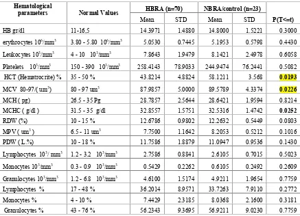
Table 1 shows the results of hematology of the blood of people in HBRA compared with controls taken from areas with levels of exposure to NBRA. It can be seen that the levels Hb, erythrocytes, leuko-cytes, lymphocytes absolute, platelets, MCH, RDW, PDW, monocytes and granulocytes did not significan-tly decline and is still within normal limits. The results of statistical tests showed that that levels of blood components in the HBRA group showed no significant difference when compared with that of NBRA as controls (p> 0.05). For other components (HCT, MCV, MCHC) the statistical test results showed a significant differences when compared with that of the control (p <0.05).
Bone marrow cells included in the cells that are actively proliferate and are susceptible to damage from exposure to ionizing radiation. It is known that a low level of radiation, i.e. 0.25 Gy, can cause changes in the blood constituent, particularly the cessation of the formation of blood cells (hematopoesis) with result in
the changes both by direct damage to tissue hematopoietic and the influence to neurohormonal mechanisms. Leukocytes, platelets and lymphocytes are blood cells that are most sensitive to radiation, whereas erythrocytes and Hb are cells that are quite resistant to radiation. From the results of measu-rements of hemoglobin levels in blood of Mamuju population presented in Table 1, it can be seen that the hemoglobin levels did not decrease significantly and still within the normal limit (11 to 16.5 g/dL). These mean values are14,397 g/dL for HBRA and 14,800 g/dL for control.
Table 1. The average level of each measurement of hematology among residents in areas of high natural radiation (HNBR) and normal levels of exposure to natural radiation (NHBRA).
Hematological
parameters Normal Values HBRA (n=70) NBRA/control (n=23)
P(T<=t)
Mean STD Mean STD
HB gr/dl 11-16,5 14.3971 1.4880 14.8000 1.5221 0.3000
erythrocytes 106/mm3 3.80 - 5.80 106/mm3 5.0530 0.7445 5.1953 0.5798 0.4430
Leukocytes 103/mm3 4 - 10 103/mm3 7.8643 1.9479 8.1421 2.4978 0.6058
Platelets 103/mm3 150 - 390 103/mm3 258.4143 78.9033 244.9474 76.2441 0.5082
HCT (Hematrocrite) % 35 - 50 % 43.8214 4.8824 58.1211 3.568 0.0193
MCV 80-97/( um3) 80 - 97 um3 87.9857 5.0000 89.5789 4.3374 0.0226
MCH ( pg) 26.5 - 35 Pg 28.7857 2.5644 28.6421 1.9594 0.8214
MCHC ( g/dl ) 31.5 - 35 g/dl 32.8557 1.5751 32.5316 1.4742 0.0252
RDW (%) 10 - 15 % 12.6786 0.9802 12.2632 0.5449 0.0803
MPV ( um3) 6.5 - 11 um3 7.7500 1.1642 8.2053 0.5212 0.1016
PDW ( L.%) 10 - 18 % 11.7586 1.8879 11.0947 0.9536 0.1430
Lymphocytes 103/ mm3 1.2 - 3.2 103/mm3 2.7586 0.8841 2.6105 0.7015 0.5023
Monocytes 103/mm3 0.3 - 0.9 103/mm3 0.5429 0.2262 0.6105 0.2492 0.2609
Granulocytes 103/mm3 1.2 - 6.8 103/mm3 4.6100 1.5174 4.9211 1.9654 0.7759
Lymphocytes % 17 - 48 % 36.2014 8.9571 33.7263 7.9110 0.2772
Monocytes % 4 - 10 % 7.4429 2.3185 8.0368 2.1600 0.3181
Granulocytes % 43 - 76 % 56.2343 9.3695 56.9211 9.0230 0.7759
Erythrocyte production is controlled by a negative feedback mechanism that is sensitive to the supply of oxygen to the body tissues. Increased production of erythrocytes can be caused by several factors, namely bleeding, heart failure, and pulmonary disease (Ethel, 2003). Results in number of blood erythrocyte in population of Mamuju (Table 1) shows that the number of erythrocytes in HBRA did not decrease compared to normal values (3.80-5.80 x 10⁶ per mm3versus 5.053- 5.195 x 10⁶per mm3). The rela-tionship between hemoglobin levels and the number of erythrocytes are known to rise and comparable between hemoglobin and erythrocytes (Figure 1). The relationship between the number of erythrocytes with hemoglobin levels following the line with equation of Y = 0.958 X + 9.555, which means that the higher hemoglobin levels would have the higher number of erythrocytes. Red Cell Distribution Width (RDW) is the coefficient of variation of erythrocytes volume (differences or variations in the size/area of erythro-cytes). This value as an estimate that useful RDW high early anemia can indicate the red cells are hetero-geneous, and are usually found in iron deficiency anemia case, a deficiency of folic acid and vitamin B12, hemolytic anemia, and sickle cell anemia. The red cells are typically 6-8μm, the higher variation in cell size indicating the abnormalities, whereas low
RDW showingthat erythrocyte have small size
variations.
Figure 1. The relationship between hemoglobin concentration with the number oferythrocyte cells/mm3
Platelets function in blood clotting mechanism. Thrombocytopenia is a condition in which the number of platelets less than normal that may caused by the initial reaction of drugs, bone marrow malignancy, or bone marrow damages caused by ionizing radiation. The opposite situation is called thrombocytosis, i.e. an increase in the number of platelets due to the bleeding,
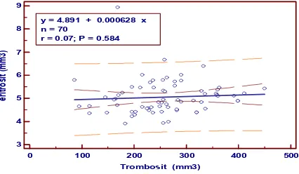
postoperative, malignancy and inflammatory diseases and as a result of a decrease in the number of platelets by radiation, the clotting time will increase due to reduced tissue factor which is usually generated by platelet G. The results of measurements of platelet count in Mamuju population are presented in Table 1. It can be seen that the platelets are not significantly decline and is still in the normal range in adults (normal value is 150-390103/mm3). The average value in HBRA was 258.4143103/mm3 and control was 244.9474103/mm3. Figure 2 shows the relation-ship between the platelet count with the number of erythro-cytes with equation of Y=0.000628 X + 4.891 which means that the more the number of platelets will be the greater the number of erythrocytes. Platelet Distribu-tion Width (PDW) PDW is the coefficient of variaDistribu-tion of platelet size. High PDW levels usually found in sickle cell disease and thrombocytosis, while the low PDW levels mean that platelets have small size. MPV is the average volume of platelets. Thrombocytopenia occurred in low MPV, while the high MPV can be used as an indicator of platelet megakaryocytes.
0 100 200 300 400 500
Figure 2. The relationship between the number of erythrocytes/mm3with the number ofplatelets/mm3
According to Campbell et al (2005), WBCs serves to protect the body against invasion of foreign substances such as bacteria and viruses. Infection or tissue damage resulting in an increase in the total number of leukocytes. Increased leukocytes can also be caused by leukemia. Leukemia is a type of cancer that is characterized by proliferation of white blood cells that are not controlled. Results show that the total number of leukocytes in the HBRA group is not signi-ficantly different the control group no (p> 0.05) as shown in Table 1. However the average leukocyte counts was lower compared to the control group (7.86 x 10³/mm3versus 8.14x10³/mm3). These levelsare not affected by environmental radiation and still within normal value (4.500-10.000 103/mm3).
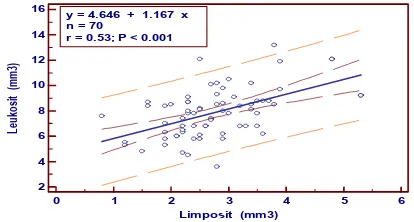
Table 1 showing that the number of blood lymphocyte cells in blood of Mamuju population does not decline (normal value is 1.2-3.2x10³/mm3). Decrease in number of absolute lymphocyte cells can be used to estimate the severity of a person such as a result of acute radiation exposure. In Figure 3 shows the relationship between the number of leukocytes with the number of lymphocytes by equation of Y = 1.167 x + 4.646 which means the number of lymphocytes increases would have results in the increase of leuko-cytes number.
Figure 3. The relationship between the number of leukocytes cells/mm3with the number of lymphocytes cells/mm3
Hematocrit is a measurement that determines the number of red blood cells in 100 ml of blood and is expressed in percent (%). The results showed that statistically HCT, MCV and MCHC in the treatment group were different compared to controls (p <0.05) and showing a normal distribution. Mean corpuscular volume (MCV) or average volume of erythrocyte that expressed in femtoliter. MCV is the erythrocyte index that equal to MCH, MCHC. All these can be used to help in diagnose the cause of anemia (a condition in which too few red blood cells). The level of MCV higher than normal means an anemia condition and known as macrocytic (cell size above normal cells). This condition is usually found in people with pernicious anemia, alcoholics, folic acid deficiency, and HIV. MCV below normal means microcytic (cell size below normal cells) and usually found in people with iron deficiency (anemia), thalassemia, and lead poisoning.
Hemoglobin is a protein binder and a carrier of oxygen. Each erythrocyte contains about 300 million hemoglobin molecules (Tavakoli, 2012). Low levels of hemoglobin in the blood is known as anemia. There are many causes of anemia, among the most frequent are bleeding, malnutrition, bone marrow disorders, chemotherapy and systemic diseases (cancer, lupus, etc.). Whereas high level of hemoglobin can be found in people who living in the highlands and smokers. Some diseases such as pneumonia, tumors, preklampsi, hemo concentration. Based on observations and
questionaires that nearly all male population in Mamuju district are smokers. Some studies found that smokers lymphocytes and DNA damage. However there are some differences about the effects of smoking on DNA damage since many studies have found no significant difference between smoking and non-smoking (Shahid, 2015). Another factor contributing to the higher DNA damage in men is caffeine. Especially in Indonesia people with smoking habits consuming lots of coffee and DNA damage can be caused by a high intake of caffeine (Schmid, 2007).
Conclusion
Results of the examination showed that exposure to high natural radiation in Mamuju did not influencing the hematopoietic system (hematologic, erythrocytes, leukocytes, lymphocytes absolute, platelets, MCH, RDW, PDW, monocytes and granulocytes) in popula-tion Mamuju, West Sulawesi and did not significantly decline and are still within normal limits. The results of statistical tests showed the treatment group when compared with controls showed no significant difference because (p> 0.05). For inspection HCT, MCV, MCHC, when compared with the control of the statistical test results showed no significant differences (p <0.05). Villagers in Botteng, Tekbong, Tangnga, Kassa, and Taludu experience anemia and immune deficiency that may because of smoking habits, consumption of food and consuming excess caffeine.
References
Ainsbury EA, Bouffler SD, Dorr W, Graw J, Muirhead CR, Edwards AA, Cooper J, 2009, Radiation Cataracto-genesis: A Review of Recent Studies. Radiat. Res., 172, 1-9.
Akleyev AV, Akushevich IV, Dimov GP, Veremeyeva GA, Varfolomeyeva TA, Ukraintseva SV, Yashin AI.,2010, Early hematopoits is inhibition under chronic radiation exposure in humans. Radiat. Environ. Bioph., 49, 281. Alatas Z, Lusiyanti Y, Purnami S, Ramadhani D, Lubis M,
Suvifan VA., 2012, Respon Sitogenetik Penduduk Daerah Radiasi Alam Tinggi Di Kabupaten Mamuju, Sulawesi Barat. Jurnal Sains Dan Teknologi Nuklir Indonesia 13(1), 13-26.
Barcellos-Hoff MH, Adams C, Balmain A, Costes SV, Demaria S, Illa-Bochaca I, Mao JH, Ouyang H, Sebastiano C, Tang J., 2014, Systems biology perspectives on the carcinogenic potential of radiation. J. Radiat. Res., 55, i145-i154.
Baskar R, Lee KA, Yeo R, Yeoh KWU., 2012, Cancer and radiation therapy: current advances and future directions, Int. J. Med. Sci., 9, 193-199.
endocrine markers predicted preterm birth in symptomatic women. J Clin Epidemiol., 58(3), 304-310.
Ethel S., 2003, Anatomi dan Fisiologi. EGC. Jakarta. Fliedner TM, GraessleDH, Meineke V, Feinendegen LE.,
2012, Hemopoietic response to low dose rates of ionizing radiation shows stem cell tolerance and adaptation. Dose-Response, 10, 644.
Hendee WR.,1990, Radiation phobia as a culturally mediated reflex. Health Phys., 59, 763-764.
Iskandar D, Bunawas and Syarbaini, 2010, Mapping radiation and radioactivity in Sulawesi island, in The Third Asian and Oceanic Congress on Radiation Protection (AOCRP-3), Toshiso Kosako (ed).
Little MP, Charles MW, Wakrford R., 1995, A review of the risks of leukemia in relation to parental pre-conception exposure to radiation, Health Physics, 68, 299-310. Liu SZ, Liu WH, Sun JB.,1987, Radiation hormoesis: its
expression in the immune system. Health Physics, 52, 579-584.
Loken MK, Feinendegen LE.,1993, Radiation hormesis its emerging significance in medical practice. Invest Radiol., 28, 446-450.
Luckey TD.,1994, Radiation hormesis in relation to radiation protection. Chinese Medical J., 107, 615-623.
Macmahon B., 1989, Some recent issues in low-exposure radiation epidemiology. Environ Health Perspect., 81, 131-135.
Miller AC., 2013, Late and low-level effects of ionizing radiation In Medical consequences of radiological and nuclear weapons. Eds. Anthony B. Mickelson MD, Borden Institute, Patricia D. Horoho, pp. 195-215. Mossman KL., 1998, The linear no-threshold debate: where
do we go from here? Med Phys., 25, 279-284.
Nel CA, Recee J Band Mitchel L G., Biologi Jilid 3 Edisi 5. Erlangga, 2002.
Nordenberg D, Yip R, Binkin NJ.,1990, The effect of cigarette smoking on hemoglobin and anemia screening. JAMA, 264, 1556-1559.
Rea ME, Gougelet RM, Nicolalde RJ., 2010, Proposed triage categories for large-scale radiation inci-dents using high-accuracy biodosimetry methods. Health Phys., 98 (February (2), 136–144.
Sawant S, Randers-Pehrson G, Geard C, Brenner D, Hall E., 2001, The by stander effect in radiation oncogenesis: I. Transformation in C3H 10T½ cells in vitro can be initiated in the unirradiated neighbors of irradiated cells.155(3), 397-401.
Schmid TE, Eskenazi B, Baumgartner A, Marchetti F, Young S, Weldon R, Anderson D and Wyrobek AJ., 2007, The effects of male age on sperm DNA damage in healthy non-smokers. Hum. Reprod., 22 (1), 180-187. Schmid E, Schrader T., 2007, Different biological
effective-ness of ionising and non-ionising radiations in mammalian cells. Advances in Radio Science, 5, 1-4. Seed TM, Meyers SM.,1993, Chronic radiation-induced
alteration in hematopoietic repair during preclinical phases of aplastic anemia and myeloproliferative disease: assessing unscheduled DNA synthesis responses. Cancer Res., 53, 4518-4527.
Shahid S, Chaudhry MN, Mahmood N. Sheikh S.,2015, Impacts of Terrestrial Ionizing Radiationon the Hematopoietic System,Pol. J. Environ. Stud., 24(4), 1783-1794.
Shapiro J.,1990, Radiation protection a guide for scientists and physicians. Boston, MA: Harvard University Press, pp. 334-429.
Shimizu Y, Kodama K. Nishi N, Kasai F, Suyama A, Soda M Grant EJ, Sugiyama H, Sakata R Moriwaki H, Hayashi M, Konda M, Shore RE.,2010, Radiation exposure and circulatory disease risk: Hiroshima and Nagasaki atomic bomb survivor data, 1950- 2003. BMJ, 340, b54349.
Syaeful H, Sukadana IG and Sumaryanto A., 2014, Radiometric Mapping for Naturally Occurring Radioactive Materials (NORM) Assessment in Mamuju, West Sulawesi. Atom Indonesia J., 40 (1), 33-39.
Tavakoli MB, Kodamoradi E and Shaneh Z., 2012, Assessment of gammadose rate in city of Kermanshah, Journal of Education and Health Promotion, 1(30), 1-7.
United Nations Scientific Committee on the Effects of Atomic Radiation. Report to the General Assembly. Sources and effects of ionizing radiation.Vol. I. New York: United Nations;2010.
Discussion
Q : Setiawan Soetopo
Why do you only assess small number of Mamuju Inhabitant and why do you not mentioned the inclusion and exclusion criteria in your presentation.
A : Teja Kisnanto