HYPOXIA AND
HUMAN DISEASES
Edited by Jing Zheng and Chi Zhou
received his PhD in Reproducive Physiology. Over the last two
mechanisms governing endothelial funcions. Dr. Zheng’s labo ratory has been coninuously funded by AHA, NIH, and private foundaions, and he has served as a regular and ad hoc member of several NIH and AHA study secions. He has also been acively involved in training students and other young scienists.
sin-Madison. She obtained her PhD in Reproducive Physiology, primarily in funcional genomics and embryonic development.
laing human placental/fetal vascular growth and development relevant to human pregnancy complicaions using funcional genomics and bio informaics approaches.
HYPOXIA AND HUMAN
DISEASES
Contributors
Raja El Hasnaoui, Veronica Carroll, Nicoletta Charolidi, Guomin Shen, Xiaobo Li, DaLiao Xiao, Jun Ke, Lei Wang, Payaningal R. Somanath, Shahrzad Movafagh, Deepak Bhatia, Mohammad Sanaei Ardekani, Qiwen Shi, Paola Casanello, Bernardo Krause, Emilio A Herrera, W. Keung Leung, Bassam Janji, Nassera Aouali, Manon Bosseler, Delphine Sauvage, Kris Van Moer, Guy Berchem, Susana Pilar Gaytán, Rosario Pasaro, Carlos Salomon, Miharu Kobayashi, Shayna Sharma, Gregory Edward Rice, Richard Kline, Mona Alharbi, Katrina Wade, Andrew Lai, Kazuhiro Kaneko, Tomonori Yano, Sergei Orlov, Yulia Birulina, Svetlana Gusakova, Liudmila Smaglii, Wahyu Widowati, Dwi Davidson Rihibiha, M Aris Widodo, Sutiman B Sumitro, Indra Bachtiar, Monica Banita, Ana Marina Andrei, Anca Berbecaru-Iovan, Felix Rares Ioan Din-Anghel, Camelia Stanciulescu, Sorin Berbecaru-Iovan, Catalina Pisoschi, Markus Mandl, Reinhard Depping, Mohamed Ghorbel, Dominga Iacobazzi, Massimo Caputo, Mitchell Huber, Yuchuan Ding, Xiaokun Geng, Hong-Lian Duan, Christopher B Wolff, Flávio Reis, Sandra Ribeiro, Luís Belo, Alice Santos-Silva, Domokos Gero
Published by InTech
Janeza Trdine 9, 51000 Rijeka, Croatia
© The Editor(s) and the Author(s) 2017
The moral rights of the editor(s) and the author(s) have been asserted.
All rights to the book as a whole are reserved by InTech. The book as a whole (compilation) cannot be reproduced, distributed or used for commercial or non-commercial purposes without InTech's written permission. Enquiries concerning the use of the book should be directed to InTech's rights and permissions department
Violations are liable to prosecution under the governing Copyright Law.
Individual chapters of this publication are distributed under the terms of the Creative Commons Attribution 3.0 Unported License which permits commercial use, distribution and reproduction of the individual chapters, provided the original author(s) and source publication are appropriately acknowledged. More details and guidelines concerning content reuse and adaptation can be found at http://www.intechopen.com/copyright-policy.html.
Notice
Statements and opinions expressed in the chapters are these of the individual contributors and not necessarily those of the editors or publisher. No responsibility is accepted for the accuracy of information contained in the published chapters. The publisher assumes no responsibility for any damage or injury to persons or property arising out of the use of any materials, instructions, methods or ideas contained in the book.
Publishing Process Manager Dajana Pemac
Technical Editor SPi Global
Cover InTech Design team
First published February, 2017
Printed in Croatia
Additional hard copies can be obtained from [email protected]
Hypoxia and Human Diseases , Edited by Jing Zheng and Chi Zhou p. cm.
BOOK CITATION
INDEX TH
OM
SON REU
TER S
IND E X E D
Interested in publishing with us?
Contact [email protected]
World’s largest Science,
Technology & Medicine
Open Access book publisher
Selection of our books indexed in the
Book Citation Index in Web of Science™
Core Collection (BKCI)
2,850+
OPEN ACCESS BOOKS
BOOKS
DELIVERED TO
151 COUNTRIES
12.2%
AUTHORS AND EDITORS FROM TOP 500 UNIVERSITIES AUTHORS AMONG
TOP 1%
MOST CITED SCIENTISTS
98,000+
INTERNATIONAL AUTHORS AND EDITORS
91+ MILLION
DOWNLOADS
Preface IX
Chapter 1 The Multifaceted Role of Hypoxia‐Inducible Factor 1 (HIF1) in
Lipid Metabolism 1
Guomin Shen and Xiaobo Li
Chapter 2 Hypoxic Upregulation of ARNT (HIF-1β): A Cell-Specific
Attribute with Clinical Implications 31
Markus Mandl and Reinhard Depping
Chapter 3 The Hypoxia-Reoxygenation Injury Model 47
Domokos Gerő
Chapter 4 Vascular Smooth Muscle as an Oxygen Sensor: Role of
Elevation of the [Na+]i/[K+]i 73
Sergei N. Orlov, Yulia G. Birulina, Liudmila V. Smaglii and Svetlana V. Gusakova
Chapter 5 Hypoxia in Mesenchymal Stem Cell 91
Wahyu Widowati, Dwi Davidson Rihibiha, Khie Khiong, M. Aris Widodo, Sutiman B. Sumitro and Indra Bachtiar
Chapter 6 Cardiovascular Adaptation to High-Altitude Hypoxia 117 Jun Ke, Lei Wang and Daliao Xiao
Chapter 7 Arterial Oxygen Saturation During Ascent to 5010 m: Heart
Rate and AMS Scores 135
Christopher B. Wolff, Annabel H. Nickol and David J. Collier
Chapter 8 Hypoxia-Induced Molecular and Cellular Changes in the
Congenitally Diseased Heart: Mechanisms and Strategies of Intervention 145
Chapter 9 Adaptations to Chronic Hypoxia Combined with Erythropoietin Deficiency in Cerebral and Cardiac Tissues 161
Raja El Hasnaoui-Saadani
Chapter 10 Hypothermia in Stroke Therapy: Systemic versus Local Application 179
Mitchell Huber, Hong Lian Duan, Ankush Chandra, Fengwu Li, Longfei Wu, Longfei Guan, Xiaokun Geng and Yuchuan Ding
Chapter 11 Hypoxia and Pulmonary Hypertension 211
Nicoletta Charolidi and Veronica A. Carroll
Chapter 12 Stage-Specific Effects of Hypoxia on Interstitial Lung Disease 227
Sandeep Artham and Payaningal R. Somanath
Chapter 13 Hypoxia Modulates the Adenosinergic Neural Network 247
Susana P. Gaytán and Rosario Pasaro
Chapter 14 The HIF System Response to ESA Therapy in CKD‐Anemia 267
Sandra Ribeiro, Luís Belo, Flávio Reis and Alice Santos‐Silva
Chapter 15 Role of the Hypoxia-Inducible Factor in Periodontal Inflammation 285
Xiao Xiao Wang, Yu Chen and Wai Keung Leung
Chapter 16 Interplay between Hypoxia, Inflammation and Adipocyte Remodeling in the Metabolic Syndrome 303
Ana Marina Andrei, Anca Berbecaru-Iovan, Felix Rareş Ioan Din-Anghel, Camelia Elena Stănciulescu, Sorin Berbecaru-Iovan, Ileana Monica Baniţă and Cătălina Gabriela Pisoschi
Chapter 17 Epigenetic Programming of Cardiovascular Disease by Perinatal Hypoxia and Fetal Growth Restriction 329
Paola Casanello, Emilio A. Herrera and Bernardo J. Krause
Chapter 18 The Critical Role of Hypoxia in Tumor-Mediated Immunosuppression 349
Chapter 19 Cross‐Talk Between Hypoxia and the Tumour via
Exosomes 365
Shayna Sharma, Mona Alharbi, Andrew Lai, Miharu Kobayashi, Richard Kline, Katrina Wade, Gregory E. Rice and Carlos Salomon
Chapter 20 A Novel Hypoxia Imaging Endoscopy System 383 Kazuhiro Kaneko, Hiroshi Yamaguchi and Tomonori Yano
Chapter 21 Hypoxia and its Emerging Therapeutics in Neurodegenerative,
Inflammatory and Renal Diseases 403
Hypoxia refers to a state in which oxygen supply to the whole body or a region of the body is inadequate. To date, after extensive and systemic research, it is clear that chronic and se‐
vere hypoxia could be detrimental to human health and is related to many pathological con‐
ditions such as cardiovascular disorders and cancers. Additionally, we should also recognize that under physiological conditions, most cells within the tissue actually reside in low O2 environments (~3–16% O2) relative to ambient O2 (~ 21% O2). This physiological low O2 is critical to many essential cellular functions.
This book aims to provide a comprehensive and most updated overview of our current un‐ derstanding of physiological (i.e., at high altitude) and pathological hypoxia’s roles in vari‐ ous aspects of human diseases. It also concludes with current advances and future directions of therapeutics of human hypoxic diseases. We hope that this book will become useful and attractive to medical students, practicing clinicians, and biomedical researchers who are working or are interested in the biology of hypoxia.
It has been an extraordinarily exciting and rewarding experience to put this book together. We wish to express our deep gratitude to all contributors for their hard work and scholarly efforts
in preparation of each individual chapter. We also would like to thank our publishing manag‐
ers, Ms. Dajana Pemac and Ms. Maja Bozicevic at InTech, for making this book available.
The Multifaceted Role of Hypoxia‐Inducible Factor 1
(HIF1) in Lipid Metabolism
Guomin Shen and Xiaobo Li
Additional information is available at the end of the chapter
http://dx.doi.org/10.5772/65340
‐
Abstract
Hypoxia‐inducible factor 1 (HIF1) is a master transcription factor and regulates expression of a large number of genes involving many aspects of biology. In addition to HIF1's roles in glucose metabolism and angiogenesis, numerous studies have revealed an emerging role of HIF1 in controlling lipid homeostasis. In this chapter, we discuss that lipid accumulation is related to HIF1's activity in several diseases and the growing evidence demonstrating the functional importance of HIF1 in controlling lipid metabolism. The functions include lipid uptake and traicking, faty acid metabolism, sterol metabolism, triacylglycerol synthesis, phospholipid metabolism, lipid droplet biogenesis, and lipid signaling. Deining the role of HIF1 in lipid metabolism is crucial to understand the pathophysiology of lipid in disease and may help us to identify additional target sites for drug development. This review would shed light on our understanding of the critical role of HIF1 in lipid metabolism.
Keywords: hypoxia‐inducible factor 1, lipid accumulation, lipid metabolism
1. Introduction
Hypoxia has been identiied as a common symptom in many diseases, such as cancer [1, 2], obesity [3], atherosclerosis [4], and ischemic heart disease (IHD) [5]. Adaptation to hypoxia involves hypoxia‐inducible factor 1 (HIF1) and requires reprogramming of essential elements of cellular metabolism [6]. HIF1 was described about 20 years ago [7]. It is a heterodimeric transcription factor that is composed of an oxygen‐regulated HIF1α subunit and a constitu‐ tively expressed HIF1β subunit [7, 8]. HIF1α is mainly regulated by protein degradation. Under normoxic conditions, HIF1α is subjected to oxygen‐dependent hydroxylation by three
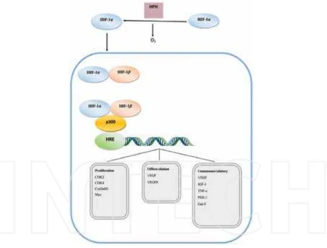
prolyl hydroxylase domain proteins (PHD1–3) on two proline residues in the oxygen‐ dependent degradation (ODD) domain [9]. The prolyl‐hydroxylated HIF1α is targeted for degradation by the tumor suppressor protein von Hippel‐Lindau (VHL), an E3 ubiquitin‐ protein ligase [10, 11]. HIF1α is also regulated in an oxygen‐dependent manner by factor inhibiting HIF1 (FIH1) [12, 13]. In this case, FIH1 mediates the hydroxylation of an asparagine residue in the C‐terminal trans‐activation domain, which prevents the binding of HIF1α with coactivators p300 and CBP [13–15]. Hydroxylation of proline and asparagine is inhibited under hypoxic conditions causing HIF1α to rapidly accumulate [12, 13]. HIF1α subsequently heterodimerizes with HIF1β, and the complex binds to hypoxic responsive elements (HREs) within the promoter regions of target genes, and allows for recruitment of coactivators and activation of transcription [16]. In addition to hypoxia, HIF1 accumulation can also be induced by growth‐factor stimulation, gene mutations, and intermediate metabolites [17] (Figure 1).
Figure 1. Regulation of HIF1 and its downstream roles related to lipid metabolism. HIF1 accumulation can be induced by hypoxia, gene mutations, intermediate metabolites, and growth factors. HIF1 plays a pivotal role in lipid metabo‐ lism. It can increase lipid uptake and traicking, faty acid synthesis, sterol synthesis, TAG synthesis, lipid droplet bio‐ genesis, and lipid signal production, and suppress faty acid β‐oxidation. Lipid droplet accumulation may be the inal result of HIF1 in lipid metabolism. It is unclear about its role in phospholipids metabolism.
plays an important role in lipid metabolism [1, 2, 18–21]. Currently, our understanding of HIF1 in regulating lipid metabolism has lagged behind that of glucose metabolism. Lipids, struc‐ turally and functionally important in all organisms, are not only one of the major components of cellular membrane systems, but also the source of energy storage. Moreover, signal molecules, such as prostaglandin E2 (PGE2), hydroxyeicosatetraenoic acid (HETE), and steroid hormones, are derived from lipids. This review would focus on the HIF1's activity related to dysregulation of lipid metabolism in several diseases, including atherosclerosis [4], faty liver disease (FLD) [19], heart failure diseases [5], obesity [3], and cancer [1, 2] as well as the involvement of HIF1 in lipid metabolism, including lipid uptake and traicking, faty acid metabolism, sterol metabolism, triacylglycerol (TAG) synthesis, phospholipid metabolism, lipid droplet (LD) biogenesis, and lipid signaling.
2. Lipid accumulation is associated with HIF1's activity in diseases
Most of the studies have demonstrated that HIF1's activity is associated with lipid accumula‐ tion positively [3, 18, 20–27], while few researches have indicated the opposite efect [28–31]. PHD2 inhibition or deletion, increasing HIF1's activity (Figure 1), decreased lipid accumula‐ tion in diferent animal models [28, 30, 31]. It indicated that the role of HIF1 in lipid metabolism may be diferent in diferent animal models. Details were described and discussed in the following sections.
2.1. Atherosclerosis
2.2. Heart failure diseases
Ischemic heart disease, systemic hypertension, and pathological cardiac hypertrophy eventu‐ ally result in heart failure. Myocardial hypoxia has been associated with these clinical condi‐ tions [25, 46]. Several studies showed a correlation between TAG accumulation and heart failure [26, 47–49]. Hypoxia promotes TAG accumulation in cardiomyocytes [48, 50]. Overex‐ pression of the constitutive active form of HIF1α in cardiomyocytes promotes intracellular lipid accumulation under normoxia [24]. The speciic deletion of VHL in mice cardiac myocytes results in lipid accumulation [25, 26]. In a pathological cardiac hypertrophy mouse model, cardiac TAG accumulation in ventricles was abolished in HIF1α knockout mice [26].
2.3. Faty liver disease (FLD)
Lipid accumulation is a common feature of faty liver disease, whether it is alcoholic (AFLD) or nonalcoholic (NAFLD) [19]. FLD initially begins with simple hepatic steatosis, but can irreversibly progress to steatohepatitis, ibrosis, cirrhosis, or hepatocellular carcinoma [19]. Hypoxia in liver has been documented in vivo in rats on a continuous ethanol diet at a constant rate for prolonged periods [51–54]. Recent studies have demonstrated that hypoxia is also observed in NAFLD [55]. Indeed, HIF1 expression is increased in faty liver diseases [19]. Nath and his colleagues found that ethanol feeding resulted in liver steatosis in wild‐type mice compared with isocaloric diet‐fed controls [27]. Constitutive activation of HIF1α in hepatocytes accelerates lipid accumulation with chronic ethanol feeding compared with wild‐type mice [27]. In contrast, hepatocyte‐speciic deletion of HIF1α protected mice from alcohol‐induced liver lipid accumulation [27]. However, another group reported that hepatocyte‐speciic HIF1α‐null mice developed severe hyper‐triglyceridemia with enhanced lipid accumulation in the liver of mice after 4 weeks of exposure to a 6% ethanol‐containing liquid diet [29]. Diferent genetic techniques used to create speciic gene expression or knockout mice in each of these studies may ofer some explanation of the diferent results each described. The other possible explanation is that the presence of inlammation may rewire the HIF‐1 pathway, which leads to a diferent gene expression proile compared to that observed in simple steatosis [19].
2.4. Obesity
litermates. In addition, serum cholesterol level and de novo lipid synthesis were decreased, and the mice were protected against hepatic steatosis in PHD2‐deicient mice [31]. It seems that HIF1 in adipocyte of obesity had diferent efect on lipid metabolism compared with other models. Thus, the efect of HIF1 in lipid metabolism of obesity has yet to be deined.
2.5. Cancer
Hypoxia in the tumor microenvironment leads to the metabolic changes in cancer cells. Over 50% cellular energy is produced by glycolysis and HIF1 plays a central role in the changes [16, 61]. Recently disorders of lipid metabolism had been demonstrated in solid tumors [62, 63], such as pancreatic cancer [64], liver cancer [1], breast cancer [65], colon cancer [66], and ovarian cancer [67]. Lipid accumulation is observed in human tumor tissue [66, 68]. Accumulation of cholesterol also has been reported in prostate cancer [69]. Indeed, recent researches had demonstrated that HIF1's activity is really involved abnormal lipid metabolism of cancer cells. Hypoxia‐induced lipid accumulation depends on HIF1's activity in cancer cells [18, 20, 21]. Under hypoxic condition, the lux from glutamine into faty acid is mediated by reductive carboxylation, and HIF1α plays an important role in this metabolic shift in tumor cells [70]. HIF1α also inhibits faty acid β‐oxidation to promote lipid accumulation in human hepato‐ cellular carcinoma [1]. Valli and his colleagues revealed that hypoxia induced many changes in lipid metabolites. Enzymatic steps in faty acid synthesis and the Kennedy pathway were modiied in an HIF1α‐dependent fashion in HCT116 cell line [2]. However, the role of HIF1 in cancer lipid metabolism has not been well addressed, so more researches should be further studied.
3. The role of HIF1 in lipid metabolism
Lipid metabolism is more complicated than glucose metabolism. Besides as major components of membrane, lipids are also a source of energy storage and signal molecules. HIF1‐induced genes involving lipid metabolism are listed in Table 1. We would discuss the role of HIF1 in lipid metabolism from the following linked aspects: lipid uptake and traicking, faty acid metabolism, sterol metabolism, TAG synthesis, phospholipids metabolism, lipid droplets biogenesis, and lipid signaling (Figure 1).
3.1. Lipid uptake and traicking
3.1.1. Free faty acid (FFA) uptake
cellular lipid accumulation in hypoxic hearts is atributable to accumulation of faty acid in the heart [92]. CD36 also can be regulated at the transcriptional level. In neonatal mouse cardiac myocytes, phenyl‐epinephrine (PE) induced free faty acid uptake in an HIF1α ‐dependent fashion while inhibition of CD36 led to decreased TAG accumulation upon PE stimulation [26]. In this model, CD36 was induced through HIF1‐PPARγ axis [26]. In human retinal pigment epithelial cells, CD36 is mediated by HIF1 binding on its promoter region [71]. Hypoxia also markedly induced CD36 mRNA in corneal and retinal tissue in in vivo [71].
Products of HIF1's target genes Functions in lipid metabolism References
CD36, PPARγ, FABP3, FABP7 Faty acid uptake [21, 26, 71] VLDLR, LRP1 LDL and VLDL uptake [18, 48, 72, 73] CAV1, RAB20 Endocytosis and lipid traicking [74, 75] PPARα*, TWIST1, Sirt2* Faty acid β‐oxidation [3, 76, 77] DEC1 Faty acid synthesis [30, 78] ABCA1* Cholesterol elux [79] PPARγ, Lipin1 TAG synthesis [20, 26] CHKA Phospholipids synthesis [80, 81] ADRP, HIG2, CAV1 Lipid droplet biogenesis [42, 74, 82–85] COX2, PTGES1 Lipid signaling [86–88]
PPARγ, peroxisome proliferator‐activated receptor gamma; VLDLR, very‐low‐density lipoprotein receptor; LRP1, low‐ density lipoprotein receptor‐related protein 1; CAV1, caveolin 1; PPARα, peroxisome proliferator‐activated receptor alpha; TWIST1, twist family bHLH transcription factor 1; SIRT2, sirtuin 2; DEC1, deleted in esophageal cancer 1; ABCA1, ATP‐binding cassete subfamily A member 1; LPIN1, lipin 1; HIG2, hypoxia inducible gene 2; CHKA, choline kinase alpha; COX2, cyclooxygenase 2; PTGES, prostaglandin E synthase 1.
“*” genes suppressed by HIF1.
Table 1. HIF1 targets genes that regulate lipid metabolism.
3.1.2. LDL and VLDL uptake
LDL and VLDL are major source of extracellular lipid, and HIF1 has been implicated in the transport of LDL and VLDL into cells. LDL receptor (LDLR) and VLDL receptor (VLDLR) are major receptors that are responsible for LDL and VLDL uptake. It had been reported that hypoxia signiicantly increased LDL uptake and enhances lipid accumulation in arterial smooth muscle cells (SMCs), exclusive LDLR activity [100]. In addition, hypoxia increased VLDL uptake in cardiac myocytes, which might be partially dependent on up‐regulating VLDLR expression [101]. Some studies had also reported that VLDLR could be induced un‐ der hypoxia [102]. In human cancer cell lines, we had demonstrated that HIF1‐mediated VLDLR induction inluenced intracellular lipid accumulation through regulating LDL and VLDL uptake under hypoxia [18]. In hepatocellular carcinoma, expression of VLDR was as‐ sociated positively with HIF1 [18]. In mice, hypoxia‐induced VLDLR expression in HL‐1 cells was dependent on HIF1α through its interaction with an HRE in the VLDLR promoter. VLDLR promoted the endocytosis of lipoproteins, and causes lipid accumulation in cardio‐ myocytes [48].
Low‐density lipoprotein receptor related protein 1 (LRP1) belongs to LDL receptor superfam‐ ily, and is a key receptor for selective cholesterol uptake in human vascular smooth muscle cells (VSMCs). Hypoxia increased LRP1 expression through HIF1α, and overexpression of LRP1 mediated hypoxia‐induced aggregated LDL (agLDL) uptake in human VSMCs [72] as well as VLDL‐cholesteryl ester (VLDL‐CE) uptake in neonatal rat ventricular myocytes (NRVMs) [73]. In contrast to the strong impact of LRP1 inhibition on VLDL‐CE uptake in hypoxic cardiomyocytes, LRP1 deiciency did not exert any signiicant efect on VLDL‐TG uptake or VLDL‐TG accumulation [73]. This indicated that VLDLR might be a key receptor for VLDL‐TG uptake. Therefore, more experiments should be done to value the precise contribution of VLDLR and LRP1 in myocardial VLDL‐CE and VLDL‐TG uptake in patho‐ physiological situation in the heart.
Taken together, HIF1 promoting lipid accumulation may increase lipid uptake and intracel‐ lular lipid traicking by inducing related genes directly. It should be further studied if there are more genes targeted by HIF1 in the process.
3.2. Faty acid metabolism
3.2.1. Faty acid β-oxidation
Hypoxia increased intracellular lipid accumulation through suppression of faty acid β‐ oxidation (FAO) in several models, and the molecular mechanism involvement of HIF1 in the process had been demonstrated (Figure 2). Under hypoxic condition, human macrophages showed in an increased TAG accumulation that was associated with a decreasing rate of FAO. The decreasing rate of FAO was shown to be partly dependent on the reduced expression of enzymes involved in FAO [42]. Peroxisome proliferator‐activated receptors (PPARs), including α, γ, and β/δ, belong to the nuclear receptor family of ligand‐activated transcription factors that were originally described as gene regulators of various metabolic pathways. PPARα and PPARβ/δ control expression of genes implicated in FAO. PPARγ, in contrast, is a key regulator of glucose homeostasis and adipogenesis [108].
Muscle carnitine palmitoyltransferase 1 (M‐CPT1), a known PPARα target gene, catalyzes the rate‐limiting step in the mitochondrial import of faty acids for the FAO cycle [109]. In cardiomyocytes, hypoxia and adenovirus‐mediated expression of a constitutively active form of HIF1α reduced the mRNA and protein levels of PPARα and M‐CPT1 [24, 50, 110] as well as the DNA binding activity of PPARα [24, 50]. CoCl2 treatment also decreased PPARα and M‐
CPT1 mRNA levels [110]. In intestinal epithelial cells, hypoxia rapidly down‐regulated PPARα mRNA and protein in an HIF1‐dependent manner in vitro and in vivo [76]. HIF1 could down‐regulate PPARα directly through binding a functional HRE in the promoter region [76]. These results suggested that the mechanism of HIF‐1 suppression of FAO involved the partial reduction of the expression of PPARα and M‐CPT1.
HIF1 also suppressed FAO by inhibition of PPARδ's activity. In a pathological cardiac hyper‐ trophy mouse model, myocardial hypoxia provoked Dnm3os activation and concomitantly mir‐199a and mir‐214 expression through the HIF1‐TWIST1 axis [49]. TWIST1 is a direct target gene of HIF1 [77]. DNM3os is a noncoding RNA transcript that harbors the mi‐RNA cluster mir‐199a∼214, for which PPARδ is a target. Increased expression of mir‐199a and mir‐214 decreased cardiac PPARδ expression and mitochondrial faty acid oxidative capacity. Reduced expression of enzymes involved in FAO, for example long‐chain acyl‐CoA dehydrogenase (LCAD) and medium‐chain acyl‐CoA dehydrogenase (MCAD), was also observed. Converse‐ ly, antagomir‐based silencing of miR‐199a∼214 in mice subjected to pressure overload de‐ repressed cardiac PPARδ, LCAD and MCAD levels, and restored mitochondrial FAO [49]. PPARγ coactivator 1α (PGC‐1α) has been prominently associated with the expression of the genes involving FAO and energy expenditure [111]. In obese mouse model, HIF1α suppressed FAO in visceral white adipocytes, in part, through transcriptional repression of sirtuin 2 (Sirt2), an NAD+‐dependent deacetylase [3]. Reduced Sirt2 function directly translated into dimin‐
ished deacetylation of PGC1α and the expression of FAO genes. HIF1α negated adipocyte‐ intrinsic pathway of faty acid catabolism by negatively regulating the Sirt2‐PGC1α regulatory axis [3].
PPARγ coactivator 1β (PGC‐1β) is a transcription factor that also plays critical roles in regulating mitochondrial function and lipid metabolism [112, 113]. PGC‐1β could regulate FAO through activating medium‐chain acyl‐CoA dehydrogenase (MCAD) and long‐chain acyl‐CoA dehydrogenase (LCAD), which catalyzes the irst step of FAO in mitochondria [1, 112]. It had been documented previously that hypoxia inhibited PGC‐1β activity through HIF1‐ dependent c‐Myc suppression in VHL‐null RCC4 renal carcinoma cells [114]. Under hypoxic condition in Hep3B and HepG2 cells, and also in PC3 prostate cancer cells, Huang and his colleagues revealed a role of the HIF1/C‐MYC/PGC‐1β regulatory axis in hypoxia‐mediated regulation of MCAD and LCAD by which HIF1 suppressed FAO [1]. This study conirmed that hypoxia inhibited FAO in an HIF1‐dependent mechanism in cancer cells [1].
In summary, it had been conirmed by diferent models that hypoxia inhibits FAO depending on HIF1's activity (Figure 2). However, HIF1 did not target FAO‐related genes directly, and it was always cross‐talk with other pathway to suppress FAO indirectly. It should be further studied if HIF1 could involve cross‐talk with more pathways to suppress FAO.
3.2.2. Faty acid synthesis
The reductive carboxylation of glutamine was part of the metabolic reprogramming associated with HIF1. Glutamine‐derived α‐ketoglutarate is reductively carboxylated by the cytosolic isocitrate dehydrogenase 1 (IDH1) [70, 115] and the mitochondrial isocitrate dehydrogenase 2 (IDH2) to form isocitrate [70, 115, 116], which could then be isomerized to citrate. The combined action of IDH1 and IDH2 was necessary and suicient to afect the reverse TCA lux [115]. Citrate was converted into Ac‐CoA by ATP citrate lyase in the cytosol. Renal cell lines deicient in the VHL preferentially used reductive glutamine metabolism for lipid biosynthesis even at normal oxygen levels [70]. Constitutive activation of HIF1 recapitulated the preferential reductive metabolism of glutamine‐derived α‐ketoglutarate even in normoxic condition [116]. This regulation by HIF1 of the reverse TCA cycle occurred partly through HIF1‐inducing PDK1. Knocking down PDK1 suppressed reductive carboxylation [70, 118]. However, more details should be studied about the role of HIF1 in TCA cycle reverse.
The irst step of faty acid synthesis is catalyzed by AcCoA carboxylase (ACC) which converts Ac‐CoA to malonyl‐CoA. Then faty‐acid synthase (FASN) catalyzes acetyl‐CoA and malonyl‐ CoA to palmitate. Further elongation and de‐saturation of newly synthesized faty acid takes place at the cytoplasmic face of the endoplasmic reticulum membrane. It had been reported that hypoxia regulated FASN expression [78, 119, 120]. However, diferent conclusions on hypoxia regulation of FASN had been reported. One group using human breast cancer cell lines found that FASN was signiicantly up‐regulated by hypoxia via activation of the Akt and HIF1 followed by the induction of the SREBP1 gene [119]. Another group, using several cell lines other than breast cancer cell lines, found that hypoxia suppressed FASN expression through HIF1‐DEC1 and/or DEC2‐SREBP1 axis. They found that HIF1 repressed the SREBP1 gene by inducing DEC1 and DEC2, and further repressing FASN expression [78]. These results might indicate that HIF1 regulated FASN in a cell‐type speciic manner. In addition, it had been reported that hypoxia could induce the expression of SCD1 which introduces a double bond in the Δ9 position of palmitic acid and stearic acid to produce mono‐unsaturated faty acid [42, 121]. It is unknown if HIF1 is involved in hypoxic‐induced SCD1.
Taken together, the role of HIF1 in de novo faty acid synthesis may depend on diferent models and conditions, and more researches should be done in the direction.
3.3. Cholesterol metabolism
Cholesterol is an essential structural component of membrane. It modulates membrane permeability and luidity and also forms microdomains named lipid rafts that integrate the activation of some signal transduction pathways [14]. Intermediates generated by the choles‐ terol biosynthesis pathway were required for the postranslational modiication of small GTPases, such as the farnesylation of Ras and the geranyl‐geranylation of Rho [15]. Finally, cholesterol also serves as a precursor for the biosynthesis of steroid hormones, bile acids, and vitamin D.
acetoacetyl‐CoA to form 3‐hydroxy‐3‐methylglutaryl (HMG)‐CoA. Then HMG‐CoA reduc‐ tase (HMGCR) reduces of HMG‐CoA to mevalonate. Early research found that Hypoxia also suppressed cholesterol synthesis in cultured rabbit skin ibroblasts [123]. However, recently research indicated that hypoxia increased sterol synthesis depending on HIF1's activity [23, 124]. In hypoxic macrophages, the increase of intracellular cholesterol content was correlated with elevated HMGCR's activity and mRNA levels [23]. In HepG2 cells, HIF1α accumulation was able to increase the level and activity of HMGCR by stimulating its transcription [124]. But it was unclear if HIF1 regulated HMGCR directly.
Hypoxia suppressed the elux of cholesterol, and this elux was substantially reversed in vitro by reducing the expression of HIF1 [23, 123]. ATP‐binding cassete transporter A1 (ABCA1) plays a major role in cholesterol elux. Hypoxia severely reduced ABCA1‐mediated choles‐ terol elux, which could be explained by subcellular redistribution of ABCA1 protein under acute hypoxia and decreased protein level under prolonged hypoxia [23]. One group reported that HIF1 could repress the transcription of ABCA1 directly [79]. Hypoxia, partly mediated by HIF1α, increased intracellular cholesterol content due to the induction of cholesterol synthesis and the suppression of cholesterol elux [23]. In addition, accumulation of cholesterol in hypoxic cells was in esteriied form [23, 100]. At 2% O2 tension, twice the total cholesteryl ester
was observed compared with that at 21% O2. At the same time, no signiicant diference was
found in the concentration of cellular‐free cholesterol [100]. Accumulation of cholesteryl ester in hypoxic cells might depend on the increased activity of AcCoA:cholesterol acyltransferases (ACATs) [123], which are important enzymes for the esteriication of cholesterol. Therefore, more studies should be done to deine the role of HIF1 involving the cholesterol metabolism in detail.
3.4. TAG synthesis and phospholipids metabolism
3.4.1. TAG synthesis
3.4.2. Phospholipids metabolism
Phospholipids are indispensable for cell growth. Phospholipids synthesis and TAG synthesis share similar steps. DAG is a precursor for phosphatidylcholine and phosphatidylethanola‐ mine. Phosphatidic acid utilizes cytidine triphosphate (CTP) as an energy source to produce a CDP‐DAG intermediate followed by conversion to phosphatidylcholine. It had been reported that the intracellular level of phosphatidic acid (PA) and DAG rose in response to hypoxia [125, 126]. However, PA accumulation in response to hypoxia was both HIF1 and VHL‐independ‐ ent [127]. Choline kinase α (ChKα) catalyzes the phosphorylation of choline, the irst step of phosphatidylcholine synthesis. In cancer cells, one group had shown that hypoxia increased ChKα expression and this was driven by HIF1 [80]. Conversely, another group had shown that choline kinase activity and choline phosphorylation were decreased, that might be mediated via HIF1α binding to the promoter of ChKα gene [81]. Thus, further studies should be done to address the role of HIF1 in phospholipids metabolism.
3.5. Lipid droplet (LD) biogenesis and lipid signaling
Lipid droplet, also named lipid body, has been largely associated with neutral lipid storage and transport in cells [106]. The internal core of the LD is rich in neutral lipids, predominantly TAGs or cholesteryl esters, that are surrounded by an outer monolayer of phospholipids and associated proteins [128]. LD was considered to be highly regulated, dynamic and functionally active organelle [106]. Proteins on the surface of lipid droplets are crucial to the droplet structure and dynamics. Currently, the complete protein composition of LD has not been deined. The best characterized LD’ proteins are the perilipin/ADRP/TIP47 (PAT) domain family. Apart from the PAT domain proteins, there are other lipid droplets associated proteins which involve the catabolism of lipids, vesicular transport, eicosanoid‐forming enzymes, protein kinases, etc. [106]. Hypoxia increased LD number and size [42, 129]. Several LD‐ associated proteins were induced by HIF1 and might also involve HIF1‐induced LD biogenesis and lipid signaling (Figure 3).
3.5.1. Lipid droplet biogenesis
that ADRP can also stimulate LCFA uptake [133]. While another research reported that ADRP did not involve LDL‐ and VLDL‐induced LD formation under hypoxia [84].
Figure 3. A hypothetical representation of molecular mechanism involving hypoxia‐induced lipid droplet biogenesis and function. HIF1‐induced structural proteins of the LD, such as ADRP, HIG2, combine with HIF1‐increased lipids to form the LD. Enzymes involving eicosanoid production are also induced by HIF1, and are recruited to the LD. These proteins can increase lipid signaling that can involve many aspects of biology, such as HIF1α's stability, angiogenesis, inlammation, cell proliferation and survival.
Hypoxia‐inducible protein 2 (HIG2), a newly identiied protein associated with LD, was up‐ regulated by hypoxia and was a direct and speciic target gene of HIF1 [85]. Overexpression of HIG2 under normoxic condition was suicient to increase LD in HeLa cells. HIG2‐driven LD might contribute to an inlammatory response. Overexpression of HIG2 stimulated cytokine expression of vascular endothelial growth factor‐A (VEGFA), macrophage migration inhibitory factor (MIF), and interleukin‐6 (IL‐6). Increasing expression of HIG2 was also detected under several conditions of pathological lipid accumulation, such as atherosclerotic arteries and faty liver disease [85]. We had mentioned that CAV1 was a target of HIF1. CAV1 could distribute to LD under several conditions [134–137] and the association with LD was reversible [134]. However, It is unknown if hypoxia can redistribute CAV1 to LD and CAV1 involves LD biogenesis under hypoxia.
3.5.2. Lipid signaling
of arachidonic acid. PGE2 is synthesized in three steps catalyzed by phospholipase (PL) A2,
COX, and terminal prostaglandin E synthase (PTGES), where each catalytic activity is repre‐ sented by multiple enzymes and/or isoenzymes. It had been reported that hypoxia could increase prostaglandins (PGI2 and PGE2) synthesis [138]. Hypoxia‐induced synthesis of PGE2
was accompanied by up‐regulation of COX2, which is a direct target gene of HIF1 [86]. Several studies had indicated that LD was reservoirs of COX2 and sites of PGE2 synthesis [66, 139,
140]. PTGES1 could also be regulated by HIF1 directly [87, 88]; however, it is unknown if PTGES1 localizes to hypoxia‐induced LD.
Lipoxygenases are a family of nonheme iron‐containing enzymes which dioxygenate polyun‐ saturated faty acid to hydroperoxyl metabolite, and mainly include 5‐lipoxygenase (5‐LO), 12‐lipoxygenase (12‐LO), and 15‐lipoxygenase (15‐LO). 5‐LO and 15‐LO were shown by immuno‐cytochemistry, immuno‐luorescence, ultrastructural postembedding immuno‐gold EM and/or western bloting from subcellular fractions to localize within lipid droplets stimulated in vitro [141–144]. Increasing level of 5‐LO was detected in lung tissue of rodent model of hypoxia‐induced pulmonary hypertension [145]. Hypoxia increased 12‐LO in rat lung and in in vitro cultured rat pulmonary artery smooth muscle cell (PASMC) and may contribute to the production of 12(S)‐hydroxyeicosatetraenoic acid (12(S)‐HETE) [146]. Increasing 12(S)‐HETE had also been demonstrated in hypoxic macrophage cells [147]. Under hypoxia, increased levels of 15‐LO had been demonstrated by diferent groups [147, 148] and its product, 15‐hydroperoxyeicosatetraenoic acid (15‐HETE), was up‐regulated [147]. Up‐ regulation of 15‐LO/15‐HETE in response to hypoxia might be partially mediated by HIF1α [149]. In addition, HIF1α was shown to be regulated by 15‐HETE in a positive feedback manner [149]. However, it is unknown if lipoxygenases are regulated by HIF1 directly.
4. Conclusions and perspectives
HIF1 plays an important role in lipid metabolism and a number of studies support the indings that HIF1 promotes lipid accumulation. Nevertheless, many questions remain. HIF1, as a master transcriptional factor, may target many genes directly or indirectly involved in lipid metabolism. HIF1 plays a pivotal role in glucose metabolism. Inhibition of GULT3, an HIF1 target gene, could signiicantly reduce both glucose uptake and hypoxia‐induced de novo lipid synthesis in human monocyte‐derived macrophages [150]. PGAM1, induced by hypoxia [151], catalyzes the reversible reaction of 3‐phosphoglycerate (3‐PGA) to 2‐phosphoglycerate (2‐ PGA) in the glycolytic pathway. Inhibition of PGAM1 led to signiicantly decreased glycolysis and de novo lipid synthesis in cancer cells [152]. Thus, it is possible that glucose metabolism might couple with lipid metabolism under hypoxia. The source of carbon for faty acid switched from glucose to glutamine under hypoxia [70]. The question thus arises. Does HIF1 induce lipid accumulation through targeting genes involving glucose metabolism, and how does glucose metabolism afect lipid metabolism under hypoxia?
lipid homeostasis in many setings, both physiological and pathological [153]. AMPK is a cellular energy sensor that normalizes lipid, glucose, and energy imbalances [154]. Inhibition of cMYC was accompanied by accumulation of intracellular LD in tumor cells as a direct consequence of mitochondrial dysfunction [155]. Recently, p53 had also been shown to regulate lipid metabolism [156]. The role of HIF1 in these pathways and the molecular mechanism will require further investigation.
Lipid accumulation in diseases, including obesity, atherosclerosis, ALD, heart failure disease and cancer, had been associated with HIF1's activity. There may be additional pathologies with lipid metabolism disorder associated with HIF1. HIF1 is an atractive target candidate for therapeutic intervention in diseases with disorder of lipid metabolism including cancer. Its involvement in the etiology of a number of diseases and its interaction with a number of regulatory genes make it an important area for further study.
Acknowledgements
This review is supported by a National Natural Science Foundation of China (Grant No. 31301076 to G. S; Grant No. 81401961 to X. L.), We thank Gerard Moskowiz, Ph. D. (Washington University in St. Louis, St. Louis, MO) for critical reading of the manuscript. We sincerely apologize to the colleagues whose works are not covered in this review due to limitations of time and space.
Author details
Guomin Shen1* and Xiaobo Li2
*Address all correspondence to: [email protected]
1 Department of Medical Genetics, Medical College, Henan University of Science and Technology, Luoyang, Henan Province, China
2 Department of Pathology & Translational Medicine Center, Harbin Medical University, Harbin, Heilongjiang Province, China
References
[2] A. Valli, M. Rodriguez, L. Moutsianas, R. Fischer, V. Fedele, H.L. Huang, R. Van Stiphout, D. Jones, M. McCarthy, M. Vinaxia, K. Igarashi, M. Sato, T. Soga, F. Bufa, J. McCullagh, O. Yanes, A. Harris, B. Kessler, Hypoxia induces a lipogenic cancer cell phenotype via HIF1alpha‐dependent and ‐independent pathways, Oncotarget 6 (2015) 1920–1941.
[3] J. Krishnan, C. Danzer, T. Simka, J. Ukropec, K.M. Walter, S. Kumpf, P. Mirtschink, B. Ukropcova, D. Gasperikova, T. Pedrazzini, W. Krek, Dietary obesity‐associated Hif1alpha activation in adipocytes restricts faty acid oxidation and energy expenditure via suppression of the Sirt2‐NAD+ system, Genes Dev 26 (2012) 259–270.
[4] E. Marsch, J.C. Sluimer, M.J. Daemen, Hypoxia in atherosclerosis and inlammation, Curr Opin Lipidol 24 (2013) 393–400.
[5] G.L. Semenza, Hypoxia‐inducible factor 1 and cardiovascular disease, Annu Rev Physiol 76 (2013) 39–56.
[6] G.L. Semenza, Hypoxia‐inducible factors in physiology and medicine, Cell 148 (2012) 399–408.
[7] G.L. Wang, B.H. Jiang, E.A. Rue, G.L. Semenza, Hypoxia‐inducible factor 1 is a basic‐ helix‐loop‐helix‐PAS heterodimer regulated by cellular O2 tension, Proc Natl Acad Sci U S A 92 (1995) 5510–5514.
[8] G.L. Wang, G.L. Semenza, Puriication and characterization of hypoxia‐inducible factor 1, J Biol Chem 270 (1995) 1230–1237.
[9] A.C. Epstein, J.M. Gleadle, L.A. McNeill, K.S. Hewitson, J. O'Rourke, D.R. Mole, M. Mukherji, E. Mezen, M.I. Wilson, A. Dhanda, Y.M. Tian, N. Masson, D.L. Hamilton, P. Jaakkola, R. Barstead, J. Hodgkin, P.H. Maxwell, C.W. Pugh, C.J. Schoield, P.J. Ratclife, C. elegans EGL‐9 and mammalian homologs deine a family of dioxygenases that regulate HIF by prolyl hydroxylation, Cell 107 (2001) 43–54.
[10] P.H. Maxwell, M.S. Wiesener, G.W. Chang, S.C. Cliford, E.C. Vaux, M.E. Cockman, C.C. Wykof, C.W. Pugh, E.R. Maher, P.J. Ratclife, The tumour suppressor protein VHL targets hypoxia‐inducible factors for oxygen‐dependent proteolysis, Nature 399 (1999) 271–275.
[11] M. Ohh, C.W. Park, M. Ivan, M.A. Hofman, T.Y. Kim, L.E. Huang, N. Pavletich, V. Chau, W.G. Kaelin, Ubiquitination of hypoxia‐inducible factor requires direct binding to the beta‐domain of the von Hippel‐Lindau protein, Nat Cell Biol 2 (2000) 423–427. [12] P.C. Mahon, K. Hirota, G.L. Semenza, FIH‐1: a novel protein that interacts with HIF‐
1alpha and VHL to mediate repression of HIF‐1 transcriptional activity, Genes Dev 15 (2001) 2675–2686.
inhibiting HIF (FIH) and is related to the cupin structural family, J Biol Chem 277 (2002) 26351–26355.
[14] D. Lando, D.J. Peet, J.J. Gorman, D.A. Whelan, M.L. Whitelaw, R.K. Bruick, FIH‐1 is an asparaginyl hydroxylase enzyme that regulates the transcriptional activity of hypoxia‐ inducible factor, Genes Dev 16 (2002) 1466–1471.
[15] D. Lando, D.J. Peet, D.A. Whelan, J.J. Gorman, M.L. Whitelaw, Asparagine hydroxyla‐ tion of the HIF transactivation domain a hypoxic switch, Science 295 (2002) 858–861. [16] G.L. Semenza, Deining the role of hypoxia‐inducible factor 1 in cancer biology and
therapeutics, Oncogene 29 (2010) 625–634.
[17] N.C. Denko, Hypoxia, HIF1 and glucose metabolism in the solid tumour, Nat Rev Cancer 8 (2008) 705–713.
[18] G.M. Shen, Y.Z. Zhao, M.T. Chen, F.L. Zhang, X.L. Liu, Y. Wang, C.Z. Liu, J. Yu, J.W. Zhang, Hypoxia‐inducible factor‐1 (HIF‐1) promotes LDL and VLDL uptake through inducing VLDLR under hypoxia, Biochem J 441 (2012) 675–683.
[19] T. Suzuki, S. Shinjo, T. Arai, M. Kanai, N. Goda, Hypoxia and faty liver, World J Gastroenterol 20 (2014) 15087–15097.
[20] I. Mylonis, H. Sembongi, C. Befani, P. Liakos, S. Siniossoglou, G. Simos, Hypoxia causes triglyceride accumulation by HIF‐1‐mediated stimulation of lipin 1 expression, J Cell Sci 125 (2012) 3485–3493.
[21] K. Bensaad, E. Favaro, C.A. Lewis, B. Peck, S. Lord, J.M. Collins, K.E. Pinnick, S. Wigield, F.M. Bufa, J.L. Li, Q. Zhang, M.J. Wakelam, F. Karpe, A. Schulze, A.L. Harris, Faty acid uptake and lipid storage induced by HIF‐1alpha contribute to cell growth and survival after hypoxia‐reoxygenation, Cell Rep 9 (2014) 349–365.
[22] G. Jiang, T. Li, Y. Qiu, Y. Rui, W. Chen, Y. Lou, RNA interference for HIF‐1alpha inhibits foam cells formation in vitro, Eur J Pharmacol 562 (2007) 183–190.
[23] S. Parathath, S.L. Mick, J.E. Feig, V. Joaquin, L. Grauer, D.M. Habiel, M. Gassmann, L.B. Gardner, E.A. Fisher, Hypoxia is present in murine atherosclerotic plaques and has multiple adverse efects on macrophage lipid metabolism, Circ Res 109 (2011) 1141–1152. [24] A.J. Belanger, Z. Luo, K.A. Vincent, G.Y. Akita, S.H. Cheng, R.J. Gregory, C. Jiang, Hypoxia‐inducible factor 1 mediates hypoxia‐induced cardiomyocyte lipid accumula‐ tion by reducing the DNA binding activity of peroxisome proliferator‐activated receptor alpha/retinoid X receptor, Biochem Biophys Res Commun 364 (2007) 567–572. [25] L. Lei, S. Mason, D. Liu, Y. Huang, C. Marks, R. Hickey, I.S. Jovin, M. Pypaert, R.S. Johnson, F.J. Giordano, Hypoxia‐inducible factor‐dependent degeneration, failure, and malignant transformation of the heart in the absence of the von Hippel‐Lindau protein, Mol Cell Biol 28 (2008) 3790–3803.
W. Krek, Activation of a HIF1alpha‐PPARgamma axis underlies the integration of glycolytic and lipid anabolic pathways in pathologic cardiac hypertrophy, Cell Metab 9 (2009) 512–524.
[27] B. Nath, I. Levin, T. Csak, J. Petrasek, C. Mueller, K. Kodys, D. Catalano, P. Mandrekar, G. Szabo, Hepatocyte‐speciic hypoxia‐inducible factor‐1alpha is a determinant of lipid accumulation and liver injury in alcohol‐induced steatosis in mice, Hepatology 53 (2011) 1526–1537.
[28] L. Rahtu‐Korpela, J. Maata, E.Y. Dimova, S. Horkko, H. Gylling, G. Walkinshaw, J. Hakkola, K.I. Kivirikko, J. Myllyharju, R. Serpi, P. Koivunen, Hypoxia‐inducible factor prolyl 4‐hydroxylase‐2 inhibition protects against development of atherosclerosis, Arterioscler Thromb Vasc Biol 36 (2016) 608–617.
[29] Y. Nishiyama, N. Goda, M. Kanai, D. Niwa, K. Osanai, Y. Yamamoto, N. Senoo‐Matsuda, R.S. Johnson, S. Miura, Y. Kabe, M. Suematsu, HIF‐1alpha induction suppresses excessive lipid accumulation in alcoholic faty liver in mice, J Hepatol 56 (2012) 441–447. [30] H. Matsuura, T. Ichiki, E. Inoue, M. Nomura, R. Miyazaki, T. Hashimoto, J. Ikeda, R. Takayanagi, G.H. Fong, K. Sunagawa, Prolyl hydroxylase domain protein 2 plays a critical role in diet‐induced obesity and glucose intolerance, Circulation 127 (2013) 2078–2087.
[31] L. Rahtu‐Korpela, S. Karsikas, S. Horkko, R. Blanco Sequeiros, E. Lammentausta, K.A. Makela, K.H. Herzig, G. Walkinshaw, K.I. Kivirikko, J. Myllyharju, R. Serpi, P. Koivu‐ nen, HIF prolyl 4‐hydroxylase‐2 inhibition improves glucose and lipid metabolism and protects against obesity and metabolic dysfunction, Diabetes 63 (2014) 3324–3333. [32] J. Niinikoski, C. Heughan, T.K. Hunt, Oxygen tensions in the aortic wall of normal
rabbits, Atherosclerosis 17 (1973) 353–359.
[33] C. Heughan, J. Niinikoski, T.K. Hunt, Oxygen tensions in lesions of experimental atherosclerosis of rabbits, Atherosclerosis 17 (1973) 361–367.
[34] J.F. Martin, R.F. Booth, S. Moncada, Arterial wall hypoxia following hyperfusion through the vasa vasorum is an initial lesion in atherosclerosis, Eur J Clin Invest 20 (1990) 588–592.
[35] T. Bjornheden, M. Evaldsson, O. Wiklund, A method for the assessment of hypoxia in the arterial wall, with potential application in vivo, Arterioscler Thromb Vasc Biol 16 (1996) 178–185.
[36] T. Bjornheden, M. Levin, M. Evaldsson, O. Wiklund, Evidence of hypoxic areas within the arterial wall in vivo, Arterioscler Thromb Vasc Biol 19 (1999) 870–876.
[38] B. Ramkhelawon, Y. Yang, J.M. van Gils, B. Hewing, K.J. Rayner, S. Parathath, L. Guo, S. Oldebeken, J.L. Feig, E.A. Fisher, K.J. Moore, Hypoxia induces netrin‐1 and Unc5b in atherosclerotic plaques: mechanism for macrophage retention and survival, Arte‐ rioscler Thromb Vasc Biol 33 (2013) 1180–1188.
[39] J.C. Sluimer, J.M. Gasc, J.L. van Wanroij, N. Kisters, M. Groeneweg, M.D. Sollewijn Gelpke, J.P. Cleutjens, L.H. van den Akker, P. Corvol, B.G. Wouters, M.J. Daemen, A.P. Bijnens, Hypoxia, hypoxia‐inducible transcription factor, and macrophages in human atherosclerotic plaques are correlated with intraplaque angiogenesis, J Am Coll Cardiol 51 (2008) 1258–1265.
[40] S. Parathath, Y. Yang, S. Mick, E.A. Fisher, Hypoxia in murine atherosclerotic plaques and its adverse efects on macrophages, Trends Cardiovasc Med 23 (2013) 80–84. [41] L.M. Hulten, M. Levin, The role of hypoxia in atherosclerosis, Curr Opin Lipidol 20
(2009) 409–414.
[42] P. Bostrom, B. Magnusson, P.A. Svensson, O. Wiklund, J. Boren, L.M. Carlsson, M. Stahlman, S.O. Olofsson, L.M. Hulten, Hypoxia converts human macrophages into triglyceride‐loaded foam cells, Arterioscler Thromb Vasc Biol 26 (2006) 1871–1876. [43] S. Akhtar, P. Hartmann, E. Karshovska, F.A. Rinderknecht, P. Subramanian, F. Gremse,
J. Grommes, M. Jacobs, F. Kiessling, C. Weber, S. Stefens, A. Schober, Endothelial hypoxia‐inducible factor‐1alpha promotes atherosclerosis and monocyte recruitment by upregulating MicroRNA‐19a, Hypertension 66 (2015) 1220–1226.
[44] J. Ben‐Shoshan, A. Afek, S. Maysel‐Auslender, A. Barzelay, A. Rubinstein, G. Keren, J. George, HIF‐1alpha overexpression and experimental murine atherosclerosis, Arte‐ rioscler Thromb Vasc Biol 29 (2009) 665–670.
[45] L. Gao, Q. Chen, X. Zhou, L. Fan, The role of hypoxia‐inducible factor 1 in atheroscle‐ rosis, J Clin Pathol 65 (2012) 872–876.
[46] G.L. Semenza, Hypoxia‐inducible Factor 1 and cardiovascular disease, Annu Rev Physiol 76 (2014) 39–56.
[47] T.S. Park, I.J. Goldberg, Sphingolipids, lipotoxic cardiomyopathy, and cardiac failure, Heart Fail Clin 8 (2012) 633–641.
[48] J.C. Perman, P. Bostrom, M. Lindbom, U. Lidberg, M. StAhlman, D. Hagg, H. Lindskog, M. Scharin Tang, E. Omerovic, L. Matsson Hulten, A. Jeppsson, P. Petursson, J. Herliz, G. Olivecrona, D.K. Strickland, K. Ekroos, S.O. Olofsson, J. Boren, The VLDL receptor promotes lipotoxicity and increases mortality in mice following an acute myocardial infarction, J Clin Invest 121 (2011) 2625–2640.
inducible MicroRNA cluster miR‐199a approximately 214 targets myocardial PPAR‐ delta and impairs mitochondrial faty acid oxidation, Cell Metab 18 (2013) 341–354. [50] J.M. Huss, F.H. Levy, D.P. Kelly, Hypoxia inhibits the peroxisome proliferator‐activated
receptor alpha/retinoid X receptor gene regulatory pathway in cardiac myocytes: a mechanism for O2‐dependent modulation of mitochondrial faty acid oxidation, J Biol Chem 56–84 (2001) 27605–27612.
[51] G.E. Arteel, Y. Iimuro, M. Yin, J.A. Raleigh, R.G. Thurman, Chronic enteral ethanol treatment causes hypoxia in rat liver tissue in vivo, Hepatology 25 (1997) 920–926. [52] G.E. Arteel, J.A. Raleigh, B.U. Bradford, R.G. Thurman, Acute alcohol produces
hypoxia directly in rat liver tissue in vivo: role of Kupfer cells, Am J Physiol 271 (1996) G494–G500.
[53] S.W. French, The role of hypoxia in the pathogenesis of alcoholic liver disease, Hepatol Res 29 (2004) 69–74.
[54] F. Bardag‐Gorce, B.A. French, J. Li, N.E. Riley, Q.X. Yuan, V. Valinluck, P. Fu, M. Ingelman‐Sundberg, S. Yoon, S.W. French, The importance of cycling of blood alcohol levels in the pathogenesis of experimental alcoholic liver disease in rats, Gastroenter‐ ology 123 (2002) 325–335.
[55] S.K. Mantena, D.P. Vaughn, K.K. Andringa, H.B. Eccleston, A.L. King, G.A. Abrams, J.E. Doeller, D.W. Kraus, V.M. Darley‐Usmar, S.M. Bailey, High fat diet induces dysregulation of hepatic oxygen gradients and mitochondrial function in vivo, Biochem J 417 (2009) 183–193.
[56] M.E. Rausch, S. Weisberg, P. Vardhana, D.V. Tortoriello, Obesity in C57BL/6J mice is characterized by adipose tissue hypoxia and cytotoxic T‐cell iniltration, Int J Obes (Lond) 32 (2008) 451–463.
[57] J. Ye, Z. Gao, J. Yin, Q. He, Hypoxia is a potential risk factor for chronic inlammation and adiponectin reduction in adipose tissue of ob/ob and dietary obese mice, Am J Physiol Endocrinol Metab 293 (2007) E1118–E1128.
[58] N. Hosogai, A. Fukuhara, K. Oshima, Y. Miyata, S. Tanaka, K. Segawa, S. Furukawa, Y. Tochino, R. Komuro, M. Matsuda, I. Shimomura, Adipose tissue hypoxia in obesity and its impact on adipocytokine dysregulation, Diabetes 56 (2007) 901–911.
[59] C. Jiang, A. Qu, T. Matsubara, T. Chanturiya, W. Jou, O. Gavrilova, Y.M. Shah, F.J. Gonzalez, Disruption of hypoxia‐inducible factor 1 in adipocytes improves insulin sensitivity and decreases adiposity in high‐fat diet‐fed mice, Diabetes 60 (2011) 2484– 2495.
[61] O. Warburg, On respiratory impairment in cancer cells, Science 124 (1956) 269–270. [62] J.A. Menendez, R. Lupu, Faty acid synthase and the lipogenic phenotype in cancer
pathogenesis, Nat Rev Cancer 7 (2007) 763–777.
[63] C.R. Santos, A. Schulze, Lipid metabolism in cancer, FEBS J 279 (2012) 2610–2623. [64] J. Swierczynski, A. Hebanowska, T. Sledzinski, Role of abnormal lipid metabolism in
development, progression, diagnosis and therapy of pancreatic cancer, World J Gastroenterol 20 (2014) 2279–2303.
[65] P.L. Alo, P. Visca, G. Trombeta, A. Mangoni, L. Lenti, S. Monaco, C. Boti, D.E. Serpieri, U. Di Tondo, Faty acid synthase (FAS) predictive strength in poorly diferentiated early breast carcinomas, Tumori 85 (1999) 35–40.
[66] M.T. Accioly, P. Pacheco, C.M. Maya‐Monteiro, N. Carrossini, B.K. Robbs, S.S. Oliveira, C. Kaufmann, J.A. Morgado‐Diaz, P.T. Bozza, J.P. Viola, Lipid bodies are reservoirs of cyclooxygenase‐2 and sites of prostaglandin‐E2 synthesis in colon cancer cells, Cancer Res 68 (2008) 1732–1740.
[67] D.K. Nomura, J.Z. Long, S. Niessen, H.S. Hoover, S.W. Ng, B.F. Cravat, Monoacylgly‐ cerol lipase regulates a faty acid network that promotes cancer pathogenesis, Cell 140 (2010) 49–61.
[68] B.K. Straub, E. Herpel, S. Singer, R. Zimbelmann, K. Breuhahn, S. Macher‐Goeppinger, A. Warth, J. Lehmann‐Koch, T. Longerich, H. Heid, P. Schirmacher, Lipid droplet‐ associated PAT‐proteins show frequent and diferential expression in neoplastic steatogenesis, Mod Pathol 23 (2010) 480–492.
[69] M.H. Hager, K.R. Solomon, M.R. Freeman, The role of cholesterol in prostate cancer, Curr Opin Clin Nutr Metab Care 9 (2006) 379–385.
[70] C.M. Metallo, P.A. Gameiro, E.L. Bell, K.R. Mataini, J. Yang, K. Hiller, C.M. Jewell, Z.R. Johnson, D.J. Irvine, L. Guarente, J.K. Kelleher, M.G. Vander Heiden, O. Iliopoulos, G. Stephanopoulos, Reductive glutamine metabolism by IDH1 mediates lipogenesis under hypoxia, Nature 481 (2011) 380–384.
[71] B.R. Mwaikambo, C. Yang, S. Chemtob, P. Hardy, Hypoxia up‐regulates CD36 expres‐ sion and function via hypoxia‐inducible factor‐1‐ and phosphatidylinositol 3‐kinase‐ dependent mechanisms, J Biol Chem 284 (2009) 26695–26707.
[72] J. Castellano, R. Aledo, J. Sendra, P. Costales, O. Juan‐Babot, L. Badimon, V. Llorente‐ Cortes, Hypoxia stimulates low‐density lipoprotein receptor‐related protein‐1 expres‐ sion through hypoxia‐inducible factor‐1alpha in human vascular smooth muscle cells, Arterioscler Thromb Vasc Biol 31 (2011) 1411–1420.
cholesteryl ester uptake and accumulation in cardiomyocytes, Cardiovasc Res 94 (2012) 469–479.
[74] Y. Wang, O. Roche, C. Xu, E.H. Moriyama, P. Heir, J. Chung, F.C. Roos, Y. Chen, G. Finak, M. Milosevic, B.C. Wilson, B.T. Teh, M. Park, M.S. Irwin, M. Ohh, Hypoxia promotes ligand‐independent EGF receptor signaling via hypoxia‐inducible factor‐ mediated upregulation of caveolin‐1, Proc Natl Acad Sci U S A 109 (2012) 4892–4897. [75] T. Hackenbeck, R. Huber, R. Schietke, K.X. Knaup, J. Monti, X. Wu, B. Klanke, B. Frey,
U. Gaipl, B. Wullich, D. Ferbus, G. Goubin, C. Warnecke, K.U. Eckardt, M.S. Wiesener, The GTPase RAB20 is a HIF target with mitochondrial localization mediating apoptosis in hypoxia, Biochim Biophys Acta 1813 (2011) 1–13.
[76] S. Narravula, S.P. Colgan, Hypoxia‐inducible factor 1‐mediated inhibition of peroxi‐ some proliferator‐activated receptor alpha expression during hypoxia, J Immunol 166 (2001) 7543–7548.
[77] M.H. Yang, M.Z. Wu, S.H. Chiou, P.M. Chen, S.Y. Chang, C.J. Liu, S.C. Teng, K.J. Wu, Direct regulation of TWIST by HIF‐1alpha promotes metastasis, Nat Cell Biol 10 (2008) 295–305.
[78] S.M. Choi, H.J. Cho, H. Cho, K.H. Kim, J.B. Kim, H. Park, Stra13/DEC1 and DEC2 inhibit sterol regulatory element binding protein‐1c in a hypoxia‐inducible factor‐dependent mechanism, Nucleic Acids Res 36 (2008) 6372–6385.
[79] P. Ugocsai, A. Hohenstat, G. Paragh, G. Liebisch, T. Langmann, Z. Wolf, T. Weiss, P. Groitl, T. Dobner, P. Kasprzak, L. Gobolos, A. Falkert, B. Seelbach‐Goebel, A. Gellhaus, E. Winterhager, M. Schmidt, G.L. Semenza, G. Schmiz, HIF‐1beta determines ABCA1 expression under hypoxia in human macrophages, Int J Biochem Cell Biol 42 (2010) 241–252.
[80] K. Glunde, T. Shah, P.T. Winnard, Jr., V. Raman, T. Takagi, F. Vesuna, D. Artemov, Z.M. Bhujwalla, Hypoxia regulates choline kinase expression through hypoxia‐inducible factor‐1 alpha signaling in a human prostate cancer model, Cancer Res 68 (2008) 172– 180.
[81] A. Bansal, R.A. Harris, T.R. DeGrado, Choline phosphorylation and regulation of transcription of choline kinase alpha in hypoxia, J Lipid Res 53 (2012) 149–157. [82] S.T. Saarikoski, S.P. Rivera, O. Hankinson, Mitogen‐inducible gene 6 (MIG‐6), adipo‐
philin and tuftelin are inducible by hypoxia, FEBS Let 530 (2002) 186–190.
[83] X. Xia, M.E. Lemieux, W. Li, J.S. Carroll, M. Brown, X.S. Liu, A.L. Kung, Integrative analysis of HIF binding and transactivation reveals its role in maintaining histone methylation homeostasis, Proc Natl Acad Sci U S A 106 (2009) 4260–4265.
tiation‐related protein is not involved in hypoxia inducible factor‐1‐induced lipid accumulation under hypoxia, Mol Med Rep 12 (2015) 8055–8061.
[85] T. Gimm, M. Wiese, B. Teschemacher, A. Deggerich, J. Schodel, K.X. Knaup, T. Hack‐ enbeck, C. Hellerbrand, K. Amann, M.S. Wiesener, S. Honing, K.U. Eckardt, C. Warnecke, Hypoxia‐inducible protein 2 is a novel lipid droplet protein and a speciic target gene of hypoxia‐inducible factor‐1, FASEB J 24 (2010) 4443–4458.
[86] A. Kaidi, D. Qualtrough, A.C. Williams, C. Paraskeva, Direct transcriptional up‐ regulation of cyclooxygenase‐2 by hypoxia‐inducible factor (HIF)‐1 promotes colorec‐ tal tumor cell survival and enhances HIF‐1 transcriptional activity during hypoxia, Cancer Res 66 (2006) 6683–6691.
[87] J.J. Lee, M. Natsuizaka, S. Ohashi, G.S. Wong, M. Takaoka, C.Z. Michaylira, D. Budo, J.W. Tobias, M. Kanai, Y. Shirakawa, Y. Naomoto, A.J. Klein‐Szanto, V.H. Haase, H. Nakagawa, Hypoxia activates the cyclooxygenase‐2‐prostaglandin E synthase axis, Carcinogenesis 31 (2010) 427–434.
[88] C. Grimmer, D. Pfander, B. Swoboda, T. Aigner, L. Mueller, F.F. Hennig, K. Gelse, Hypoxia‐inducible factor 1alpha is involved in the prostaglandin metabolism of osteoarthritic cartilage through up‐regulation of microsomal prostaglandin E synthase 1 in articular chondrocytes, Arthritis Rheum 56 (2007) 4084–4094.
[89] G.S. Hotamisligil, D.A. Bernlohr, Metabolic functions of FABPs—mechanisms and therapeutic implications, Nat Rev Endocrinol 11 (2015) 592–605.
[90] A. Chabowski, J. Gorski, J.J. Luiken, J.F. Glaz, A. Bonen, Evidence for concerted action of FAT/CD36 and FABPpm to increase faty acid transport across the plasma membrane, Prostaglandins Leukot Essent Faty Acids 77 (2007) 345–353.
[91] R.W. Schwenk, G.P. Holloway, J.J. Luiken, A. Bonen, J.F. Glaz, Faty acid transport across the cell membrane: regulation by faty acid transporters, Prostaglandins Leukot Essent Faty Acids 82 (2010) 149–154.
[92] A. Chabowski, J.C. Chatham, N.N. Tandon, J. Calles‐Escandon, J.F. Glaz, J.J. Luiken, A. Bonen, Faty acid transport and FAT/CD36 are increased in red but not in white skeletal muscle of ZDF rats, Am J Physiol Endocrinol Metab 291 (2006) E675–E682. [93] R.L. Smathers, D.R. Petersen, The human faty acid‐binding protein family: evolution‐
ary divergences and functions, Hum Genomics 5 (2011) 170–191.
[94] V.L. Spitsberg, E. Matitashvili, R.C. Gorewit, Association and coexpression of faty‐ acid‐binding protein and glycoprotein CD36 in the bovine mammary gland, Eur J Biochem 230 (1995) 872–878.
[96] B. Binas, H. Danneberg, J. McWhir, L. Mullins, A.J. Clark, Requirement for the heart‐ type faty acid binding protein in cardiac faty acid utilization, FASEB J 13 (1999) 805– 812.
[97] L.Z. Xu, R. Sanchez, A. Sali, N. Heinz, Ligand speciicity of brain lipid‐binding protein, J Biol Chem 271 (1996) 24711–24719.
[98] A. Slipicevic, K. Jorgensen, M. Skrede, A.K. Rosnes, G. Troen, B. Davidson, V.A. Florenes, The faty acid binding protein 7 (FABP7) is involved in proliferation and invasion of melanoma cells, BMC Cancer 8 (2008) 276.
[99] G. Kaloshi, K. Mokhtari, C. Carpentier, S. Taillibert, J. Lejeune, Y. Marie, J.Y. Delatre, R. Godbout, M. Sanson, FABP7 expression in glioblastomas: relation to prognosis, invasion and EGFR status, J Neurooncol 84 (2007) 245–248.
[100] Y. Wada, A. Sugiyama, T. Yamamoto, M. Naito, N. Noguchi, S. Yokoyama, M. Tsujita, Y. Kawabe, M. Kobayashi, A. Izumi, T. Kohro, T. Tanaka, H. Taniguchi, H. Koyama, K. Hirano, S. Yamashita, Y. Matsuzawa, E. Niki, T. Hamakubo, T. Kodama, Lipid accu‐ mulation in smooth muscle cells under LDL loading is independent of LDL receptor pathway and enhanced by hypoxic conditions, Arterioscler Thromb Vasc Biol 22 (2002) 1712–1719.
[101] J. Castellano, J. Farre, J. Fernandes, A. Bayes‐Genis, J. Cinca, L. Badimon, L. Hove‐ Madsen, V. Llorente‐Cortes, Hypoxia exacerbates Ca(2+)‐handling disturbances induced by very low density lipoproteins (VLDL) in neonatal rat cardiomyocytes, J Mol Cell Cardiol 50 (2011) 894–902.
[102] N. Loewen, J. Chen, V.J. Dudley, V.P. Sarthy, J.R. Mathura, Jr., Genomic response of hypoxic Muller cells involves the very low density lipoprotein receptor as part of an angiogenic network, Exp Eye Res 88 (2009) 928–937.
[103] W.A. Prinz, Lipid traicking sans vesicles: where, why, how?, Cell 143 (2010) 870–874. [104] Y. Li, M.P. Marzolo, P. van Kerkhof, G.J. Strous, G. Bu, The YXXL motif, but not the two NPXY motifs, serves as the dominant endocytosis signal for low density lipoprotein receptor‐related protein, J Biol Chem 275 (2000) 17187–17194.
[105] L. Auderset, L.M. Landowski, L. Foa, K.M. Young, Low density lipoprotein receptor related proteins as regulators of neural stem and progenitor cell function, Stem Cells Int 2016 (2016) 1–16.
[106] P.T. Bozza, K.G. Magalhaes, P.F. Weller, Leukocyte lipid bodies – biogenesis and functions in inlammation, Biochim Biophys Acta 1791 (2009) 540–551.
[108] N. Marx, H. Duez, J.C. Fruchart, B. Staels, Peroxisome proliferator‐activated receptors and atherogenesis: regulators of gene expression in vascular cells, Circ Res 94 (2004) 1168–1178.
[109] J.M. Brandt, F. Djouadi, D.P. Kelly, Faty acids activate transcription of the muscle carnitine palmitoyltransferase I gene in cardiac myocytes via the peroxisome prolifer‐ ator‐activated receptor alpha, J Biol Chem 273 (1998) 23786–23792.
[110] P. Razeghi, M.E. Young, S. Abbasi, H. Taegtmeyer, Hypoxia in vivo decreases peroxi‐ some proliferator‐activated receptor alpha‐regulated gene expression in rat heart, Biochem Biophys Res Commun 287 (2001) 5–10.
[111] C. Handschin, B.M. Spiegelman, Peroxisome proliferator‐activated receptor gamma coactivator 1 coactivators, energy homeostasis, and metabolism, Endocr Rev 27 (2006) 728–735.
[112] D.O. Espinoza, L.G. Boros, S. Crunkhorn, H. Gami, M.E. Pati, Dual modulation of both lipid oxidation and synthesis by peroxisome proliferator‐activated receptor‐gamma coactivator‐1alpha and ‐1beta in cultured myotubes, FASEB J 24 (2010) 1003–1014. [113] J. Lin, R. Yang, P.T. Tarr, P.H. Wu, C. Handschin, S. Li, W. Yang, L. Pei, M. Uldry, P.
Tontonoz, C.B. Newgard, B.M. Spiegelman, Hyperlipidemic efects of dietary saturated fats mediated through PGC‐1beta coactivation of SREBP, Cell 120 (2005) 261–273. [114] H. Zhang, P. Gao, R. Fukuda, G. Kumar, B. Krishnamachary, K.I. Zeller, C.V. Dang, G.L.
Semenza, HIF‐1 inhibits mitochondrial biogenesis and cellular respiration in VHL‐ deicient renal cell carcinoma by repression of C‐MYC activity, Cancer Cell 11 (2007) 407–420.
[115] F.V. Filipp, D.A. Scot, Z.A. Ronai, A.L. Osterman, J.W. Smith, Reverse TCA cycle lux through isocitrate dehydrogenases 1 and 2 is required for lipogenesis in hypoxic melanoma cells, Pigment Cell Melanoma Res 25 (2012) 375–383.
[116] D.R. Wise, P.S. Ward, J.E. Shay, J.R. Cross, J.J. Gruber, U.M. Sachdeva, J.M. Plat, R.G. DeMateo, M.C. Simon, C.B. Thompson, Hypoxia promotes isocitrate dehydrogenase‐ dependent carboxylation of alpha‐ketoglutarate to citrate to support cell growth and viability, Proc Natl Acad Sci U S A 108 (2011) 19611–19616.
[117] A. Le, A.N. Lane, M. Hamaker, S. Bose, A. Gouw, J. Barbi, T. Tsukamoto, C.J. Rojas, B.S. Slusher, H. Zhang, L.J. Zimmerman, D.C. Liebler, R.J. Slebos, P.K. Lorkiewicz, R.M. Higashi, T.W. Fan, C.V. Dang, Glucose‐independent glutamine metabolism via TCA cycling for proliferation and survival in B cells, Cell Metab 15 (2012) 110–121. [118] P.A. Gameiro, J. Yang, A.M. Metelo, R. Perez‐Carro, R. Baker, Z. Wang, A. Arreola, W.K.
Rathmell, A. Olumi, P. Lopez‐Larrubia, G. Stephanopoulos, O. Iliopoulos, In vivo HIF‐ mediated reductive carboxylation is regulated by citrate levels and sensitizes VHL‐ deicient cells to glutamine deprivation, Cell Metab 17 (2013) 372–385.
Watabe, Faty acid synthase gene is up‐regulated by hypoxia via activation of Akt and sterol regulatory element binding protein‐1, Cancer Res 68 (2008) 1003–1011. [120] S.Y. Jung, H.K. Jeon, J.S. Choi, Y.J. Kim, Reduced expression of FASN through SREBP‐
1 down‐regulation is responsible for hypoxic cell death in HepG2 cells, J Cell Biochem 113 (2012) 3730–3739.
[121] R.M. Young, D. Ackerman, Z.L. Quinn, A. Mancuso, M. Gruber, L. Liu, D.N. Giannou‐ kos, E. Bobrovnikova‐Marjon, J.A. Diehl, B. Keith, M.C. Simon, Dysregulated mTORC1 renders cells critically dependent on desaturated lipids for survival under tumor‐like stress, Genes Dev 27 (2013) 1115–1131.
[122] J.R. Krycer, A.J. Brown, Cholesterol accumulation in prostate cancer: a classic observa‐ tion from a modern perspective, Biochim Biophys Acta 1835 (2013) 219–229.
[123] J. Mukodani, Y. Ishikawa, H. Fukuzaki, Efects of hypoxia on sterol synthesis, acyl‐CoA: cholesterol acyltransferase activity, and elux of cholesterol in cultured rabbit skin ibroblasts, Arteriosclerosis 10 (1990) 106–110.
[124] V. Pallotini, B. Guantario, C. Martini, P. Tota, I. Filippi, F. Carraro, A. Trentalance, Regulation of HMG‐CoA reductase expression by hypoxia, J Cell Biochem 104 (2008) 701–709.
[125] E. Temes, S. Martin‐Puig, J. Aragones, D.R. Jones, G. Olmos, I. Merida, M.O. Landazuri, Role of diacylglycerol induced by hypoxia in the regulation of HIF‐1alpha activity, Biochem Biophys Res Commun 315 (2004) 44–50.
[126] J. Aragones, D.R. Jones, S. Martin, M.A. San Juan, A. Alfranca, F. Vidal, A. Vara, I. Merida, M.O. Landazuri, Evidence for the involvement of diacylglycerol kinase in the activation of hypoxia‐inducible transcription factor 1 by low oxygen tension, J Biol Chem 276 (2001) 10548–10555.
[127] S. Martin‐Puig, E. Temes, G. Olmos, D.R. Jones, J. Aragones, M.O. Landazuri, Role of iron (II)‐2‐oxoglutarate‐dependent dioxygenases in the generation of hypoxia‐induced phosphatidic acid through HIF‐1/2 and von Hippel‐Lindau‐independent mechanisms, J Biol Chem 279 (2004) 9504–9511.
[128] S. Martin, R.G. Parton, Lipid droplets: a uniied view of a dynamic organelle, Nat Rev Mol Cell Biol 7 (2006) 373–378.
[129] L.M. Scarfo, P.F. Weller, H.W. Farber, Induction of endothelial cell cytoplasmic lipid bodies during hypoxia, Am J Physiol Heart Circ Physiol 280 (2001) H294–H301. [130] L.M. Pawella, M. Hashani, P. Schirmacher, B.K. Straub, Lipid droplet‐associated
[131] D.A. Ostler, V.G. Prieto, J.A. Reed, M.T. Deavers, A.J. Lazar, D. Ivan, Adipophilin expression in sebaceous tumors and other cutaneous lesions with clear cell histology: an immunohistochemical study of 117 cases, Mod Pathol 23 (2010) 567–573.
[132] A. Paul, B.H. Chang, L. Li, V.K. Yechoor, L. Chan, Deiciency of adipose diferentiation‐ related protein impairs foam cell formation and protects against atherosclerosis, Circ Res 102 (2008) 1492–1501.
[133] J. Gao, G. Serrero, Adipose diferentiation related protein (ADRP) expressed in transfected COS‐7 cells selectively stimulates long chain faty acid uptake, J Biol Chem 274 (1999) 16825–16830.
[134] A. Pol, S. Martin, M.A. Fernandez, C. Ferguson, A. Carozzi, R. Lueterforst, C. Enrich, R.G. Parton, Dynamic and regulated association of caveolin with lipid bodies: modu‐ lation of lipid body motility and function by a dominant negative mutant, Mol Biol Cell 15 (2004) 99–110.
[135] A. Pol, R. Lueterforst, M. Lindsay, S. Heino, E. Ikonen, R.G. Parton, A caveolin dominant negative mutant associates with lipid bodies and induces intracellular cholesterol imbalance, J Cell Biol 152 (2001) 1057–1070.
[136] D.L. Brasaemle, G. Dolios, L. Shapiro, R. Wang, Proteomic analysis of proteins associ‐ ated with lipid droplets of basal and lipolytically stimulated 3T3‐L1 adipocytes, J Biol Chem 279 (2004) 46835–46842.
[137] D. Marchesan, M. Rutberg, L. Andersson, L. Asp, T. Larsson, J. Boren, B.R. Johansson, S.O. Olofsson, A phospholipase D‐dependent process forms lipid droplets containing caveolin, adipocyte diferentiation‐related protein, and vimentin in a cell‐free system, J Biol Chem 278 (2003) 27293–27300.
[138] A.J. North, T.S. Brannon, L.B. Wells, W.B. Campbell, P.W. Shaul, Hypoxia stimulates prostacyclin synthesis in newborn pulmonary artery endothelium by increasing cyclooxygenase‐1 protein, Circ Res 75 (1994) 33–4