Pathological
fi
ndings in premolar and molar teeth in 100 horses
during routine clinical examinations
Nika Brkljača Bottegaro1*, Josip Kos1, Ozren Smolec1, Dražen Vnuk1,
Dražen Matičić1, Boris Pirkić1, Berislav Radišić1, Zoran Vrbanac2,
and Jelena Selanec3
1Clinic of Surgery, Orthopaedics and Ophthalmology, Faculty of Veterinary Medicine University of Zagreb, Zagreb, Croatia
2Department of Radiology, Diagnostic Ultrasound and Physical Therapy, Faculty of Veterinary Medicine University of Zagreb, Zagreb, Croatia
3Clinic of Internal medicine, Faculty of Veterinary Medicine University of Zagreb, Zagreb, Croatia ________________________________________________________________________________________
BRKLJAČA BOTTEGARO, N., J. KOS, O. SMOLEC, D. VNUK, D. MATIČIĆ,
B. PIRKIĆ, B. RADIŠIĆ, Z. VRBANAC, J. SELANEC: Pathological fi ndings
in premolar and molar teeth in 100 horses during routine clinical examinations. Vet. arhiv 82, 143-153, 2012.
ABSTRACT
The purpose of this study was to document abnormalities in premolar and molar teeth detected in a group of 100 equine patients. The oral cavities of standing, sedated horses were examined under fi eld conditions. Clinical dental examinations revealed a high prevalence of premolar and molar tooth lesions, with dental overgrowths present in all the animals. The high prevalence of buccal soft tissue lesions (38%) present in this population was believed to be due to the high prevalence of moderate (72%) and severe (8%) dental overgrowths. Other abnormalities in premolar and molar teeth detected were: focal dental overgrowth (85%), wolf teeth (27%), ramps (12%), deciduous caps (8%), diastemata (4%), worn teeth (7%), shear mouth (1%), wave mouth (2%), step mouth (2%), missing teeth (1%) and fractured teeth (1%). This study showed that many horses had dental disorders without showing any clinical signs.
Key words: horse, dental examination, premolar and molar teeth
________________________________________________________________________________________
Introduction
Dental disorders are very common in equine practice (DIXON and DACRE, 2005). In the USA, equine dentistry is currently accepted as the third most common clinical treatment *Corresponding author:
Nika Brkljača Bottegaro, PhD, DVM, junior research assistant, Clinic of Surgery, Orthopaedics and Ophthalmology, Faculty of Nika Brkljača Bottegaro, PhD, DVM, junior research assistant, Clinic of Surgery, Orthopaedics and Ophthalmology, Faculty of Veterinary Medicine, Heinzelova 55, 10000 Zagreb, Croatia, Phone: +385 1 2390 390; E-mail:
Materials and methods
Clinical cases. Medical records were reviewed from routine dental examinations performed by the authors over a period of 3 years (2007-2009). Routine dental examinations were performed on 100 horses; 98 were examined in the fi eld and 2 as outpatients at a clinic. There were 42 mares, 36 stallions and 22 geldings, aged 2 to 27 (median 11.5 years). The study group consisted of 86 warm-blooded horses, 6 Arabian and Arab crosses, 5 thoroughbreds and 3 draft-breed horses. All horses were evaluated for a body condition score (BCS) by observation and palpation. A grade scale from 1 (emaciated) to 9 (obese) was used (HENNEKE, 1985). The following indices were also evaluated: age, breed, gender, use, reason for dental examination and previous occlusal adjustment (dental fl oating).
Clinical examination. Each horse was sedated with detomidine-hydrochloride
(Domosedan®, Pfi zer, Kent, UK) at 0.01-0.015 mg/kg bodyweight i.v., or with a
combination of detomidine-hydrochloride (Domosedan®, Pfi zer, Kent, UK) 0.01-0.015
mg/kg bodyweight i.v. and butorphanol-tartrate (Butomidor®, Richter Pharma AG, Wels,
Austria) at 0.02-0.03 mg/kg bodyweight i.v.
After inserting a Haussman full-mouth speculum, the oral cavity was rinsed with water and examined using a bright head lamp. When necessary, a periodontal probe was used. Both visual and manual examinations of the oral cavity were performed. Findings were documented using dental charts and dental identifi cation using the Triadan-system (FLOYD, 1991). Specifi c dental abnormalities were recorded and graded according to their severity. The presence and extent of soft tissue lesions on the buccal mucosa were recorded. Lesions <0.5 cm diameter were classifi ed as small and those >0.5 cm diameter as large. Sharp tooth edges, on the buccal aspect of the maxillary premolar and molar teeth and lingual aspect of the mandibular premolar and molar teeth secondary to incomplete dental attrition, were defi ned as mild if 1 to 3 mm above the occlusal surface, overgrowths of 3 to 5 mm as moderate and those of 5 mm or more as severe (SIMHOFER et al., 2008). Focal dental overgrowths were found when there was an overgrowth of 1 mm or more of part of a premolar and molar occlusal surface, especially of the mesial (rostral) aspect of the second premolar teeth and the distal (caudal) aspect of the third molar teeth (SIMHOFER et al., 2008). Ramps were noted as the presence of more crown substance on the mandibular second premolar teeth (JOHNSON, 2003). Wave mouth was used as a term to describe a series of convex and concave changes of the dental crowns and occlusal surfaces with reciprocal concave and convex changes on the opposing quadrant (BAKER, 2000). Step mouth was defi ned as a change in the conformation of the occlusal surfaces caused by the mesial and distal movement of teeth due to the absence of a tooth or teeth (BAKER, 2000). Shear mouth was noted as an extreme form of premolar and molar malocclusion, with reduction of the lingual surfaces of the maxillary teeth and
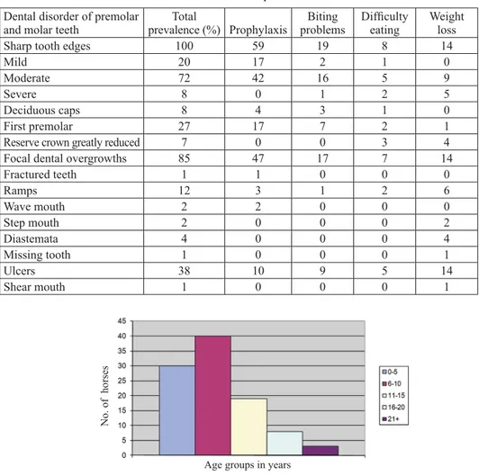
Table 2. Prevalence of disorders of premolar and molar teeth Dental disorder of premolar
and molar teeth prevalence (%) ProphylaxisTotal problemsBiting Difeatingfi culty Weight loss
Sharp tooth edges 100 59 19 8 14
Mild 20 17 2 1 0
Moderate 72 42 16 5 9
Severe 8 0 1 2 5
Deciduous caps 8 4 3 1 0
First premolar 27 17 7 2 1
Reserve crown greatly reduced 7 0 0 3 4
Focal dental overgrowths 85 47 17 7 14
Fractured teeth 1 1 0 0 0 Ramps 12 3 1 2 6 Wave mouth 2 2 0 0 0 Step mouth 2 0 0 0 2 Diastemata 4 0 0 0 4 Missing tooth 1 0 0 0 1 Ulcers 38 10 9 5 14 Shear mouth 1 0 0 0 1
Fig. 1. Number of horses in each age group
Sharp tooth edges were found in all horses and their severity and connection with previous occlusal adjustment (fl oating) are presented in Fig. 3. Among horses with mild sharp tooth edges, 35% had had regular dental examinations and occlusal adjustments, among those with moderate sharp tooth edges, 18% had had regular dental examinations and occlusal adjustments, but none of the horses with severe sharp tooth edges had had a regular dental examination.
No. of horses
Deciduous caps were found in 8% of the horses. In all, 14 deciduous teeth were found. Two horses had 3 deciduous caps, 2 had 2 caps and 4 had 1 deciduous cap each. All the deciduous teeth found were third or fourth premolar teeth; 4 belonged to the upper and 10 to the lower quadrants.
Teeth with greatly reduced reserve crown (“worn teeth”) were recorded in 7 horses aged between 15 and 25. Of these, 2 horses had 6, 3 had 3, 1 had 4 and 1 had 1 tooth with a greatly reduced reserve crown. The most commonly affected teeth were the fi rst maxillary molar teeth (14), followed by the second maxillary molar teeth (6). In addition, greatly reduced reserve crowns were recorded twice on the third maxillary molar teeth, as well as on the second mandibular premolar teeth and fi rst mandibular molar teeth.
Diastemata were found in 4 horses (4%) aged between 16 and 27, most commonly affecting the caudal premolar and molar teeth, especially the interproximal space between the second and third molar teeth. All cases presented clinical signs of weight loss.
Discussion
Most of the horses in our study were presented for a routine examination. Although they did not show any clinical signs of dental disease, we found almost half of them had focal dental overgrowths and moderate sharp tooth edges, while 10% also had ulcerations on the mucosa adjacent to the maxillary premolar and molar teeth. This shows that signs of dental disease were often not apparent to the owner. EASLEY (1996) also reported a high incidence and comparatively low clinical diagnosis of dental disease. Clinical signs of dental disease are often not specifi c and may be refl ected in other body systems. We found that 14% of horses were presented because of weight loss. All of them had buccal
Fig. 3. Association of the severity of sharp tooth edges and the frequency of dental examinations Fig. 3. Association of the severity of sharp tooth edges and the frequency of dental examinations
and fl oating and fl oating
No. of horses
Sharp tooth edges
In our study, we noted 14 persistent deciduous cheek teeth (“caps”) in 8 horses aged between 3 and 5. These deciduous teeth normally exfoliate at 2.5, 3 and 4 years of age, respectively, for the second, third and fourth premolar teeth (SISSON and GROSSMAN, 1962). Occasionally, compression between adjacent teeth results in the retention of these deciduous teeth. We found that the majority (10 out of 14) of the persistent deciduous cheek teeth were the mandibular third and the fourth premolar teeth.
Most of the old horses in this study were thin or moderately thin, and this may have been caused by their advanced dental disorders. The youngest horse with a tooth with the reserve crown greatly reduced was 19 years old, while the same problem was not found in a horse 26 years of age. Teeth with signifi cantly reduced reserve crowns due to attrition may be seen in horses as young as 16 or as old as 25 (LOWDER and MUELLER, 1998; BAYARD, 2006). The most common teeth were the maxillary fi rst molar teeth, the same teeth BAYARD (2006) found exfoliated fi rst.
In our study, diastemata were diagnosed in 4% of the horses, the same frequency found by DIXON et al. (1999). In contrast to these low levels, SIMHOFER et al. (2008) recorded 24.3% of horses with diastemata. In the latter study, endoscopic visualisation was used, which may be superior to conventional examination methods. As in SIMHOFERS (2008)
study, we also detected most diastemata affecting the molar teeth, especially the maxillary second to third molar interproximal spaces.
The results of the current study showed that dental abnormalities are common in horses, but may go unnoticed by horse owners. However, the fact that many horses had dental disorders without showing obvious eating diffi culties, biting problems, or weight loss does not mean that the lesions did not cause discomfort. Most of the horses in our study were young or young adults, contradicting previous recommendations that only older horses need dental care.
References
BAKER, E. (1945): Das Ergebnis von 30000 Zahnuntersuchungen bei Truppenpferden. Zentralblatt Veterinärkunde 1, 32.
BAKER, G. J. (2000): Abnormalities of wear and periodontal disease. In: Equine Dentistry. (Baker, G. J., J. Easley, Eds.). Saunders. London. pp. 70-78.
BAYARD, A. R. (2006): Dental conditions affecting the geriatric horse. Proceedings of the Focus Meeting of AAEP, July 30-August 1. Indianapolis, USA. pp. 1-8.
BENNETT, D. (1992): The evolution of the horse. In: Horse Breeding and Management. (Evans, J.W., Ed.). Elsevier. Amsterdam. pp. 1-37.
BRIGHAM, E. J., G. DUNCANSON (2000): Case study of 100 horses presented to an equine dental technician in the UK. Equine Vet. Educ. 12, 63-67.
SISSON, S., J. D. GROSSMAN (1962): Horse teeth. In: Anatomy of Domestic Animals. Poljoprivredni nakladni zavod. Zagreb. pp. 453-462.
TELL, A., A. EGENVALL, T. LUNDSRŐM, O. WATTLE (2008): The prevalence of oral ulceration in Swedish horses when ridden with bit and bridle and when unridden. Vet. J. 178, 405-410. TRAUB-DARGATZ, J. L., M. D. SALMAN, J. L. VOSS (1991): Medical problems of adult horses,
as ranked by equine practitioners. J. Am. Vet. Med. Assoc. 198, 1745-1747. Received: 3 March 2011 Accepted: 14 July 2011
________________________________________________________________________________________ BRKLJAČA BOTTEGARO, N., J. KOS, O. SMOLEC, D. VNUK, D. MATIČIĆ, B. PIRKIĆ, B. RADIŠIĆ, Z. VRBANAC, J. SELANEC: Rezultati kliničkih pretraga pretkutnjaka i kutnjaka u 100 konja. Vet. arhiv 82, 143-153, 2012.
SAŽETAK
Svrha ovog istraživanja bila je prikupiti rezultate kliničkih pretraga pretkutnjaka i kutnjaka u grupi od 100 kliničkih pacijenata. Pretrage usnih šupljina obavljene su u terenskim uvjetima na sediranim konjima u stojećem stavu. Kliničkim pretragama zubala ustanovljeni su različiti poremećaji pretkutnjaka i kutnjaka. Najučestaliji nalaz, zabilježen u svih konja, bili su oštri rubovi zuba. Visok postotak čireva obrazne sluznice (38%) pripisao se velikoj učestalosti umjerenog (72%) i jakog (8%) stupnja oštrih rubova zuba. Osim toga, zabilježena su žarišna preraštavanja griznih površina (85%), vučji zubi (27%), kosine zuba (12%), zaostali mliječni pretkutnjaci (8%), dijasteme (4%), istrošeni zubi (7%), koso zubalo (1%), valovito zubalo (2%), stepeničasto zubalo (2%), nedostatak zuba (1%) i lom zuba (1%). Ovo je istraživanje pokazalo da veći broj konja nije pokazivao jasne kliničke znakove usprkos prisutnosti patoloških procesa na pretkutnjacima i kutnjacima.
Ključne riječi: konj, pretraga zubala, pretkutnjaci, kutnjaci