Internat. J. Waste Resources, Vol. 3(1)2013:9-12, Bui Thi Thrang at al. ISSN : 2252-5211
9 © IJWR – ISSN: 2252-5211, 15th March 2013, All rights reserved
International Journal of Waste Resources (IJWR
)
Journal homepage: http://www.ijwr.co
Screening
of
Natural Rubber-Degrading Microorganisms
from Rubber Processing Factory Waste in Vietnam
Bui Thi Trang
1, Dao Viet Linh
3, Nguyen Lan Huong
1, To Kim Anh
1,
Phan Trung Nghia
2and Masao Fukuda
31School of Biotechnology and Food Technology, Hanoi University of Science and Technology (HUST),
1 Dai Co Viet, Hanoi, Vietnam
e-mail: [email protected]; [email protected]; [email protected]
2School of Chemical Engineering, HUST, 1 Dai Co Viet, Hanoi, Vietnam
e-mail: [email protected]
3Department of Bioengineering, Nagaoka University of Technology,
Nagaoka, Niigata 940-2188, Japan
e-mail: [email protected]; [email protected]
Paper history:
Submission : January 20, 2013 Correction recieved: February 28, 2013
Accepted : March 5, 2013
doi: 10.12777/ijwr.3.1.2013.9-12
Abstract — Natural rubber-degrading microorganisms were isolated from waste of rubber processing factory in Cam Thuy of Vietnam. Four of them belong to Streptomyces sp. that showed the higher abilities for natural rubber degradation than the others. They are able to use both deproteinised natural rubber (DPNR) and synthetic rubber cis-1,4-polyisoprene (SR) as a sole source of carbon. Gel permeation chromatography (GPC) analysis revealed that these strains degraded DPNR and SR to low-molecular-weight products. The growth of isolates occurs essentially on the latex glove pieces after one month of incubation in mineral salt medium. The total nitrogen contents of glove pieces were determined using Kjeldahl method, which were 10-20 times higher than that in un-inoculated sample. Moreover, the degradation was also confirmed by observing the growth of isolates on glove’s surface using scanning electron microscopy (SEM).
Keywords — degradation, isolation, natural rubber, poly(cis-1,4-isoprene), Streptomyces sp.
I. INTRODUCTION
Natural rubber is a biopolymer containing poly(cis -1,4-isoprene). It is an important material for tires, latex gloves, catheters, and so on. The typical method for isolation degrading- microorganisms is using a clearing zone technique. Two district groups were found. Member of the first group form clearing zones around their colonies on latex overlay agar plates. Most of them belong to the
Actinomycetes [3], [4].Member of the second group do not
produce clearing-zones on latex overlay agar plate like
Gordonia, Mycobaterium,and Nocardia. They are supposed
to grow adhesively on the surface and extensively disintegrate of rubber particles [5], [6]. In this study, we screened and isolated natural rubber-degrading microorganisms from waste of Cam Thuy rubber
processing factory including Streptomyces sp.
Characterization of these strains revealed transformation
of poly(cis-1,4-isoprene), DPNR and also the growth of them on the latex glove pieces.
II. MATERIALSANDMETHODS
2.1 Isolation of Natural Rubber-Degrading Microorganism Waste rubber and waste water were collected from Cam Thuy rubber processing factory in Thanh Hoa province in Viet Nam. Three grams of waste rubber or one milliliter with waste water was suspended in 9ml sterile distilled water, and 100µl of the supernatant after standing still was spread on a latex overlay agar plate, 0,4% (v/v) latex was included in minimal salt medium (MSM) [5] to prepare the overlay layer of each plate. Colonies with clearing-zones formed after incubation at 300C for 3 days
Internat. J. Waste Resources, Vol. 3(1)2013:9-12, Bui Thi Thrang at al. ISSN : 2252-5211
10 © IJWR – ISSN: 2252-5211, 15th March 2013, All rights reserved
2.2 Aldehyde Staining of Colonies on Overlay Agar
Aldehyde groups in latex overlay agar were stained for 30 minutes with Schiff’s reagent. Afterwards, the reagent was removed, and the plates were washed with 10%(w/v) sodium hydrogen sulfite solution, following by distilled water.
2.3 Determination of 16S rRNA Gene Sequences
Toal DNA of these strains were prepared as described by [1] with modifications were describled by [6]. 16S rRNA gene amplication, and nucleotide sequencing analysis were performed as described in previous studies [4], [6]. Sequences were aligned with published sequence in Genbank database using the basic local alignment search tool (BLAST) at the National Center of Biotechnology Information.
2.4 Degradation of Poly(cis-1,4-isoprene) andDPNR in Liquid Culture
The method of this test was decribed by [4].
2.5 Degradation of Poly(cis-1,4-isoprene) of a Latex Glove Piece
The latex glove pieces were cut from latex glove and washed in distilled water after being dried to constant weights. Initial dry weight of each latex glove piece added to each test tube was 37 to 40 mg. Each test tube filled 5ml MSM was inoculated with 500µl of the cell suspension at 30oC. After 4 weeks incubation, latex glove pieces were
taken to scanning electron microscopy (SEM). The rubber weight loss (%) was calculated according to the following equation, 100 x (initial weight of rubber - residual rubber after one month incubation)/initial weight. The cell suspended in the medium were collected by centrifugation and weighed after being dried. The nitrogen content of latex glove pieces and cell suspensions were determined following the Kjeldahl method. scanning electron microscope S-4800 (Hitachi, Japan).
III.RESULTS AND DISCUSSION
3.1 Isolation of Natural Rubber-Degrading Microorganism The bacterial colonies with clearing zone on latex overlay agar plates were picked up and transferred to fresh latex overlay agar plates. The reproduced colonies with clearing zone on latex overlay agar plates were subjected to repeat single-colony isolation on LB (Luria-Bertani Broth) medium three times. Four isolates with
large clearing zone were selected for further
investigations.
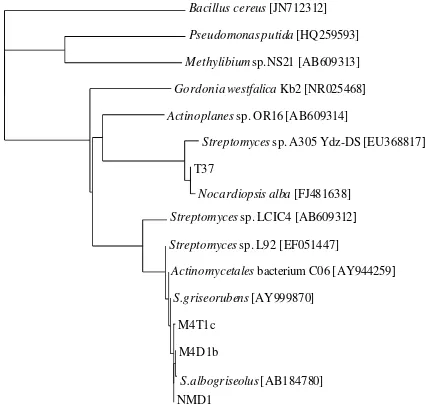
The 16S rRNA gene sequences of four isolates were determined, and showed the highest similarity to those of Streptomyces griseorubens [AY999870] (99% identity), S. albogriselus [AB184780] (99% identity), and Nocardiopsis
alba [FJ4811638] (99% identity) (Fig. 1). However,
Nocardiopsis alba belongs to Streptomyces group, therefore these isolates were designated as Streptomyces sp. strain NMD1, strain M4D1b, strain M4T1c, and strain T37. To analyze the phylogenetic relationship among them and the other known rubber-degrading microorganisms, a phylogenetic tree was constructed by the neighbor-joining method (Fig. 1).
Fig. 1 Phylogenetic tree of 16S rRNA gene sequences of isolates (NMD1, M4D1b, M4T1c, and T37) with the other rubber-degrading microorganisms. The tree was constructed by the neighbor-joining
method, and the scale corresponds to a genetic distance of 0.1 nucleootide substitutions per position (10% difference).
In order to determine whether the rubber-degradation products contain aldehyde group, formation of aldehydes on latex overlay agar plates was examined by staining with Schiff’reagent. When Schiff’s reagent was poured into latex overlay agar plates after inocubation for 3 days at 300C,
purple color developed around the colony of NMD1,
M4D1b, and M4T1c. In contrast, no color development was detected around the colony of T37.
3.2 GPC Analysis of Poly(cis-1,4-isoprene) and DPNR Degradation
To examine the rubber-degradation ability of isolates, these strains were incubated in mineral salt medium (MSM) containing 0.25% (w/v) poly(cis-1,4-isoprene) or 0.5% (v/v) DPNR in flask. The whole culture broth after 30 days incubation was extracted with the equal volume of n-pentane at low temperature. Along with the poly(cis-1,4-isoprene) incubation at 30oC, SR-GPC profiles of isolates
show a large peak area at about 27th minute of retention
time deriving from rubber degradation product about 10kDa (Fig. 2). Interestingly, in the case of strain M4D1b, this peak lightly shifts to the higher retention time demonstrating for the lower degree of polymerization. This data clearly indicated that poly(cis-1,4-isoprene) was degraded by our isolates except T37. Among them, M4D1b is most potential strain for synthetic rubber degradation. In the case of DPNR-GPC profile (Fig. 3), also a significant peak was observed at 26 th minute of retention time by
Internat. J. Waste Resources, Vol. 3(1)2013:9-12, Bui Thi Thrang at al. ISSN : 2252-5211
11 © IJWR – ISSN: 2252-5211, 15th March 2013, All rights reserved
by isolates is confirmed by further GPC analysis considering more longer incubation time.
Fig. 2 GPC elution profiles for the residual polymers after incubation of isolates (NMD1; M4D1b; M4T1c; and T37) for 30 days (solid line) and no
inoculum (dashed line) as a control; SR, Poly(cis-1,4-isoprene).
Fig. 3 GPC elution profiles for the residual polymers after incubation of isolates (NMD1; M4D1b; M4T1c; and T37) for 30 days (solid line) and no inoculum (dashed line) as a control; DPNR, Deproteinised natural rubber.
3.3 Degradation of Latex Glove Piece
The weight loss of latex glove pieces as well as cell mass and nitrogen content during incubation with isolates is shown in Table 1.
After one month incubation, the degradation of latex glove piece seems weakly, it was around 2-4%, however in control sample without inoculation the weight loss of the glove piece was 0.26%. The nitrogen contents of glove pieces were 10-20 times higher than that in control sample. During incubation, the isolates cells were growing on the surface of the latex pieces and in the culture liquid. The nitrogen contents in cell suspensions were higher than that in latex glove pieces, indicating soluble latex degradation products that may serve as carbon sources for the isolates. In case of strain NMD1, its weight loss and nitrogen total were the highest than other strains, suggesting this strain was the strongest degrader than other isolates. Nevertheless, the degradation of latex glove by strain NMD1 was lower than those of Streptomyces sp. strain La7 [2]. After 32 days incubation without shaking at 30oC, the weight loss of latex glove was 13.5%. In another
study Tsuchii et al. 1996 showed the degradation of natural rubber by a strain of Nocardia after 21 days of incubation, 60-70 mg of latex glove strips were almost completely degraded [5].
Table 1. Degrdation of latex glove piece by isolates through determination weight loss, cell mass and nitrogen content after 30 days incubation at
30oC
After one month incubation, the degradation of latex glove piece seems weakly, it was around 2-4%, however in control sample without inoculation the weight loss of the glove piece was 0.26%. The nitrogen contents of glove pieces were 10-20 times higher than that in control sample. During incubation, the isolates cells were growing on the surface of the latex pieces and in the culture liquid. The nitrogen contents in cell suspensions were higher than that in latex glove pieces, indicating soluble latex degradation products that may serve as carbon sources for the isolates. In case of strain NMD1, its weight loss and nitrogen total were the highest than other strains, suggesting this strain was the strongest degrader than other isolates. Nevertheless, the degradation of latex glove by strain NMD1 was lower than those of Streptomyces sp. strain La7 [2]. After 32 days incubation without shaking at 30oC, the weight loss of latex glove was 13.5%. In another
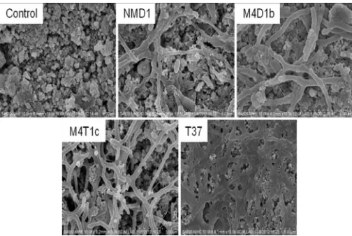
study Tsuchii et al. 1996 showed the degradation of natural rubber by a strain of Nocardia after 21 days of incubation, 60-70 mg of latex glove strips were almost completely degraded [5]. Colonization of the surface of latex glove pieces by isolates was visualized by scanning electron microscopy (Fig. 4).
Fig. 4 SEM of isolates (NMD1; M4D1b; M4T1c; and T37) growing on latex gloves and surface of latex glove as a control sample after one month
incubation in MSM medium
Obtained SEM clearly indicated the growth as well as the colonization of each isolate on the glove piece. In case of strain NMD1 and M4D1b, it seems these strains colonized not only on the surface but also deeply inside. The colonization and/or growth on the rubber surface shown by SEM micrographs have been observed for
Streptomyces sp. [2].
Sample
Latex glove piece Suspended cells
Weight loss (%)
Nitrogen content (%)
Cell mass (mg)
Nitrogen content
(%)
Control 0.26 ±0.02 0.01 ±0.01 0 0
Internat. J. Waste Resources, Vol. 3(1)2013:9-12, Bui Thi Thrang at al. ISSN : 2252-5211
12 © IJWR – ISSN: 2252-5211, 15th March 2013, All rights reserved
IV.CONCLUSIONS
In the present study, four rubber-degrading bacteria, Streptomyces sp. strain NMD1, strain M4D1b, strain M4T1c, and strain T37, which produced clearing-zones around their colonies on latex overlay agar plates, were isolated.
These strains degraded poly(cis-1,4-isoprene) to
low-molecular-weight products. The results of the weight loss, the increase in nitrogen contents, the detection of growth by SEM confirmed the degradation of natural rubber by these strains.
ACKNOWLEDGMENT
This research is supported by grants of ESCANBER project from JICA and JST, Ministry of Science and
Technology of Vietnam (10/2012-HĐ-NĐT), and Ministry
of Education and Training of Vietnam (06/2012/HTQTSP).
REFERENCES
[1] F.M. Ausubel, R. Brent, R. E. Kingston, D. D. Moore, Current rotocols in molecular biology, John Wiley & Sons, New York, N.Y.1, 1987.
[2] C. Gallert, "Degradation of latex and of natural rubber by Streptomyces strain La7", System. Appl. Microbiol, vol. 23, pp. 433-441, 2000. Doi:
http://dx.doi.org/10.1016/S0723-2020(00)80075-2
[3] K. Rose, and A. Steinbuchel. 2005. "Biodegradation of natural rubber and related compounds: recent insights into a hardly understood catabolic capability of microorganisms", Appl. Environ. Microbiol, vol. 71, pp. 2803-2812, 2005. Doi:
http://dx.doi.org/10.1128/AEM.71.6.2803-2812.2005
[4] S. Imai, K. Ichikawa, Y. Muramatsu, D. Kasai, E. Masai, and M. Masao Fukuda. 2011. "Isolation and characterization of Streptomyces, Actiniplanes, and Methylibium strains that are involved in degradation of natural rubber and synthetic poly(cis-1,4-isoprene)", Enzyme and Microbial Technology, vol. 49, pp. 526-531, 2011. Doi:
http://dx.doi.org/10.1016/j.enzmictec.2011.05.014
[5] A. Tsuchii, K. Takeda, T. Suzuki, and Y., Tokiwa, "Colonization and degradation of rubber pieces by Nocardia sp.", Biodegradation, vol. 7, pp. 41-48, 1996. http://dx.doi.org/10.1007/BF00056557