March 2009
Protein expression on Cr resistant microorganism using electrophoresis
method
UMI FATMAWATI1,♥, SURANTO², SAJIDAN1,²
¹Biology Education Program, Department of Mathematics and Natural Sciences Education, Faculty of Teacher Training and Education Science, Sebelas Maret University Surakarta 57 126, Central Java, Indonesia
²Bioscience Program, School of Graduates, Sebelas Maret University, Surakarta 57126, Central Java, Indonesia
Manuscript received: 19 Augustus 2008. Revision accepted: 17 November 2008.
Abstract. Fatmawati U, Suranto, Sajidan. 2009. Protein expression on Cr resistant microorganism using electrophoresis method. Nusantara Bioscience 1: 31-37. Hexavalent chromium (Cr(VI)) is known as toxic heavy metals, so the need is reduced to Cr(III) is much less toxicity. Pseudomonas aeruginosa, Pseudomonas putida, Klebsiella pneumoniae, Pantoea sp. and Saccharomyces cerevisiae
are resistant Cr(VI) microorganism and have ability to reduce Cr(VI). The aim of this research is to know ability of microorganism to reduce Cr(VI) and to know protein band pattern between Cr(VI) resistant microorganism and non resistant microorganism which inoculated on LB broth. SDS-PAGE was used to indentify protein expression. While, Cr(VI) concentration was identified by 1.5 diphenylcarbazide method. The quantitative data was analyzed by two factorial ANOVA that continued with DMRT at 1% level test. The qualitative data i.e. protein expression analyzed by relative mobility (Rf). The results showed that the ability of microorganisms to reduce Cr(VI) at initial concentration of 0.5 ppm, 1 ppm, 5 ppm and 10 ppm may vary, the average percentage of the ability of each microorganism in reducing Cr(VI) is P. putida (65%) > S. cerevisiae (64.45%) >. P. aeruginosa (60.73%) > Pantoea sp. (50.22%) > K. pneumoniae (47.82%) > without microorganisms (34.25%). The adding microorganisms have significantly influenced toward reduction of Cr(VI). The SDS-PAGE shows that protein expression between resistant and not resistant microorganisms are no different, but resistant microorganisms have more protein (protein band is thicker).
Key words: Cr heavy metal, microorganism, protein, electrophoresis.
Abstrak. Fatmawati U, Suranto, Sajidan. 2009. Ekspresi protein pada mikroorganisme resisten Cr dengan metode elektroforesis. Nusantara Bioscience 1: 31-37. Krom heksavalen (Cr(VI)) dikenal sebagai logam berat beracun, sehingga perlu direduksi menjadi Cr(III) yang lebih rendah toksisitasnya. Pseudomonas aeruginosa, Pseudomonas putida, Klebsiella pneumoniae, Pantoea sp. dan
Saccharomyces cerevisiae adalah mikroorganisme resisten dan mampu mereduksi Cr(VI). Tujuan penelitian ini adalah mengetahui kemampuan mikroorganisme dalam mengurangi Cr(VI) dan mengetahui pola pita protein antara mikroorganisme resisten Cr(VI) dan mikroorganisme tidak resisten yang diinokulasi pada medium kaldu LB. SDS-PAGE digunakan untuk mengetahui ekspresi protein, sementara konsentrasi Cr(VI) diidentifikasi dengan metode 1,5 difenilkarbazid. Data kuantitatif dianalisis dengan ANAVA dua faktorial dilanjutkan dengan uji jarak berganda Duncan pada taraf 1%. Data kualitatif yaitu ekspresi protein dianalisis dengan mobilitas relatif (Rf). Hasil penelitian menunjukkan bahwa kemampuan mikroorganisme dalam mereduksi Cr(VI) pada konsentrasi awal 0.5 ppm, 1 ppm, 5 ppm dan 10 ppm berbeda-beda, persentase rata-rata kemampuan masing-masing mikroorganisme dalam mereduksi Cr(VI) adalah: P. putida (65%) > S. cerevisiae (64,45%) > P. aeruginosa (60,73%) > Pantoea sp. (50,22%) > K. pneumoniae (47,82%) > tanpa mikroorganisme (34,25%). Penambahan mikroorganisme secara nyata mempengaruhi reduksi Cr(VI). SDS-PAGE menunjukkan bahwa ekspresi protein antara mikroorganisme resisten dan tidak resisten tidak berbeda, tetapi mikroorganisme resisten memiliki lebih banyak protein (pita protein lebih tebal).
Kata kunci: logam berat Cr, mikroorganisme, protein, elektroforesis.
INTRODUCTION
Chromium (Cr) as one of heavy metal contaminants can potentially become a pollutant as a result of coloring fabrics in the textile industry, paints, leather tanning, metal plating, batteries or industrial chromium (Ackerley et al. 2004). Through the food chains chromium can be deposited in a living body part which at a certain size it can cause toxicity (Mulyani 2004). Generally chromium in open nature is in the valence of 3 (Cr3+) and valence 6 (Cr 6 +). Cr6+ is more toxic than Cr3+. The toxicity of Cr6+ is due to its high solubility and mobility in the environment (Palar
1994; Lowe et al. 2002; Uprati et al. 2003; Rahman et al. 2007). If it enters into cells, then it can cause DNA structural damage or further it can generate mutations (Larashati 2004).
Several microorganisms species such as Pseudomonas putida, Pseudomonas aeruginosa, Klebsiella pneumoniae,
Pantoea sp. and Saccharomyces cerevisiae are
of 6.5-7. While in acidic condition (pH = 2.1) the corps of
S. cerevisiae is still able to reduce the level of Cr(VI) by 70%. Jianlong et al. (2003) tested the tolerance level of S. cerevisiae at a concentration of Cr(VI) 5 uM and it was found that it did not affect microbial growth, while at the concentration of Cr(VI) 15 uM it inhibited microbial growth by 30%.
Ganguli and Tripathi (2002) reports that the bacteria P. aruginosa is able to reduce Cr(VI) by 96% of the initial concentration of Cr(VI) at 10 ppm. But the bacteria P aeruginosa also has a limited ability to reduce the concentration of 50 ppm which is only reduced by 16%. P. putida is a bacterium that is resistant to Cr and Cd so that it can be used in reducing Cr in a certain medium (Timothy et al. 1989; Lowe et al. 2002; Ackerley et al. 2004; Rahman et al. 2007). Bacteria P. putida can reduce Cr(VI) with a speed of 6 ppb min-1 which was previously tested on gelatin containing Cr (IV) (Lowe et al. 2002). K.
pneumoniae which was inoculated in BHI medium is
capable of reducing Cr(VI) by 27% (Mardiyono 2005). Obraztsova et al. (2002) suggested that the reduction of Cr(VI) 150 ppm by Pantoea sp was optimum with the addition of sulfate (SO4-2) which took 20 hours.
Knowing the potential for some microorganisms such as P aeruginosa, P. putida, K. pneumoniae, Pantoea sp. and S. cerevisiae in reducing heavy metal Cr, then a research was conducted on the ability of these microorganisms to reduce Cr(VI) in liquid media containing heavy metal Cr, to know the genetic changes in microorganisms through the expression of protein bands by polyacrylamide gel electrophoresis detection in SDS-PAGE. Electrophoresis is the ideal analysis to purify the protein component of the mixture sample by the adding the medium that can bind proteins during electrophoresis process. The best method in the purification of proteins by electrophoresis is by using polyacrylamide gel electrophoresis (PAGE). Polyacrylamide gel is a solution of acrylamide and bisakrilamid (Davis and Heywood 1963; Hames and Rickwood 1990; Matsudaira 1993).
The aim of this research was to know ability of microorganism to reduce Cr(VI) and to know protein band pattern between Cr(VI) resistant microorganism and non resistant microorganism which inoculated on LB broth.
MATERIALS AND METHODS
The microorganism used. Microorganisms such as K. pneumoniae, P. aeruginosa, P. putida, Pantoea sp., and S. cerevisiae are used in testing the reduction of Cr(VI), and the expression pattern of protein bands is obtained from the University of Gadjah Mada University, Yogyakarta. Bacterial isolates are multiplied in LB liquid medium with the composition of each isolate was 100 mL of 1 g Tryptone, 0.5 g yeast extract, and 0.5 g NaCl, and for the proliferation of molds liquid PDA (potato dextrose agar) medium was used.
Preparation of liquid media which contains heavy metal Cr. A total of 0.1414 g K2Cr2O7 was reconstituted
with distilled water in a 1 liter measuring flask and diluted until it reached mark boundaries to obtain solution concentrations of 0.05 mg/mL to prepare supply of standard Cr. Then chromium standard solution was made by diluting 1 mL of Cr stock solution into 100 mL liquid medium, until we obtained liquid media containing concentrations of heavy metals Cr 0.5 ppm. For concentrations of 1 ppm, 5 ppm and 10 ppm Cr preparations was done by diluting standard solution into a liquid medium (LB or PDA) as much as 100 mL.
Counting the number of microorganism cells. To determine the ability of living microorganisms to survive in media containing heavy metals, then the number of cells inoculated in microorganisms was calculated in liquid media with Cr(VI) 0 ppm and 10 ppm for 16 hours. The growing culture was diluted several times by 10-5 and 10-6. The result of dilution was grown on 100 µL of solid LB media, and then it was incubated again for 16 hours at 37ºC. Colonies that were formed then were calculated by
colony counter and the number of microorganisms cells was also counted in units of cells/mL (Hadioetomo 1993). Cultures which grew on both Cr(VI) were resistant microorganisms, while the cultures which only grew on the control were not resistant microorganisms.
Inoculation of microorganisms in liquid LB media. To determine the ability to reduce Cr(VI), each microorganism was taken with an ose needle and was grown in erlenmeyer containing liquid LB media (Luria-Bertani media) with Cr(VI) 0 ppm, 0.5 ppm, 1 ppm, 5 ppm and 10 ppm concentration. Each species of microorganism was grown in 100 mL of LB liquid medium on five different initial concentrations of Cr(VI) above and then they were incubated in an incubator with a temperature of 30-36ºC for 16 hours.
Hexavalent chromium test. A total of 50 mL liquid bacterial culture medium containing chromium was put into Eppendorf tubes and centrifuged at 3000 rpm for 30 minutes, the supernatant was collected and filtered with Whatman filter paper of 0.2 μm and then the heavy metal content was analyzed (Lowe et al. 2002). The solution was then neutralized by adding H2SO4 (1+1) or NH4OH, and
then it was added with 1 mL of H2SO4 (1+1) and 0.3 mL of
85% H3PO4. The solution was then quantitatively
transferred into 100 mL measuring flask and 2 mL diphenylcarbazide solution was added, diluted to mark boundaries and whipped until it was well mixed. After 5-10 minutes, it was measured with UV-VIS spectrophotometer with a wavelength of 540 nm.
Preparation of standard solution of chromium. Standard supply chromium solvent as much as 20-20 mL was taken with pipette (2, 4, 6, 8, and 10, and so on in stages) into several pieces of measuring 100 mL flask. Put 25 mL of distilled water into the other measuring flask a blank. Into each measuring flask, add 1 mL of H2SO4 (1
+1), 0.3 mL 85% H3PO4 and 2 mL of solution
SDS-PAGE electrophoresis. A total of 5 mL bacterial culture was poured into 1.5 mL an eppendorf tube and the cells was sedimented by centrifuging in for 5 minutes at speed of 13,000 rpm. The cell sediment then was cleaned from liquid LB medium by removing its supernatant. The pellet cell which settled then was suspended with Phosphate Buffered Saline Solution (PBS) twice, then it was again disentrifused back, and the PBS supernatant was discarded. One mL of PBS was added into the pellets. To break the cells sonication was used for 30 seconds for 4 times. Redisentrifuse it and take the supernatan for running by adding sample buffer (4:1/v: v). Before being placed in wells, the mixture of sample and sample buffer was boiled in boiling water for 2 minutes, then put it in ice for + 5 minutes, after which the samples are ready for running. The gel that has been formed (discontinuous gel 10% and 3%
stacking gel), was transferred into the electrophoresis tank (Hames and Rickwood 1990).
Furthermore, electrophoresis tank was filled with
running buffer (0.19 M Glycine, 10 mL SDS 10%, and 0.0248 M Tris in 1 L) until it is full. As much as 10 µL of mikropippete of mixture of the sample and the sample buffer was poured into the electrophoresis wells carefully Then close the tank lid and set the voltage (100V, 90 minutes). To identify the molecular weight of proteins, use protein markers that have a molecular weight range 212-11.3 kDa.
Comassie Blue staining. Solution was made with the composition of 1 g Comassie Blue coloring that was dissolved in 1 L of destaining solution (100 mL acetic acid, 400 mL of methanol, then diluted with the addition of distilled water until it reaches the volume of 1 L) (Hames and Rickwood 1990). After running, the gel was soaked in dye solution Comassie blue for 12 hours, then it was washed with destaining for 3-4 times for 2 hours until the protein band pattern was formed.
Data analysis. Differences in the ability of each microorganism to reduce the heavy metals Cr(VI) at each concentration were analyzed by analysis of variance (ANOVA) followed by further test of Duncan Multiple Range Test (DMRT). The differences in protein expression between the microorganisms were described descriptively.
RESULTS AND DISCUSSION
The growth of microorganisms in media containing Cr Preliminary test results in the form of counting the number of colonies, or Colony Form Unit (CFU) of five species of microorganisms which were grown in an gel medium containing 10 ppm Cr(VI) indicated that the five species of microorganisms were able to live in a media containing Cr(VI) ( Table 1). The highest number of cells contained in S. cerevisiae was 460x106 cells/mL at a concentration of 0 ppm Cr(VI), while the concentration of 10 ppm Cr(VI) S. cerevisiae cells also produce most of 317x106 cells/mL. The number was obtained from the calculation of the average number of colonies of two types of dilution: 10-5 and 10-6. The gel medium that was used
to grow S. cerevisiaea was Potato Dextrosa Agar (PDA), because S. cerevisiae is a type of yeast that can ferment and it needs a lot of substrate fermentation of glucose that will be converted into ethanol. The ability of S. cerevisiae to survive in an gel medium containing Cr(VI) indicates that this microorganisms are resistant or tolerant to Cr heavy metals (Jianlong et al. 2003; Mulyani 2004; Gao et al. 2006). P. aeruginosa also has the ability to live in a media containing heavy metal Cr(VI) 10 ppm, where it is proven by the growth of colonies on LB agar media containing Cr(VI) 10 ppm with an average cell amount of 287x106 cells/mL. Other microorganisms also have the ability to live in a media containing heavy metal Cr(VI) 10 ppm. The fewest number of cells observed on Pantoea sp. with the number of cells of 177x106 cells/mL.
Table 1. The number of cells inoculated microorganisms on solid LB medium with the addition of Cr(VI) 0 ppm and 10 ppm.
Five species of microorganisms in general have a decrease in the number of cells in media containing heavy metal Cr(VI). The lowest decrease in the number of cells is present in K. pneumoniae by 20.7%, while the highest decrease is in P. putida at 34.5%. The decrease in number of cells of inoculated microorganisms in media containing heavy metal Cr(VI) shows that these microorganisms select the tolerant variant toward the heavy metals. Based on Table 1, the decrease in the number of cells of microorganisms after being inoculated into the gel medium with the addition 10 ppm of Cr(VI), generally as much as 20-30%, a decline that is not too extreme and indicates that the five species of microorganisms are resistant to the environment containing metal weight of Cr(VI).
The microorganisms that are capable of living in media containing Cr(VI) can also serve as a reductor of heavy metal Cr(VI) to Cr(III). Several studies have proven that the presence of Cr(VI) at levels of 0-50 ppm in the microorganisms cells does not interfere cell growth microorganisms (Jianlong et al. 2003; Gao et al. 2006; Rahman et al. 2007) because besides growth, the microorganisms will make side product of H2S. The increase in the number of cells of microorganisms will increase the speed of H2S production that will accelerate the reduction of Cr(VI). H2S which is produced by the
bacteria will react with chromium to form chromium sulfides that are not stable in solution and will more quickly be deposited to form Cr (OH)3 namely Cr with a
Reduction of Cr(VI) in liquid media
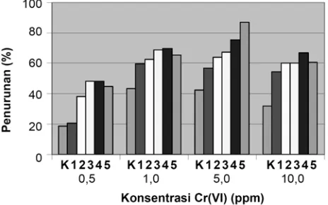
Based on the result of the preliminary research it is proven that the five species of microorganisms are capable of living in an gel medium containing heavy metal Cr(VI) then test was conducted to determine the changes in levels of Cr(VI) before and after treatment. The treatment was done with five species of microorganisms namely: K. pneumoniae, Pantoea sp., P. aeruginosa, P. putida, and S. cerevisiae which were inoculated in LB liquid media containing Cr(VI) 0.5 ppm, 1 ppm, 5 ppm and 10 ppm with the initial inoculum concentration of 1% (Mardiyono 2005) and was incubated for 16 hour at room temperature on a shaker. Decrease percentage in Cr(VI) by five species of microorganisms at different concentrations are shown in Figure 1.
Figure 1. Percentage decrease in Cr(VI) by five species of microorganisms. Note: K = without microorganisms or control, 1 = K. pneumoniae, 2 = Pantoea sp., 3 = P. aeruginosa, 4 = P. putida, 5 = S. cerevisiae.
Reduction ability of Cr(VI) by the five species of microorganisms is tested on LB liquid medium with a solution of 1 ppm Cr(VI). On visual observation there is a difference between liquid LB media that are not inoculated microorganisms and LB liquid medium which are inoculated with five species of microorganisms, and that the liquid media which are inoculated by microorganisms look more cloudy. This indicates that there are growth and proliferation of cells in the media. In addition to the growth activity, some microorganisms also have the ability to reduce Cr(VI) (Suzuki et al. 1992; Ganguli and Tripathi 2002; Krauter and Krauter 2002; Jianlong et al. 2003; Mulyani 2004; Upreti et al. 2004; Mardiyono 2005).
Saccharomyces cerevisiae has the highest ability to reduce among other microorganisms. The decrease percentage in
S. cerevisiae is as much as 87% at the initial concentration of 5 ppm. At the initial concentration of this 5 ppm the lowest percentage decline occurred in K. pneumoniae that is as much as 56.7%. In the treatment without bacteria, decrease also happens by 42.5%. Several other studies state that Saccharomyces is a microorganism that has the highest effectiveness in reducing Cr(VI). Krauter and Krauter (2002) states that S. cerevisiae can reduce Cr(VI) 100% at
pH 6.5-7, while at acidic pH its bioremoval capabilities are less effective.
One of the benefits for Saccharomyces as an agent of biosorbtion of Cr(VI) is that its character is not pathogenic compared to other bacterial pathogens such as P. aeruginosa and K. pneumoniae. S. cerevisiae also has the ability to reduce other types of heavy metals such as Mo, Co, Ca, Zn, Sr, Hg and Cu in water (Krauter and Krauter 2002; Mulyani 2004).
Pseudomonas putida has the second largest percentage in reducing Cr(VI) that is 66.8% in the initial concentration of Cr(VI) initial 5 ppm. The lowest ability is bacteria K. pneumoniae at 20.4% on the initial concentration of 0.5 ppm. In the treatment without addition of microorganisms there was also a decrease as much as 31%. Several other studies also use bacterium P. putida to reduce Cr(VI) into Cr(III); among others are Ackerley et al. (2004), Lowe et al. (2002), Timothy et al. (1989), and Rahman et al. (2007). The results shows that P. putida is able to reduce Cr(VI).
The highest initial concentration of Cr(VI) that was used in this study is 10 ppm. At this concentration the microorganisms still can grow and reproduce, it is seen from the turbidity of liquid LB medium inoculated with five species of microorganisms. Logically, with higher survival ability, certainly there must be more microorganisms that live so as to increase the ability to reduce Cr(VI) into Cr(III) in the environment where Cr(VI) is also greater. The highest percentage in decreasing Cr(VI) occurrs at concentration of 1 ppm and 5 ppm.
Cr(VI) on five species of microorganisms
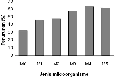
Based on Figure 2 we can see the order of the ability of microorganisms in reducing Cr(VI). The highest ability is in bacteria P. putida with an average percentage of reduction as much as 65%, while the lowest is bacteria K. pneumoniae with an average percentage reduction of 47.8%. The sequence of comparison of the ability to reduce Cr(VI) among the microorganisms are as follows: P. putida
(65%)> S. cerevisiae (64.45%)> P. aeruginosa (60.73%)>
Pantoea sp. (50.22%)> K. pneumoniae (47.82%)> without microorganisms (34.25%).
The analysis results of variance calculations of the influence of initial concentration of heavy metals Cr(VI) and species of microorganisms toward the decrease in Cr(VI) is shown in Table 2. Based on the calculation of two-factorial ANOVA where the main factor is the species of bacteria and the subplot factor is the concentration, it can be concluded that the species of microorganism have real effects on decreasing the concentration of Cr(VI) at level 1%. Initial concentration of Cr(VI) has real effect on decreasing the concentration of Cr(VI) at level 1%, and the interaction of species of microorganisms with initial concentration of Cr(VI) has significant effect on decreasing the concentration of Cr(VI) at level 1%.
The presence of Cr(VI) in the environment can interfere the organism but also can result in selection of resistant bacteria. Compounds of Cr(VI) are really more dangerous than the Cr(III) due to its high solubility in water, rapid permeability and subsequent interaction with intracellular proteins and nucleic acids (Upreti et al. 2004). Microorganisms can develop resistance mechanisms to select the next resistant variants.
70
Figure 2. The average reduction capability. Note: M0 = without microorganisms (control), M1 = K. pneumoniae, M2 = Pantoea
sp., M3 = P. aeruginosa, M4 = P. putida, M5 = S. cerevisiae
As noted previously, according to Rahman et al. (2007) reduction of Cr(VI) happens because not only of growth, but also the fact that microorganism produces byproducts in the form of H2S. The increasing number of cells of microorganisms will increase the speed of H2S production that will accelerate the reduction of Cr(VI). H2S that is produced by the bacteria will react with chromium to form chromium sulfides which are not stable in solution and will more quickly deposited to form Cr (OH)3 which is Cr with
a valence of three who has a lower toxicity of Cr with a valence of six. Meanwhile, according to Suhendrayatna (2001) reduction of Cr(VI) to Cr(III) by microorganisms called bioremoval has two kinds of mechanisms, namely passive and active. Passive absorption is known as biosorbsi. This process occurs when heavy metal ions bind to the cell wall in two different ways, namely (i) ion exchange of monovalent and divalent ions which like Na, Mg, and Ca on the cell wall was replaced by heavy metal ions, and (ii) complex formation between heavy metal ions with functional groups such as carbonyl, amino, thiol, hydroxyl, phosphate, hydroxyl, carboxyl located on the cell wall.
Biosorbsion process can occur back and forth and quickly. The process of alternating bond of heavy metal ions on the surface of these cells can occur in both dead cells and living cells from a biomass. The biosorbtion process can also be more effective at certain pH and the presence of other ions in the medium in which heavy metals can be deposited as salt which is not dissolved. Heavy metal absorption can also occur actively, which occurs in various types of living cells. This mechanism simultaneously occurs in line with the consumption of metal ions for the growth of microorganisms or the accumulation of intracellular heavy metal ions. Heavy metals can also be deposited in the metabolism and excretion process. This process depends on the energy contained in it and the sensitivity of different parameters such as pH, temperature, ionic strength, and light. This process can also be inhibited by low temperatures, lack of energy sources and inhibition of cell metabolism (Suhendrayatna 2001).
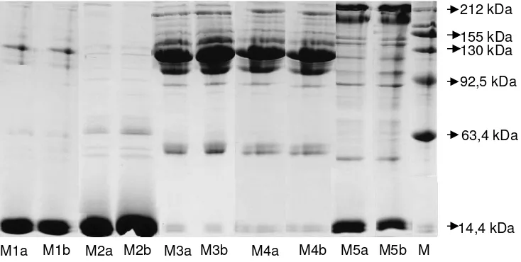
Protein expression in microorganisms resistant Cr in Figure 3 shows the results of running the pattern of protein bands on SDS-PAGE of the five species of microorganisms namely K. pneumoniae, Pantoea sp., P. aeruginosa, P. putida, and S. cerevisiae, each sample of which is extracted from the cells of microorganisms that are grown in LB liquid culture with concentrations of Cr(VI) 0 ppm and 10 ppm. This two kinds of concentration are chosen to distinguish between populations of microorganisms that are resistant and that is not resistant.
Protein markers can be used to identify the molecular weight of a mixture of polypeptides (Hames and Rickwood 1990). In this study, markers of protein that are used have a molecular weight range 212-11.3 kDa. From the results of electrophoresis there is a number of protein bands which have different thicknesses. The protein that has a more thickness and greater color intensity than any other proteins and is always there in every variety is called major protein (Wijaya and Rahman 2005).
Table 2. Results of analysis of variance the variation of initial concentration of Cr(VI) and species of microorganisms to the decrease of Cr(VI).
Source of diversity Degrees of freedom
Microorganisms (A) 5 8626.752 1752.350 50.858 3.330* 5.640
Error (a) 10 339.249 33.925
Initial conc. of Cr (VI) (B) 3 8988.810 2996.270 101.644 2.860* 4.380
AxB 15 1350.517 90.034 3.054 1.960* 2.580
Error (b) 36 1061.207 29.478
General 71 20474.792
Note: kk (a) = 10.631%; kk (b) = 9.910%; * = significant difference at level 1%
Table 3. Further test results significantly different from Duncan's to the decline of Cr (VI) concentrations.
Species of microorganisms Initial concentrations of Cr (VI)
0.5 ppm 1 ppm 5 ppm 10 ppm
Figure 3. Protein expression in five species of microorganisms are grown on LB liquid medium with Cr(VI) 0 ppm and 10 ppm. Note: M = marker proteins, M1a, M1b = K. pneumoniae, M2A, M2b = Pantoea sp., M3a, M3b = P. aeruginosa, M4A, M4b = P. putida, M5a, M5b = S. cerevisiae. a = 0 b = 10 ppm ppm
In the electrophoresis results, there are several major proteins on the species of P. aeruginosa (M3) and P. putida
(M4). This major protein molecular weight ranges from 148.7 kDa, 121.6 kDa, and 105 kDa. The two major proteins of these type have same color intensity and thickness because this two microorganisms belong to the same genus namely Pseudomonas. Bacteria K. pneumoniae
(M1) has several protein bands, but because proteins from the sample that is running is only a few, then the formation of bands on polyacrylamide gel is still less than optimal. From Figure 3 we can note that there are three major protein bands of resistant microorganisms and not resistant microorganisms with the molecular weight of 121.6 kDa, 14.4 and 14 kDa, 3 kDa. Generally, the pattern of protein bands from two samples that running do not have a dramatic difference, because they are actually of one type.
Protein running of S. cerevisiae (M5) shows the same banding pattern among the microorganisms that are resistant and which are not resistant. These microorganisms have three pairs of major proteins with molecular weight of 212 kDa, 188.5 kDa, 14.5 kDa, and 14.4 kDa. In addition, there are some bands with less color intensity, due to less protein concentration. The lower the location of protein bands are, the smaller the molecular weight. This happens because the low molecular weight have a greater speed to migrate in the matrix medium polyakrilamid.
The reason of why electrophoresis was used in this study is because it has a very important role in the process of separating biological molecules, especially proteins. This method does not affect the structure of biopolymers, as well as very sensitive to the difference in charge and in small molecular weight (Bachrudin 1999). Proteins that run in a medium that contain electric field causes the charged compounds will move in the solution as a result of the nature of the opposite polarity, so that the mobility of a
molecule is a function of shape, molecule size, and large content type.
The use of SDS and merkaptoetanol accompanied by heating will break three-dimensional structure of proteins, particularly the disulfide bonds into polypeptides sub units individually. SDS also wraps the chain of proteins that are not bound by the same negative charge to form SDS-protein complex. SDS-SDS-protein complexes have an identical charge density and move on the gel based on the size of the protein (Wijaya and Rohman 2005). Therefore, the greater SDS-protein complexes have slower mobility than the smaller SDS-protein complexes.
Methods for extracting proteins in microorganisms are done with Phosphate Buffer Saline Solution (PBS) solution followed by breaking the cell by using sonification (cell-breaking equipment using sound waves that produce high frequency). The purpose of the cell breaking is to provide opportunities to extract the protein that will be purified. The cells that have been broken must be preserved from the influence of oxygen, because oxygen can cause the protein to be inactive, denatured, and compact (Bachrudin 1999).
CONCLUSION
The species of microorganisms Klebsiella pneumoniae, Pseudomonas aeruginosa, Pseudomonas putida, Pantoea
sp., and Saccharomyces cerevisiae have a very significant effect on the percentage reduction of Cr(VI). The variation of initial concentration of Cr(VI) also has a very significant effect on the percentage reduction of Cr(VI) and interaction species of microorganism with initial concentration of Cr(VI) which have significant effect on decreasing the concentration of Cr(VI). Microoganisms ability in reducing Cr(VI) to Cr(III) can be sorted as follows: P. putida
(65%)> S. cerevisiae (64.45%)> P. aeruginosa (60.73%)>
M1a M1b M2a M2b M3a M3b M4a M4b M5a M5b
212 kDa
155 kDa
14,4 kDa 92,5 kDa
63,4 kDa 130 kDa
Pantoea sp. (50.22%)> K. pneumoniae (47.82%)> without microorganisms (34.25%). Expression of proteins that are formed on each of the microorganisms that are resistant and are not resistant have almost the same pattern of protein bands.
REFERENCES
Ackerley DF, Gonzales CF, Park CH, Blake R Keyhan M, Martin A. 2004. Chromat reducing properties of soluble flavoprotein from
Pseudomonas putida and Escherichia coli. Appl Environ Biol 70 (2): 873-882.
Bachrudin Z. 1999. Laboratory manual: isolation, identification, and coloring protein. Inter University Center of Bioteknologi, Gadjah mada University. Yogyakarta. [Indonesia]
Davis PH, VH Heywood. 1963. Basic methods in molecular biology. 2nd ed. Appleton & Lange. Conecticut.
Ganguli A, Tripathi A. 2002. Bioremediation of toxic chromium from electroplating effluent by chromate-reducing Pseudomonas aeruginosa A2Chr in two bioreactors. Appl Microbiol Biotechnol 58: 416-420.
Gao J, Zhang Y, Ntoni J, Begonia MFT, Lee KS, Hicks L, Hwang WW, Hwang H-M. 2006. Effects of selected by-products of an acid hydrolyzate on cell growth and ethanol fermentation by
Saccharomyces cerevisiae. J Mississippi Acad Sci. October 01, 2006 www.accessmylibrary.com/article-1G1-156274382/effects-selected-products-acid.html
Hadioetomo RS. 1993. Microbiology basis in practice. UI Press. Jakarta. [Indonesia]
Hames BD, Rickwood D. 1990. Gel electrophoresis of proteins: a practical approach. Oxford University Press. London.
Jianlong W, Zeyu M, Xuan Z. 2003. Response of Saccharomyces cerevisiae to chromium stess. Process Biochem 39 (10): 1231-1235. Krauter PAW, Krauter GW. 2002. Water treatment process and system for
metals removal using Saccharomyces cerevisiae. The Regents of the
University of California, United States Patent and Trademark Office Granted Patent. www.uspto.gov/patft/index.html
Larashati S. 2004. Reduction of chromium (Cr) in vitro by mixed culture of bacteria isolated from landfill leachate (landfill). [Thesis]. Bandung Institute of Technology. Bandung. [Indonesia]
Lowe KL, Fliflet RE, Ly T, Little BJ, Jones-Meehan J. 2002. Chromium tolerant microbial communities from the Chesapeake Bay watershed. Virginia J Sci 53(3): 142-155.
Mardiyono. 2005. Reduction of Cr(VI) textile industrial waste by a bacterium Pseudomonas aeruginosa, Ecsherichia coli, and Klebsiella pneumoniae. Thesis. Environmental Science, School of Graduates, Sebelas Maret University. Surakarta. [Indonesia]
Matsudaira P. 1993. A practical guide to protein and peptide purification for microsequencing. 2nd ed. Academic Press. San Fransisco, CA. Mulyani B. 2004. Variation analysis of biomass of Saccharomyces
cerevisiae to chromium metal uptake. Sains dan Matematika 2 (4): 1-9. [Indonesia]
Obraztsova AY, Francis CA, Tebo BM. 2002. Sulfur disproportionation by the facultative anaerob Pantoea aglomerans sp-1 as a mechanisme for chromium (VI) reduction. Geomicrobil J 19: 121-132
Palar H. 1994. Analisis variasi biomassa Saccharomyces cerevisiae
terhadap serapan logam krom. Rineka Cipta. Jakarta. [Indonesia] Rahman MU, Gul S, UlHaq MZ. 2007. Reduction of chromium (VI) by
locally isolated Pseudomonas sp. C171. Turkey J Biol 31: 161-166 Suhendrayatna. 2001. Bioremoval of heavy metals using microorganisms:
a literature review. Biotechnology Seminar,Sinergy Forum-Institute of Technology. Tokyo
Suzuki T, Miyata N, Horitsu H, Kawai K, Takamizawa K, Tai Y, Okazaki M. 1992. NAD(P)H dependent chromium (VI) reductase of
Pseudomonas ambigua G-1:a Cr(V) intermediated is formed during the reductin of Cr(VI) to Cr(III). J Bacteriol 174 (16): 5340-5345 Timotius KH, Widianarko B, Laksmani S. 1989. Interaction between
bacteria and heavy metals. Scientific Seminar on the Soil Ecology and Ecotoxicology. Faculty of Biology, Satya Wacana Christian University. Salatiga. [Indonesia]
Upreti RK, Srivastha R, Chaturvedi UC. 2004. Gut microflora, toxic metal: chromium as a model. Indian J Med Res 119: 49-59.