THE EVALUATION OF CURRENTLY USED HOME-MADE
ANTIMICROBIAL LINKED TO POLYMETHYLMETHACRYLATE
(PMMA) BEADS FOR THE PREVENTION AND TREATMENT OF
CHRONIC BONE INFECTION
Rasyid N.H*., Kuspriyanto*., Mengko L.T*., Soegijoko S*.,
Neut D**., Van Horn J.R**. and Busscher H.J**
*Biomedical Engineering Program, Department of Electrical Engineering, Institut Teknologi Bandung
** Dept. of Biomedical Engineering, University of Groningen, The Netherlands
Abstract: Infection of bone tissue, or osteomyelitis, is very difficult to cure a reason why stringent prevention is indicated. Currently antibiotic-impregnated polymethylmethacrylate (PMMA) beads are widely used. Placed in the surgical wound, they function as a slow release system to obtain a high local concentration of antibiotic while systemic concentrations remain low. This delivery system is clinically accepted as the method of choice for the treatment of chronic osteomyelitis as well as ITB beads by definition is an antibiotic loaded beads in home-made, mixtures antibiotic (fosfomycin sodium) with the artificial resin PMMA. The purpose of the study is to evaluate currently used home-made antibiotic-loaded beads. First, under sterile conditions, eight different types of hand-made fosfomycin sodium-loaded bone cement bead sets are built in spherical shape. The sizes of beads are varies between 10 and 20 mm. A first evaluation will include a laboratory study on the antibiotic release kinetics, as well as a scanning electron miscroscopic (SEM) evaluation of the porosity and a quantification of the porosity using mercury-intrusion porosimetry. Personal Computer (PC) is used to simulate the porosity of the beads. We may conclude that beads having 10 mm in diameter revealed to more homogeneous pores. The concept of BME-ITB beads by using bone cement as a depot for antibiotic make sense, as it allows delivery of antibiotics directly to the site of infection.
Introduction
Buccholz and Engelbrecht 1 initiated the addition of
antibiotics to polymethylmethacrylate bone cement in the beginning of the 1970s. By mixing gentamycin in acrylic bone cement, they succeeded in reducing the infection rate. The assumption underlying the incorporation of antibiotics in bone cements was that the
antibiotic would gradually be released to yield higher local concentrations than can be achieved by systemic therapy. Since the introduction, a number of clinical studies on the use of antibiotic-loaded bone cements have been conducted. The results of these studies have shown that local administration of antibiotics through antibiotic-loaded bone cement yields good results and cost effective prevention of infection in total joint athroplasty 2,3,4.
In Indonesia, orthopaedic surgeons use bone cement, mixed it themselves with antibiotic in home-made molds and apply them in their patients. This is despite the fact that the release mechanisms of antibiotic from bone cements is poorly understood and controlled, with some reports claiming the release of antibiotic from bone cements for up to five years after implantation 5.
The concept of using the bone cement as a depot for antibiotics is reliable, as it allows delivery of antibiotics directly to the site of (imminent) infection. The implantation of non-compatible materials in the human body is limited by a so-called foreign body reaction. This is an inflammatory response due to the foreign body that may lead to migration and rejection of the implanted material. Therefore, any material applied in the human body should be able to perform with an appropriate host response in a specific application. In other words, it should show a high degree of biocompatibility. Bone cement mainly consists of polymethylmethacrylate (PMMA). Pieces of the PMMA, due to its excellent biocompatibility and ease of manipulation.
All commercially available bone cements have two components. The first is a powder that consists mainly of a polymer based on methylmethacrylate (MMA). The second component is mainly liquid MMA. The dough phase resulting from mixing these components is important for bone cements for a variety of reasons. To start, it offers the possibility of molding and being used to support a prosthesis, while allowing its insertion. Furthermore, the initial presence of PMMA enables the use of less monomer for obtaining the same amount of end product. Finally, this mix of PMMA and MMA means that undesirable effects of polymerization are reduced. Firstly, use of less monomer leads to less heat
production. Secondly, polymerization from a monomer necessarily leads to a decreased volume and increased density due to the fact that the molecules take up less space in a polymer than in their liquid monomer. If less polymerization occurs, the volumetric shrinkage is proportionally lower. Apart from these main components, other substances are required to achieve a controlled polymerization at body temperature. N,N dimethyl-p-toluidine dissolved in the monomer is a tertiary amine, enabling cold curing of the polymer, instead of the required preheating to 100°C. It reacts with the benzoyl peroxide in the powder to create free radicals that can break part of the C=C bond and start the addition polymerization. Furthermore hydroquinone and chlorophyll are additives that prevent premature polymerization under exposure to light or elevated temperatures. The presence of barium sulphate and zirconium (IV) oxide in the powder is necessary for a clinical reason. The first bone cements not containing these could not be visualized on radiographs. These agents are so-called radio pacifiers and make the bone cement visible on radiographs.
BME-ITB beads
The use of polymethylmethacrylate (PMMA) bone cement loaded with antibiotics has become increasingly common in the treatment of infected bone (chronic bone infection), infected knee and hip arthroplasties and also as prophylaxis in primary joint replacement. We proposed Fosfomycin Sodium is used antibiotic in bone cement. Fosfomycin sodium is an antibiotic with extremely low molecular weight of 138, produced by strains of Streptomyces 6, and is characterized by
structural features of an epoxide ring and a carbon-phosphorus bond. It is also characterized by its action, which inhibits the first step of peptidoglycan biosynthesis, and is synergistic in combination with many other anti-microbial agents 7. Moreover,
fosfomycin sodium is antibiotic that remains stable when exposed to the high temperatures generated during the curing of the bone cement. Antibiotic is released from bone cement in a biphasic manner, meaning high elution in the first hours to days post surgery and after a few days it slows down considerably.
In BME-ITB beads, mixture of fosfomycin sodium antibiotic and bone cement were used. These beads constitute an effective drug delivery systems for local antibiotic therapy in bone and soft-tissue infections and at the site of the infection can exceed the minimal inhibitory concentration of the infecting organism .
Research objective
1. Evaluate currently used home-made loaded beads and compare their antimicrobial efficiency with the efficiency of an existing (patented) product.
2. Develop an improved concept for the local production by orthopaedic surgeons of antibiotic-loaded beads for the treatment of orthopaedic application.
The new concept is expected to be applicable worldwide and will allow surgeons to locally develop their own effective beads.
Materials and Methods
A number of orthopaedic surgeons are requested to make an overview of different local concepts developed to prepare antibiotic-loaded beads. Orthopaedic bone cements are being used, amongst others in combination with a variety of different antibiotics. Based on this overview, five concepts are selected for further research and the participating surgeons will be asked to submit an extensive protocol of their concept. To evaluate the porosities of the beads scanning electron microscopy (SEM) type 6301F was used. Personal Computer (PC) is used to simulate the porosity of the beads.
Commercially available Surgical Simplex® P
Radiopaque Bone Cement (a product of Stryker Howmedica OSTEONICS, Howmedica International S, de R.L. Raheen Business Park, Limerick, Ireland) was used. Bone cement was prepared by mixing the powdered polymethylmethacrylate with the liquid monomer in a bowl with a spatula. Manual mixing was done according to the manufacturer’s instructions and resulted in doughy cement, then, the antibiotic stainless steel wire string. All procedures were carried out under sterile conditions.
Results
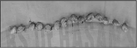
Under sterile conditions, eight different types of hand-made fosfomycin sodium-loaded bone cement bead sets are built in spherical shape. It consists of three hand made spherical shape beads. The sizes of beads are varies between 10 and 20 mm and the beads are which can usually be easily seen on scanning electron micrographs of bone cements (see Fig. 1).
Figure 1: A string of hand-made BME-ITB beads.
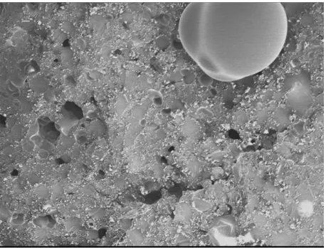
To evaluate the amount of porosities scanning electron microscopy (SEM) was used. In the same 80 times magnification at 2.0 KV demonstrated that size of 20 mm beads showed little porous comparing small diameter (see Fig 2). Some “white dots” of fosfomycin sodium can be seen closed to the pores. Figure 3 revealed at 120 times magnification the “white dots” become homogen in particular at the small size (10 mm in diameter). In this evaluation, to obtain good porosities the size of beads is very important.
We may conclude that beads having 10 mm in diameter revealed to more homogeneous pores. BME-ITB beads, however, release much more fosfomycin sodium than solid bone cement plugs, mainly due to the greatly increased surface area of the many, relatively small beads
Figure 2: This picture showed little porosities in large diameter beads. It was evaluated in 80 times magnification at 2.0 KV.
Figure 3: In 120 times magnification at 1.0 kV revealed “white dots” of antibiotics around the pores
Discussion
Bone cements are made up of two primary components: a powder containing copolymers based on the substance polymethylmethacrylate, and liquid monomer, methylmethacrylate. The powder contains a starter, di-benzoil peroxide; while the liquid contains the initiator, N,N-dimethyl-p-toluoidin. Both substances together start the polymerisation process and enable a reaction at room temperature. A special radio-opaque agent (barium sulphate) is added to the powder to
provide X-ray contrast. The polymer from which the bone cements are made is capable of taking up very small quantities of dissolution fluid into its outermost layers. This dissolution fluid then slowly transport the antibiotic molecules contained within the polymer out into the surrounding tissue. At the beginning, when the infection is still very active, large amounts of antibiotic are released, because it is particularly easily available at the surface of bone cement. The sustained release of antibiotics from bone cements is largely influenced by the penetration of dissolution fluids into the polymer matrix, which requires a certain porosity of the cement
8. A point on which all studies have agreed is that the
period of maximum antibiotic release is limited to the first few hours or days after implantation. The maximum effectiveness of the released antibiotic might thus be expected to occur during this period of time9.
Most, if not all, of the antibiotic is released from the superficial regions of the cement and fails to be released from the centre10.
The porosity of the polymer matrix depends on air entrapment during the wetting and stirring of the cement powder during transfer to the cement gum ad on effects of monomer boiling11 . Baker and Greenham, concluded
that bone cement with greater porosity would be expected to allow more antibiotic release than one with less porosity. Therefore, the methods of cement preparation that are designed to improved mechanical properties by decreasing the porosity could have deleterious effect on elution characteristics.
Conclusions
The concept of BME-ITB beads by using bone cement as a depot for antibiotic make sense, as it allows delivery of antibiotics directly to the site of infection.
The beads having 10 mm in diameter revealed to more homogeneous pores.
References
[1] BUCCHOLZ, H.W. and ENGELBRECHT, H. (1970): ‘Uber die Depotwirkung einiger Antibiotics bei Vermischung mit dem Kunstharz Palacos’. Chirurg, 40, pp. 511-515
[2] JOSEFFSON, G. and KOLMERT, L. (1993): ‘Prophylaxis with systematic antibiotics versus gentamycin bone cement in total hip arthroplasty’. A ten-year survey of 1,688 hips. Clin Orthop, 292, pp. 210-214
[3] ESPEHAUG, B., ENGESAETER,L.B., VOLLSET, S.E., HAVELIN, L.I., and LANGELAND N. (1987): ‘Antibiotic prophylaxis in total hip arthroplasty. Review of 10,905 primary cemented total hip replacements reported to the Norwegian arthroplasty register, 1987 to 1995’. J Bone Joint Surg Br, 79, pp. 590-595
[4] SCULCO, T.P. (1995): ‘The economic impact of infected joint arthroplasty’. Orthopaedics, 18, pp. 871-873
[5] WAHLIG, H. and DINGELDEIN E. (1980): ‘Antibiotics and bone cements. Experimental and clinical long term observations’. Acta Orthop Scand, 51, p. 495
[6] HENDLIN, D., STAPLEY, E.O., JACKSON, M. and WALLICK, H. (1969): ’Phosphonomycin, a new antibiotic produced by strains of Streptomyces’. Science, 166, pp. 122-123
[7]BERGERON, M., ROBERT, J. and BEAUCHAMP, D.(1993): ’Pharmacodynamics of antibiotics in fibrin clot’. J Antimicrob. Chemother, 31, pp. 113-136
[8] BAKER, A.S. and GREENHAM LW. (1998): ‘Release of gentamicin from acrylic bone cement’. J Bone Joint Surg Am, 70, pp. 1551-1557
[9] TRIPPLE, S.B. (1986): ‘Antibiotic-impragnated cement in total joint arthroplasty’. J Bone Joint Surg Am, 68, pp.1297-1302
[10]SCHURMAN, D.J., TRINDALE, C., HIRSCHMAN, H.P., MOSER, K., KAJIYAMA, G., STEVENS, P. (1978): ‘Antibiotic-loaded acrylic bone cement composites’. J Bone Joint Surg Am, 60, pp.978-8=984
[11]WIXSON, R.L., LAUTENSCHLAGER, E.P. and NOVAK MA. (1987): ’Vacuum mixing of acrylic bone cement’. J Arthroplasty, 2, pp.141-149