Asian Online Journals (www.ajouronline.com) 429
Evaluation of Five Methods Used to Extract Deoxyribonucleic Acid
(DNA) From Human Malaria Parasitized Blood Spotted on the Filter
Paper
Mukh Syaifudin1*, Dyan Hera Wiluyaningtias2, Yuliwati3
1
Center for Technology of Radiation Safety and Metrology, National Nuclear Energy Agency, Jl. Lebakbulus Raya No. 49 Jakarta, Indonesia
2
Biology Study Program, Semarang State University, Gunungpati, Semarang, Central Java, Indonesia Email : [email protected]
3
Clinical Laboratory, Jayapura General Hospital, Jl, Kesehatan No. 1 Dok II, Jayapura, Papua, Indonesia
*Corresponding author’s email: mukh_syaifudin [AT] batan.go.id
_________________________________________________________________________________
ABSTRACT— Efficient deoxyribonucleic acid (DNA) extraction is critical for a good polymerase chain reaction (PCR) performance in molecular diagnostic, genomic and epidemiological studies of malaria. Blood collected on filter paper is a pratical method for this puposes in high parasitemic individuals. In this paper we evaluated five different methods (Commercial kit, Chelex-saponin, microwave, methanol and Tris-EDTA) for DNA extraction. Blood specimens from six asymptomatic Plasmodium sp.-infected patients in Jayapura General Hospital were spotted onto filter paper, dried, and transported for processing. Evaluation of the results of DNA extraction was carried out by measuring yields using spectrophotometry and PCR amplification for mitochondrial gene. Results showed that the best results of PCR obtanied only with commercial kit method, whereas very low quality of PCR by microwave and no amplified DNA for three other methods. The purity of DNA for all methods was low. The sensitivity of detection for kit was approximately 100% for P. falciparum, P. malariae and P. vivax, whereas microwave method, even though resulted in high concentration of DNA, did not or slightly show the effectiveness in DNA amplification that may due to high inhibitor contents. Even though chelex method was widely used by many malaria laboratories, in this experiment we did not found its effectiveness. We concluded that commercial extraction kit method is well-suited for the DNA extraction in PCR-based assays of malaria.
Keywords —malaria, blood spot, extraction, purity, PCR.
_________________________________________________________________________________
1.
INTRODUCTIONMalaria is a mosquito-borne disease responsible for approximately 250 million human infections and about a million deaths annually [1,2]. Therefore, the research and development of additional malaria control methods such as vaccine are crucial [3]. According to the World Health Organization [4], half of the world’s population is at risk of contracting malaria where most of this population resides in endemic countries in Africa and Southeast Asia. In Indonesia, the prevalence rate of malaria remains high in spite of eradication efforts that are kept being done [5].
Malaria is a debilitating disease, compounded by the problems of drug resistance and accessibility to effective new and combination drugs. On the other hand, a malaria vaccine has been difficult to make because of the genetic variation or polymorphisms between parasites. This genetic complexity of the parasite means that each infection presents thousands of antigens to the human immune system [6]. Most vaccine development is based on gamma ray attenuated or inactivated whole pathogens or materialderived directly from the infectious agent. It is well known that immunization with radiation-attenuated P. falciparum sporozoites provides sterile protective immunity in >90% of immunized individualsfor at least 10.5 months against multiple isolates of Pf fromthroughout the world [7,8].
The ability to detect individuals with asymptomatic low density parasitaemia, i.e., below detection limit of
microscopy (~100 parasites/μL blood), in low endemic settings has been increasingly acknowledged as a challenge to
Asian Online Journals (www.ajouronline.com) 430
and spread of tolerance/resistance to artemisinin-based combination therapy before clinical treatment failures are apparent.
Collection of blood samples on filter paper is a suitable alternative for malaria diagnostic screening, drug resistance monitoring, genetic analysis and other molecular studies in patients in distant areas where collection, storage and transport are problematic [10,11]. Thus a comprehensive study is needed to investigate whether wide-scale collection of blood spotted on the filter paper can provide the basis for modern molecular surveillance of malaria.
Since its introduction in the mid-80s, PCR technology has been recognised as a rapid, sensitive and specific molecular diagnostic tool for the analysis of micro-organisms in clinical, environmental and food samples [12-14]. Several techniques based on DNA fingerprinting for microorganisms such as restriction fragment length polymorphism (RFLP), microsatellites and single stranded confirm polymorphism (SSCP) have also been used for genotyping of P. falciparum [15]. In our research that focused on the developing malaria vaccine, the extraction method is important tool to know the effectiveness of gamma irradiation in attenuating the Plasmodium berghei as parasite model such as for molecular detection of the parasites in blood, liver and spleen of mouse after injected with irradiated parasites.
The aim of this study was to evaluate five published DNA extraction methods and to assess the field applicability of DNA extraction from filter paper for PCR amplification in molecular diagnosis or genomic and epidemiological studies of malaria.
2. MATERIALS AND METHODS
2.1 Sample collection.
Whole blood samples of around 100 μL obtained from malaria suspected outpatient in Dok II Municipal Hospital in Jayapura Papua were collected on 3MM Whatman® (Brentford, United Kingdom) filter papers. Each sample was dried at room temperature and stored in separate plastic packets to avoid cross contamination. A drop of blood was also prepared for thick blood smear on the glass slide to microscopy diagnose of Plasmodium species. It was done in the laboratory of Emergency Installation of Hospital in Jayapura by experienced microscopist.
2.2 DNA extraction.
Two punch out of filter paper (3-mm diameter of circle) were transferred to each five sterile 1.5 mL microcentrifuge tube and then was weighted with digital balance to determine the weight of spotted blood. The paper punch was cleaned between samples by rigorous manual wiping with 70% ethanol. It was then used to punch 3-5 holes from a separate clean, unused blood sample collection card, prior to performing a single sample punch. Each sample spots were then processed according to either Commercial kit, Chelex-saponin, microwave, methanol and Tris-EDTA methods.
2.3. Commercial kit.
The procedure wasfollowed the protocol of manufacturer. Briefly, into a tube containing punched out paper 200 μL AL buffer was added, and then homogenized and incubated for 10 min at room temperature. Two-hundred μL of 96% ethanol (Merck) was added. The mixture was transferred to a QIAamp column and centrifuged for 1 minute at 13,000 rpm. The column was put in a new collection tube, 500 μL AW1 buffer was added and centrifuged for 1 minute at 11,000 rpm. This procedure was repeated with 500 μL AW2 buffer and the column was centrifuged for 1 min. at 14,000 rcf. To remove all ethanol from the column it was put in a new collection tube and then subjected to a dry spin for 1 minute at 14,000 rpm. Elution was performed by adding 75 μL EL buffer, incubating for 1 minutes at room temperature followed by centrifugation for 1 minute at 11,000 rpm.
2.4. Chelex-saponin protocol.
Two punch out of filter paper in the tube was incubated overnight at 4 °C in one mL of 0.5% saponin in phosphate buffered saline (PBS). The punches were washed for 30 minutes in PBS at 4 °C, transferred into new tubes containing 25
μL of 20% Chelex® and vortexed for 30 seconds. Then the tubes were heated at 99 °C for 15 minutes to elute the DNA, vortexed, and centrifuged at 10,000 × g for two minutes. The supernatants (± 65 μL) were transferred into new tubes. The
DNA extract was kept at 4 °C for use within a few hours or at -20 °C for long time storage.
2.5. Microwave.
Asian Online Journals (www.ajouronline.com) 431 2.6. Methanol protocol.
Each filter paper punch in tube was soaked in 125 μL of methanol and incubated at room temperature for 15 minutes.
Then the methanol was removed and the samples were dried before adding 65 μL of distilled water. Each punch was
mashed using a new pipette tip and heated at 97 °C for 15 minutes to elute the DNA4. The DNA extract was kept at 4 °C
for use within a few hours or at −20 °C for long time storage. 2.7. Tris-EDTA protocol.
Two filter paper punch in tube was soaked in 65 μL of TE buffer (10 mM Tris base plus Tris- HCl (pH 8.0) and 0.1 mM EDTA in distilled water, conserved at room temperature) and incubated at 50 °C for 15 minutes. The punches were pressed gently at the bottom of the tube several times, using a new pipette tip for each punch and heated at 97 °C for 15 minutes to elute the DNA. The liquid condensing on the lid and the wall of the tubes was removed by a short centrifugation (2-3 seconds). The supernatant as DNA extract was decanted into new tubes was kept at 4 °C for use within a few hours or at -20 °C for long time storage.
2.8. DNA concentration and its purity determination.
Using a nanodrop spectrophotometer, the amount of DNA yield and its purity was measured according to the standard protocol recommended by the manufacturer. The quality of DNA yields was evaluated using the PCR amplification with one pair of primers, and PCR method. The quantity of nucleic acids in solution is determined based on the absorbance of light at a wavelength of 260 nm. An A260 of 1.0 correlates roughly with a double stranded DNA content of 50 mg/ml. A260 values between 0.10 and 1.00 are thought to correlate in a linear fashion with nucleic acid content. The A280 is traditionally taken as a measure of protein content in a solution (though nucleic acids absorb a considerable amount of light at 280 nm) and the A260/A280 ratio as a measure of the purity of the nucleic acid extract. A260/A280 ratios of 1.8–2.0 are generally considered relatively free of protein contamination, though in reality a solution with an A260/A280 of 1.8 may represent a 60/40 mixture of protein and nucleic acids [16]. A pure nucleic acid solution should have an A260/280 of 2.0.
2.9. Polymerase chain reaction.
The DNA obtained from each method was used for amplification of a 432 base pair (bp) fragment of the mitochondrial gene. PCR was performed according to Snounou et al.[17] protocol with some minor modifications. A region of the mitochondria of Plasmodium was amplified. In brief, PCR amplifications (Termocycler GeneAmp PCR Systems 9600, Applied, USA) were performed in 25 μL volume containing 1.5 mM MgCl2, 0.12 mM dNTPs, 240 nM of each oligonucleotide and 0.5 U of Taq polymerase (Invitrogen®, USA). In the first reaction, 5 μL of extracted DNA was added, using pairs of primers targeting a region specific to the Plasmodium genus. The primers used were: PfF 5’-CAG TGC TCC ATT CAA GGC ATA GA-3’, and PfR: 5’-CAT TGG AAT GAG AGT TCA CCG T-3’. The PCR products were visualized under UV light after 2% agarose gel electrophoresis in 0.5X Tris borate EDTA buffer and ethidium bromide staining. A sample was considered positive if a 432 base-pair product (for P. falciparum) was detected. In the set of reactions, negative and positive controls were used (DNA extracted from patients presenting clinical malaria and patent parasitemia with P. falciparum and P. vivax).
2.10. Ethics.
This study is part of a larger research project of malaria vaccine research and development with ionizing radiation in the Center. The protocol was submitted and approved by the Ethics Committee of Health Research, National Institute of Health Research and Development, Indonesian Ministry of Health (protocol number LB02.01/5.2/KE.162/2014, date of 25 April 2014).
3.
RESULTS
Here we present the results on the effective and practical method used to extract DNA from blood spotted on the filter paper for diagnostic tool that are also urgently needed to control the spread of malaria and as a baseline of vaccination program. Thus, DNA extraction plays an important role in estimating diversity and identification of genotypes in an organism prevalent in an area with high transmission of malaria such as in Jayapura. Out of the 6 individuals (age between 16 and 60 years old) infected with human parasites examined with microscope, four had a positive thick smear (mean: 225 parasites/μL, range: 5-1000) of P. falciparum and one P.vivax and one of P. malariae
Asian Online Journals (www.ajouronline.com) 432
Figre 1. Microscopic observation of malaria parasites species of P. falciparum (left) and P. malariae (right) on the Giemsa stained thick blood smear found in the blood of patients under study.
In this study four samples are infected with P. falciparum, of which this species is found in the most parts of Africa, Asia and elsewhere in the tropics area including Indonesia and is the greatest killer in these regions. Microscopically the species of P. falciparum is characterizded by delicate cytoplasm with one to two small chromatin dots, occasional appliqué forms, comma-shaped, red dots called "Maurer's dots" that are seen on the red cell surface. One of sample was infected with P. vivax that characterized by occasionally fine Schüffner's dots and large cytoplasm with occasional pseudopods as well as large chromatin dot. One other sample is infected with P. malariae which characterized by the appearance of parasite as sturdy cytoplasm and large chromatin.
Table 1. Clinical data of malaria suspected patients enrolled in the study and microscopic observation of malaria species.
Note: WBC, white blood cells; RBC, red blood cells; HGB, hemoglobin; P. fal, P. falciparum; P. viv, P. vivax; P. mal, P. malariae.
The content and purity of DNA determined by spectrophotometer are varied for the different extraction methods and the results are shown in Table 2. The contents of DNA as measured byabsorbance at 260 nm (A260) were ranged from 17.10 to 27.40 for Invitrogene commercial kit, 2.10 to 14.00 for microwave (MAE), 176.30 to 244.10 for Chelex, 2.00 to 4.80 for methanol, and 0.90 to 2.00 for Tris-EDTA extractions. The purity as measured by A260/A280 ratios were ranged from 0.38 to 0.72 for Invitrogene, 0.64 to 1.47 for MAE, 0.39 to 0.48 for Chelex, 0.75 to 1.62 for methanol, and 0.43 to 1.22 for Tris-EDTA extractions.
Table 2. Concentration of DNA (µg/ml) extracted by five methods.
Sample
ID
DNA concentration (µg/ml)
Invitrogene MAE Chelex Methanol Tris-EDTA
Ant 27.40 5.30 176.30 2.00 1.90
Alf 24.60 7.90 244.10 2.60 1.40
Sil 22.70 5.50 211.50 4.80 2.00
No.
Sample ID WBC x10
3
cells/mm3
RBC x106 cells/mm3
HGB g%
Microscopic observation
Code Age P. fal P.
viv P. mal
1 Ant 40 5.14 4.36 11.5 √
2 Alf 16 7.04 2.79 7.7 √
3 Sil 36 5.63 5.13 1.2 √
4 Zad 26 5.29 4.37 11.3 √
5 Aku 39 5.76 3.39 9.4 √
Asian Online Journals (www.ajouronline.com) 433
Zad 23.00 5.90 237.50 2.40 0.50
Aku 17.10 14.00 240.30 2.00 1.60
Her 27.30 2.10 214.40 2.80 0.90
Mean±S.D 23.68±3,80 6.78±3,99 220.68±25,72 2.76±1.05 1.38±0.58
Table 3. Purity (A260/A280) of DNA extracted by five methods.
Sample
ID
Purity (A260/A280)
Invitrogen MAE Chelex Methanol Tris-EDTA
Ant 0.39 0.64 0.4 0.75 0.57
Alf 0.46 1.17 0.48 1.05 0.43
Sil 0.38 0.66 0.4 1.11 1.22
Zad 0.72 0.74 0.41 1.62 0.49
Aku 0.53 1.02 0.41 1.31 0.56
Her 0.51 1.47 0.39 1.16 0.55
Mean ±S.D 0.49±0.12 0.95±0.33 0.415±0.03 1.16±0.29 0.64±0.29
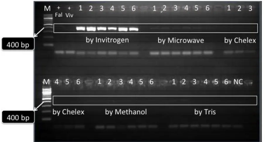
Results showed that not all samples were positive in the PCR for DNA amplification after extracted with five methods examined. PCR products of around 432 bp in size can be generated consistently only with commercial kit, which in the majority of cases did not permits further genetic analysis to be performed. Only commercial kit that resulted in very sharp bands of PCR, whereas microwave method showed very thin bands. Other three methods (Chelex, methanol and Tris-EDTA) did not resulted in any PCR product. Results of these observations are presented in Figure 2. Chelex, methanol and Tris-EDTA methods for DNA extractions resulted in no DNA, whereas microwave method resulted in DNA that appears as thin threads. Although DNA that strands is the most impressive, DNA that has sheared still shows that DNA is present.
Figure 2. PCR amplification results for all methods used to extract DNA that target Plasmodium mitochondrial gene of ~432 bp expected band. M, 100 bp ladder marker; +Fal, positive control for P. falciparum (3d7 strain); +Viv, positive
control for P. vivax (field positive sample), no of samples (gDNA were extracted by different method); NC, negative control.
Asian Online Journals (www.ajouronline.com) 434
these different methods were influenced by several factors such as duration of storage, and parasite densities that are potentially influencing sensitivity [18]. Beside that, PCR inhibitors are other factor which prevent the amplification of nucleic acids through PCR. Inhibitors usually affect it through interaction with DNA or interference with the DNA polymerase. Inhibitors can escape removal during the DNA purification procedure by binding directly to single or double-stranded DNA [19]. Inhibitors may be present in the original sample, such as blood, fabrics, tissues and soil or as a result of the sample processing and DNA extraction techniques used.
4. DISCUSSION
In the context of malaria elimination, the development of molecular diagnostic approach capable to detect low parasit infection for mass screening is essential. Moreover routine analysis of DNA from malaria parasite infected blood samples using PCR require procedures for rapid DNA preparation with the minimum number of steps and a reduced possibility of contamination.
Here we evaluated the sensitivity of PCR detection of human malaria parasites using DNA templates extracted by five different methods. We compared these methods using blood samples infected with three species of plasmodium (P. falciparum, P. vivax and P. malariae) that spotted on filter paper, which is one of the most practical way to store blood samples. The selected extraction methods are representative of the diverse approaches that are commonly employed in many laboratories covering commercial and non commercial extraction methods and already published elsewhere. The endpoints compared were proportion of extractions producing amplifiable DNA as measured by PCR amplification of a fragment of the mitochondrial gene.
In our research, DNA samples extracted using the kit have consistently provided high-quality results. In general, even though more expensive, commercial kits are recommended because the associated reagents have been subjected to quality control before use and are not likely to introduce problems. The procedure is also time and cost effective and eliminates several sources of possible contamination associated with many other isolation procedures. On the other hand, we also have used microwave irradiation of the parasites obtained from blood which was then directly subjected to a PCR technique to accurately and rapidly identify the presence of mitochondrial gene which characterise human malaria species. The microwave lysis method followed by PCR has been found to be less time consuming, only 0.5 hours, as compared to 2-3 hours by kit, methanol and TE techniques, and to 13 hours for chelex method. Use of this strategy would enable early identification and early implementation of control measures [20].
Here we also use Chelex-100 in the extraction process that function to bind ions that may be inhibitory to polymerase and that catalyze DNA degradation. Actually this technique represents a sensitive and practical field method for the determination of genetic variation within parasit species such as P. falciparum and the study of molecular epidemiology [21]. But in our research this fact was not seen.
PCR has greatly facilitated the diagnosis and genetic analysis of microorganisms. However, in the case of plasmodia and other pathogens found in whole blood, identification and genetic characterization are limited by the presence of iron and other metals, which may inhibit PCR amplification of DNA. It can be avoid by erythrocyte lysis, proteinase K digestion, phenol-chloroform extractions, ethanol precipitation, or cesium banding [22]. The other main limitations of the PCR are related to the DNA extraction step and mainly to the volume of blood analysed.
We also described methanol method used to extract DNA. Sixty five percent methanol is not enough to precipitate small plasmid size DNA efficiently, but will increase in efficiency with the addition of salt. Some DNA, especially high molecular weight DNA, will precipitate in 65% methanol, especially as nucleoprotein complexes at the chloroform methanol junction. My opinion is if it is DNA you want, you may want to consider a protease digestion step prior to protein extraction using phenol chloroform and then DNA precipitation with ethanol/isopropanol and salt .
The TE buffer-based DNA extraction method described in this report has not shown good results, compared with four standard methods for extraction of blood samples blooted on the filter paper, that are independent of parasite density and duration of storage. The described method may therefore represent a useful tool in molecular epidemiological studies [23]. Different with this report, Bereczky et al. [10] found that Tris EDTA method had shown superior results for DNA extraction compared with two other methods (methanol and chelex) of archived blood samples spotted on filter paper, and that are independent of parasite density and duration of storage and may represent a useful tool in molecular epidemiologic studies.
Asian Online Journals (www.ajouronline.com) 435
5. SUMMARY
A simple and rapid method is needed that possibly used in analysing a large number of preserved samples at the molecular level. The simplicity of this method combined with overall time and cost saving extends the life of the biological sample and increases the beneficial value of the filter paper system. Using the method describe herein, it is possible to obtain amplification products reproducibly from DNA isolated from dried blood spotted on filter paper. From results above, we concluded that commercial extraction kit method is well-suited for the DNA extraction in PCR-based assays of malaria.
6. ACKNOWLEDGEMENTS
We greatly appreciate Ms. Anggi from Eijkman Institute for Molecular Biology for kind help in providing technical assistance during experiment. Work of our laboratory presented herein has been supported by DIPA (Annual Research Projects) of the Center for 2014 fiscal year.
7. REFERENCES
[1] CDC, Malaria Parasites, 2010.
[2] World Health Organization. World Malaria Report 2005. WHO, Geneva, 2005.
[3] Malkin E, Dubovsky F, Moree M, ―Progress towards the development of malaria vaccines‖, Trends in Parasitology, Vol. 22, No.7, pp.292–295, 2006.
[4] World Health Organization, Malaria Fact Sheet, Geneva, 2011.
[5] Simanjuntak CH and Arbani PR, ―Situasi Malaria di Indonesia‖, Cermin Dunia Kedokteran, Vol. 55, pp. 3-4, 1994.
[6] Bojang KA, Milligan PJ, Pinder M, Vigneron L, Allouche A, Kester KE, Ballou WR, Conway DJ and Reece WH,
―Efficacy of RTS,S/AS02 malaria vaccine against Plasmodium falciparum infection in semi-immune adult men in The Gambia: a randomised trial‖,Lancet, Vol. 358, pp. 1927-1934, 2001.
[7] Hoffman SL, Goh LM, Luke TC, Schneider I, Le TP, Doolan DL, Sacci J, de la Vega P, Dowler M, Paul C. et al.,
―Protection of humans against malaria by immunization with radiation-attenuated Plasmodium falciparum
sporozoites‖,J. Infect. Dis. Vol. 185, pp.1155 -1164, 2002.
[8] Luke TC and Hoffman SL, ―Rationale and plans for developing a non-replicating, metabolically active, radiation-attenuated Plasmodium falciparum sporozoite vaccine‖,The Journal of Experimental Biology, Vol. 206, pp. 3803-3808, 2003.
[9] Okell LC, Bousema T, Griffin JT, Ouedraogo AL, Ghani AC, Drakeley CJ, ―Factors determining the occurrence of submicroscopic malaria infections and their relevance for control‖,Nat Commun Vol. 3, pp. 1237, 2012. [10] Bereczky S, Martensson, A, Gil P, Färnert A, ―Short report: rapid DNA extraction from archive blood spots on
filter paper for genotyping of Plasmodium falciparum‖,Am J Trop Med Hyg., Vol. 72, pp. 249-51, 2005.
[11] Cox- Singh J, Mahayet S, Abdullah MS, Singh B, ―Increased sensitivity of malaria detection by nested polymerase chain reaction using simple sampling and DNA extraction‖,Int J Parasitol., Vol. 27, pp. 1575-1577, 1997.
[12] Yang S and Rothman RE, ―PCR-based diagnostics for infectious diseases:uses, limitations, and future applications inacute-care settings‖, The Lancet Infectious Disease, Vol. 4, pp. 337-348, 2004.
[13] Fredericks DN and Relman DA, ―Application of polymerase chain rection to the diagnosis of infectious diseases‖,
Clin Infect Dis, Vol. 29, pp. 475-486, 2000.
[14] Louie M, Louie L, Simor AE, ―The role of DNA amplification technology in the diagnosis of infectious diseases‖,
CMAJ, Vol. 163, pp. 301-309, 2000.
[15] Farooq U,Dubey ML,Shrivastava SK,and Mahajan RC, ―Genetic polymorphism in Plasmodium falciparum: Differentiation of parasite isolates of high & low virulence by RAPD‖,Indian J Med Res., Vol. 136, No. 2, pp. 292–295, 2012.
[16] Glasel JA, ―Validity of nucleic acid purities monitored by 260 nm/280 nm absorbance ratios‖, Biotechniques, Vol. 18, pp. 62–63, 1995.
[17] Snounou G, Viriyakosol S, Zhu XP, Jarra W, Pinheiro L, Do Rosario VE, Thaithong S, Brown KN, ―High sensitivity of detection of human malaria parasites by the use of nested polymerase chain reaction”, Mol Biochem Parasitol, Vol. 61, pp. 315-320, 1993.
[18] Farnert A, Arez AP, Correia AT, Bjorkman A, Snounou G, do Rosario V, ―Sampling and storage of blood and the detection of malaria parasites by polymerase chain reaction‖, Trans R Soc Trop Med Hyg, Vol. 93, pp. 50–53, 1999.
[19] Reza A, ―Forensic implications of PCR inhibition—A review‖,Forensic Science International: Genetics, Vol. 6, No. 3, pp. 297–305, 2012.
Asian Online Journals (www.ajouronline.com) 436
[21] Kain KC and Lanar DE, ―Determination of genetic variation within Plasmodium falciparum by using enzymatically amplified DNA from filter paper disks impregnated with whole blood‖, Journal of Clinical Microbiology, Vol. 29, No. 6, pp. 1171-1174, 1991.
[22] Canier L, Khim N, Kim S, Sluydts V, Heng S, Dourng D, Eam R, Chy S, Khean C, Loch K, Ken M, Lim H, Siv S, Tho S, Masse-Navette P, Gryseels C, Uk S, Van Roey K, Grietens KP, Sokny M, Thavrin B, Chuor CM, Deubel V, Durnez L, Coosemans M, Ménard D, ―An innovative tool for moving malaria PCR detection of parasite reservoir into the field‖, Malaria Journal, Vol. 12, p. 405, 2013.
[23] Morris U, Aydin-Schmidt B, Shakely D, Mårtensson A, Jörnhagen L, Ali AS, Msellem MI, Petzold M, Gil JP,
Ferreira PE and Björkman A, “Rapid diagnostic tests for molecular surveillance of Plasmodium falciparum