The journal homepage www.jpacr.ub.ac.id ISSN : 2302 ‐ 4690
94
Polymeric Switch on Lysozyme Activity: Role of Hydrophobic
1
and Electrostatic Interactions
2 3
Sumon Ganguli1*, Md. Bellal Hossain2
4 5
1
Department of Applied and Environmental Chemistry, Faculty of science, University of Chittagong, 6
Chittagong-4331, Bangladesh. 7
2
Department of Nutrition and Food Engineering, Allied Health Science, Daffodil International University, 8
Dhanmondi R/A, Dhaka-1207, Bangladesh. 9
10
*
Corresponding Contact: To whom correspondence should be addressed: Sumon Ganguli, Ph.D., Department of 11
Applied and Environmental Chemistry, Faculty of Science, University of Chittagong, Chittagong-4331, 12
Bangladesh. Phone: +88-01757-808239. E-mail: [email protected] 13
14
Received 13 July 2013; Accepted 16 August 2013 15
This article was retracted because author requesting 16
17
ABSTRACT 18
Enzymes have attracted potential applications in both medicine and biotechnology. In our 19
present study, we show a strategy for switching the enzymatic activity of lysozyme by the 20
complex formation with a cationic smart copolymer. PAMA-g-PEG graft copolymers 21
suppressed the enzymatic activity of lysozyme without any conformational change, 22
indicating the formation of complex and covering the active site of lysozyme by 23
copolymers. The addition of polyanion, poly(acrylic acid) (PAAc), recovered the 24
suppressed enzymatic activity of the lysozyme/polymer complex efficiently. These finding 25
suggest that that hydrophobic interaction coupled with electrostatic interactions has a great 26
role for the complexation and decomplexation of the lysozyme/polymer complex. Circular 27
dichroism (CD) spectral analysis indicated that the conformation of the enzymes 28
maintained largely during the course of the complexation. 29
. 30
Key word: PAMA-g-PEG copolymer, lysozyme, enzymatic activity, hydrophobic and 31
Enzymes have attracted great attention in biomaterials education because they offer 35
very important industrial applications. In nature, the application of enzymes would increase 36
drastically if we can able to regulate their enzymatic activity. There are several techniques 37
have been developed such as metal dependent switch for regulating the enzymatic activity,1 38
by introducing temperature2 and photo3 responsive polymers to the active site of enzymes, 39
whose activity was then switched by changes in temperature and light conditions, enzyme-40
polymer hybrids, which are created by nonspecific4 or specific5 site-directed modifications of 41
synthetic polymers, etc. However, metal binding site or polymers into the enzymes at the 42
active site or to the enzyme surface is required for these technologies. Tailored nanoparticle 43
surface is another way to control the enzymatic activity6 although tailoring of the 44
nanoparticle or conjugation with polymers is required which makes a complex process such 45
as combination of the several preparative steps. It is noteworthy to note that the cost effective 46
and easy procedure is long awaited. As reported on several previous studies6,7,8 attractive and 47
The journal homepage www.jpacr.ub.ac.id ISSN : 2302 ‐ 4690
95
switching the enzymatic activity. In addition, polymeric compounds carrying charged are 49
particularly smart agents for complexation with enzymes.9,10 Based on the recent 50
development of complexation between enzyme and charged polymer, for the first time, we 51
succeeded in regulating the enzymatic activity of lysozyme11 through a simple process, the 52
external addition of a polycationic polyamine-poly(ethylene glycol) (PEG) graft copolymer, 53
poly(N,N-diethylaminoethyl methacrylate)-graft-PEG (PEAMA-g-PEG). They might form a 54
complex despite the fact that both are positively charged. In the present study, effect of 55
another polyamine-PEG copolymers, polycationic poly(N,N-dimethylaminoethyl 56
methacrylate)-graft-PEG (PAMA-g-PEG) on the enzymatic activity of lysozyme was 57
investigated. It should be noted that PAMA-g-PEG was used in this work due to the presence 58
of low hydrophobic factor (di-ethyl to di-methyl group) (Figure 1) in PAMA-g-PEG as 59
compared with PEAMA-g-PEG to investigate the mechanism of interaction between enzyme 60
and polymer. These finding suggest that complexation between lysozyme and polyamine-61
PEG copolymers might driven not only by electrostatic interaction but also by hydrophobic 62
PAMA-g-PEG was synthesized as described in the previous article and was a kind gift 67
from Mr. Shinya Matsumoto12. PAMA-g-PEG was used before HPLC purification. 68
Micrococcus lysodeikticus for activity assay and PAAc (Mn = 5,000 g/mol) were purchased
69
from Wako (Osaka, Japan). Sodium dihydrogen phosphate dihydrate (NaH2PO4.2H2O) was
70
obtained from Nacalai Tesque Inc. (Kyoto, Japan). All chemicals used were of high-quality 71
analytical grade. The water used in this study was purified using the Milli-Q system (Nihon 72
Millipore Co., Tokyo, Japan). 73
74
Enzymatic Activity Measurements 75
The enzymatic activity of lysozyme was measured based on bacteriolysis reaction with 76
M. lysodeikticus.8 Lysozyme concentration was determined by measuring the absorbance at 77
280 nm with an appropriate blank, using an extinction coefficient of 2.63 mL mg-1 cm-1.13 M. 78
lysodeikticus suspension (substrate solution) was prepared by mixing 0.5 mg/mL of M. 79
lysodeiktictus with 50 mM sodium phosphate buffer at pH 7.0. All the stock solution for 80
additives and protein were dissolved in 50 mM phosphate buffer solution (pH 7.0). A 10 μL 81
aliquot of the sample was added to a 1490 μL of M. lysodeikticus solution and decrease in 82
turbidity of the solution was monitored at 600 nm for 60 s using a UV-vis spectrophotometer 83
model V-550 (Japan Spectroscopic Co., Tokyo, Japan) at room temperature. The absorbance 84
decay plots from 10 to 20 sec were fitted to a linear equation and then the enzymatic 85
activities were determined from the slope of the fitted line.14 86
87
CD Spectra Measurements 88
Far-ultraviolet CD spectra were monitored using a Jasco spectropolarimeter (JASCO J-89
720W, Jasco Ltd., Tokyo, Japan). A cuvette with 0.1 cm path length was used and 90
photomultiplier voltage did not exceed 600 V in the measurements. The results are directly 91
obtained from CD spectrophotometer. 92
93
The journal homepage www.jpacr.ub.ac.id ISSN : 2302 ‐ 4690
96
Hen egg white lysozyme (pI=9.2015) was used as the model enzyme in this study. In our 95
previous study, it was found that the enzymatic activity of lysozyme was suppressed by 96
PEAMA-g-PEG (pKa 7.4Error! Bookmark not defined.) owing to capping of the active site of
97
lysozyme, which involved an electrostatic interaction between the negatively charged active 98
site of lysozyme and the positively charged amine moiety of PEAMA-g-PEG at neutral pH.11 99
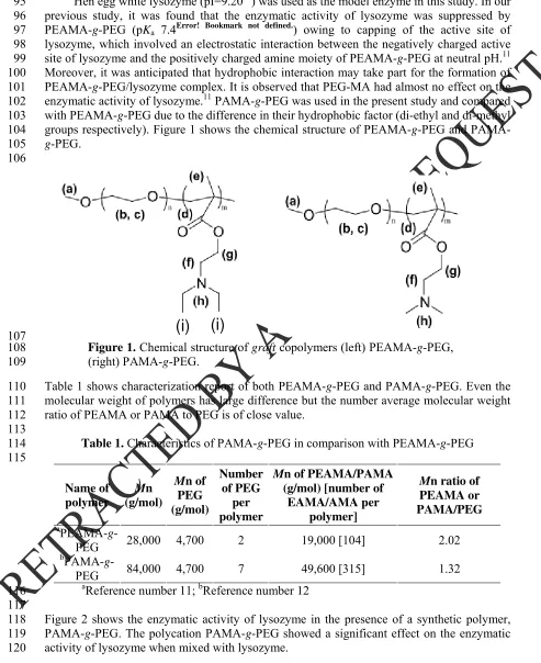
Moreover, it was anticipated that hydrophobic interaction may take part for the formation of 100
PEAMA-g-PEG/lysozyme complex. It is observed that PEG-MA had almost no effect on the 101
enzymatic activity of lysozyme.11 PAMA-g-PEG was used in the present study and compared 102
with PEAMA-g-PEG due to the difference in their hydrophobic factor (di-ethyl and di-methyl 103
groups respectively). Figure 1 shows the chemical structure of PEAMA-g-PEG and PAMA-104
g-PEG. 105
106
107
Figure 1. Chemical structure of graft copolymers (left) PEAMA-g-PEG, 108
(right) PAMA-g-PEG. 109
Table 1 shows characterization report of both PEAMA-g-PEG and PAMA-g-PEG. Even the 110
molecular weight of polymers has large difference but the number average molecular weight 111
ratio of PEAMA or PAMA to PEG is of close value. 112
113
Table 1. Characteristics of PAMA-g-PEG in comparison with PEAMA-g-PEG 114
115
Name of polymer
Mn
(g/mol)
Mn of
PEG (g/mol)
Number of PEG
per polymer
Mn of PEAMA/PAMA
(g/mol) [number of EAMA/AMA per
polymer]
Mn ratio of
PEAMA or PAMA/PEG
a
PEAMA-g-PEG 28,000 4,700 2 19,000 [104] 2.02
b
PAMA-g-PEG 84,000 4,700 7 49,600 [315] 1.32
a
Reference number 11; bReference number 12 116
117
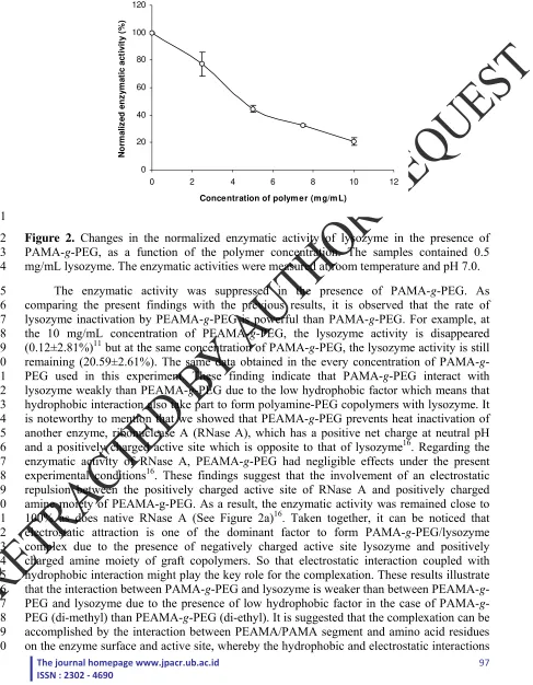
Figure 2 shows the enzymatic activity of lysozyme in the presence of a synthetic polymer, 118
PAMA-g-PEG. The polycation PAMA-g-PEG showed a significant effect on the enzymatic 119
activity of lysozyme when mixed with lysozyme. 120
The journal homepage www.jpacr.ub.ac.id
Concentration of polym er (m g/m L)
No
Figure 2. Changes in the normalized enzymatic activity of lysozyme in the presence of 122
PAMA-g-PEG, as a function of the polymer concentration. The samples contained 0.5 123
mg/mL lysozyme. The enzymatic activities were measured at room temperature and pH 7.0. 124
The enzymatic activity was suppressed in the presence of PAMA-g-PEG. As 125
comparing the present findings with the previous results, it is observed that the rate of 126
lysozyme inactivation by PEAMA-g-PEG is powerful than PAMA-g-PEG. For example, at 127
the 10 mg/mL concentration of PEAMA-g-PEG, the lysozyme activity is disappeared 128
(0.12±2.81%)11 but at the same concentration of PAMA-g-PEG, the lysozyme activity is still 129
remaining (20.59±2.61%). The same data obtained in the every concentration of PAMA-g-130
PEG used in this experiment. These finding indicate that PAMA-g-PEG interact with 131
lysozyme weakly than PEAMA-g-PEG due to the low hydrophobic factor which means that 132
hydrophobic interaction also take part to form polyamine-PEG copolymers with lysozyme. It 133
is noteworthy to mention that we showed that PEAMA-g-PEG prevents heat inactivation of 134
another enzyme, ribonuclease A (RNase A), which has a positive net charge at neutral pH 135
and a positively charged active site which is opposite to that of lysozyme16. Regarding the 136
enzymatic activity of RNase A, PEAMA-g-PEG had negligible effects under the present 137
experimental conditions16. These findings suggest that the involvement of an electrostatic 138
repulsion between the positively charged active site of RNase A and positively charged 139
amine moiety of PEAMA-g-PEG. As a result, the enzymatic activity was remained close to 140
100% as does native RNase A (See Figure 2a)16. Taken together, it can be noticed that 141
electrostatic attraction is one of the dominant factor to form PAMA-g-PEG/lysozyme 142
complex due to the presence of negatively charged active site lysozyme and positively 143
charged amine moiety of graft copolymers. So that electrostatic interaction coupled with 144
hydrophobic interaction might play the key role for the complexation. These results illustrate 145
that the interaction between PAMA-g-PEG and lysozyme is weaker than between PEAMA-g-146
PEG and lysozyme due to the presence of low hydrophobic factor in the case of PAMA-g-147
PEG (di-methyl) than PEAMA-g-PEG (di-ethyl). It is suggested that the complexation can be 148
accomplished by the interaction between PEAMA/PAMA segment and amino acid residues 149
The journal homepage www.jpacr.ub.ac.id ISSN : 2302 ‐ 4690
98
take place on the polymer-enzyme interface even though other noncovalent interactions can 151
not be ruled out at this moment. 152
-60 -50 -40 -30 -20 -10 0
210 220 230 240 250
Wavelength (nm )
C
D
(m
de
g)
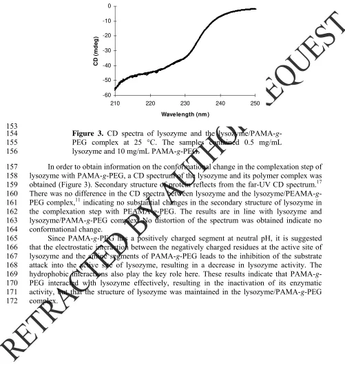
153
Figure 3. CD spectra of lysozyme and the lysozyme/PAMA-g-154
PEG complex at 25 °C. The samples contained 0.5 mg/mL 155
lysozyme and 10 mg/mL PAMA-g-PEG. 156
In order to obtain information on the conformational change in the complexation step of 157
lysozyme with PAMA-g-PEG, a CD spectrum of the lysozyme and its polymer complex was 158
obtained (Figure 3). Secondary structure of protein reflects from the far-UV CD spectrum.17 159
There was no difference in the CD spectra between lysozyme and the lysozyme/PEAMA-g-160
PEG complex,11 indicating no substantial changes in the secondary structure of lysozyme in 161
the complexation step with PEAMA-g-PEG. The results are in line with lysozyme and 162
lysozyme/PAMA-g-PEG complex. No distortion of the spectrum was obtained indicate no 163
conformational change. 164
Since PAMA-g-PEG has a positively charged segment at neutral pH, it is suggested 165
that the electrostatic interaction between the negatively charged residues at the active site of 166
lysozyme and the amine segments of PAMA-g-PEG leads to the inhibition of the substrate 167
attack into the active site of lysozyme, resulting in a decrease in lysozyme activity. The 168
hydrophobic interactions also play the key role here. These results indicate that PAMA-g-169
PEG interacted with lysozyme effectively, resulting in the inactivation of its enzymatic 170
activity, but that the structure of lysozyme was maintained in the lysozyme/PAMA-g-PEG 171
The journal homepage www.jpacr.ub.ac.id
Concentration of PAAc (m g/m L)
N
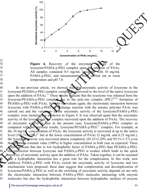
Figure 4. Recovery of the enzymatic activity of the 174
lysozyme/PAMA-g-PEG complex upon the addition of PAAc. 175
All samples contained 0.5 mg/mL lysozyme with 10 mg/mL 176
PAMA-g-PEG, and measurements were carried out at room 177
temperature and pH 7.0. 178
179
In our previous article, we showed that the enzymatic activity of lysozyme in the 180
lysozyme/PEAMA-g-PEG complex completely recovered to the level of the native lysozyme 181
upon the addition of PAAc.11 These results indicate that the lysozyme was released from the 182
lysozyme/PEAMA-g-PEG complex due to the poly-ion complex (PIC)18,19 formation of 183
PEAMA-g-PEG with PAAc. In order to evaluate again, the electrostatic interaction between 184
lysozyme with PAMA-g-PEG, an exchange reaction with the anionic polymer PAAc was 185
carried out and the variations in the enzymatic activity of the lysozyme/PAMA-g-PEG 186
complex were monitored is as shown in Figure 4. It was observed again that the enzymatic 187
activity of the lysozyme in the complex recovered upon the addition of PAAc. The recovery 188
of enzymatic activity is fast in the present case, lysozyme/PAMA-g-PEG complex as 189
compared with the previous results, lysozyme/PEAMA-g-PEG11 complex. For example, at 190
the 10 mg/mL concentration of PAAc, the lysozyme activity is recovered at up to the native 191
level (100.7±3.17%)11 but at the lower concentration of PAAc (5 mg/mL and 6.25 mg/mL), 192
the lysozyme activity was recovered almost completely (83.25±2.20% and 95.51±1.57) even 193
though attain constant value (100%) at higher concentration in both case as expected. These 194
results indicate that due to low hydrophobic factor of PAMA-g-PEG than PEAMA-g-PEG, 195
the interaction between lysozyme and PAMA-g-PEG is weaker which leads to the quick 196
recovery of enzymatic activity upon the addition of PAAc. Taken together, it can be assumed 197
that a hydrophobic interaction has a great role for the complexation. In this work, new 198
additives PAMA-g-PEG with PAAc switch the enzymatic activity of lysozyme and two 199
mechanisms were proposed; these data suggest that complexation and decomplexation of 200
lysozyme/PAMA-g-PEG as well as the switching of enzymatic activity depends on not only 201
the electrostatic interaction between PAMA-g-PEG molecules interacting with enzyme 202
The journal homepage www.jpacr.ub.ac.id ISSN : 2302 ‐ 4690
100
molecules with hydrophobic factor of PAMA-g-PEG. Complexation and decomplexation 204
mechanism between enzyme and polymer were depicted in scheme 1. 205
206
207
Scheme 1. Schematic illustration of complexation and decomplexation of lysozyme and 208
PAMA-g-PEG. 209
210
CONCLUSION 211
The smart copolymer, PAMA-g-PEG and polyanion PAAc, switched the enzymatic 212
activity of lysozyme dramatically. It was interesting to note that PAMA-graft-PEG polymers 213
suppressed the enzymatic activity of lysozyme, and that PAAc easily restored the lysozyme 214
activity by means of electrostatic and hydrophobic interactions. Binding behavior between 215
polymer and enzyme in addition to maintaining their secondary structure offers new 216
prospects. Such switching of enzyme activity using a polymer-enzyme system may able to 217
expand the application of enzymes in the industrial field including enzyme delivery, enzyme 218
separations, biosensor, and bio-nanoreactors etc. This same strategy might be extended to 219
regulate the enzymatic activity of other enzymes or the binding affinity of enzymes to other 220
polymers, DNA or other proteins. 221
222
ACKNOWLEDGMENT 223
The authors gratefully acknowledge the financial support of the Department of Applied 224
and Environmental Chemistry, University of Chittagong, Chittagong-4331, Chittagong, 225
Bangladesh. The author also would like to express sincere gratitude to Dr. Parimal Talukder, 226
Manager, Research and Development, Square Pharmaceuticals Ltd. Bangladesh for technical 227
assistance and Institute of Applied Physics, University of Tsukuba, Japan for CD 228
measurements. 229
230
REFERENCES 231
1 Corey, D. R. and Schultz, P. G., J. Biol. Chem., 1989, 264, 3666-3669.
2 T. Shimboji, T., Larenas, E., Fowler, T., Kulkarni, T., Hoffman, A. S. and Stayton, P. S., Proc. Natl. Acad. Sci. U.S.A., 2002, 99, 16592-16596.
3 Shimboji, T., Ding, Z. L., Stayton, P. S. and Hoffman, A. S., Bioconju. Chem., 2003, 14, 517-525.
The journal homepage www.jpacr.ub.ac.id ISSN : 2302 ‐ 4690
101
5 Shao, H., Crnogorac, M. M., Kong, T., Chen, S., Willams, J. M., Tack, J. M.,
Gaerigaian, V., Cagle, E. N., Carnevail, M., Tumelty, D., Paliard, X., Miranda, L. P., Bradburne, J. A. and Kochendoerfer, G. G., J. Am. Chem. Soc, 2005, 127, 1350-1351. 6 A. Verma, A., Simrad, J. M. and Rotello, V. M., Langmuir., 2004, 20, 4178-4181. 7 B. S. Sandanaraj, B. S., Vutukori, D. R., Simrad, J. M., Klaikherd, A., Hong, R.,
Rotello, V. M. and Thayumanavan, T, J. Am. Chem. Soc, 2005, 27, 10693-10698. 8 Hirano, A., Hamada H. and Shiraki, K., Protein J., 2008, 27, 253-257.
9 Pispas, S., J. of Polymer Science: Part A: Polymer Chem., 2007, 46, 509-520. 10 Monien, B. H., Cheang, K. L. and Desai, U. R., J. Med. Chem., 2005, 48, 5360-5368. 11 Ganguli, S., Yoshiomoto, K., Tomita, S., Sakuma, H., Matsuoka, T., Shiraki, K. and
Nagasaki, Y., J. Am. Chem. Soc., 2009, 131 (18), 6549-6549-6553.
12 Yoshimoto, K., Nozawa, M., Matsumoto, S., Echigo, T., Nemeto, S., Hatta, T. and Nagasaki, Y., Langmuir, 2009, 25, 12243-12249.
13 Wetlaufer, D. B. and Saxena, V. P.,Biochemistry,1970, 9, 5015-5023.
14 T. Matsuoka, T., Tomita, S., Hamada, H. and Shiraki, K., J. Biosci.Bioeng, 2007, 103, 440-443.
15
Kudou, M., Shiraki, K., Fujiwara, S., Imanaka, T. and Takagi, M., Eur. J. Biochem., 2003, 270, 4547-4554
16 Ganguli, S., Yoshiomoto, K., Tomita, S., Sakuma, H., Matsuoka, T., Shiraki, K. and Nagasaki, Y., Macromol. Biosci., 2010, 10 (8), 849-853.
17 Sreerama, N. and Woddy, R. W., Analytical Biochemistry, 2000, 287, 252-260. 18 Harada, A. and Kataoka, K., Macromolecules, 1995, 28, 5294-5299.