PAPER
Differential impact of cerebral white matter changes, diabetes,
hypertension and stroke on cognitive performance among
non-disabled elderly. The LADIS study
Ana Verdelho, Sofia Madureira, Jose´ M Ferro, Anna-Maria Basile, Hugues Chabriat, Timo Erkinjuntti,
Franz Fazekas, Michael Hennerici, John O’Brien, Leonardo Pantoni, Emilia Salvadori, Philip
Scheltens, Marieke C Visser, Lars-Olof Wahlund, Gunhild Waldemar, Anders Wallin, Domenico
Inzitari, on behalf of the LADIS Study
. . . .
See end of article for authors’ affiliations . . . . Correspondence to: Dr Ana Verdelho, Department of Neurosciences, Hospital Santa Maria, 1649-035 Lisbon, Portugal; [email protected]
Received 28 November 2006 Revised 27 March 2007 Accepted 31 March 2007
Published Online First 30 April 2007
. . . .
J Neurol Neurosurg Psychiatry2007;78:1325–1330. doi: 10.1136/jnnp.2006.110361
Background and purpose:Age related white matter changes (ARWMC) are frequent in non-demented old subjects and are associated with impaired cognitive function. Our aim was to study the influence of vascular risk factors and ARWMC on the neuropsychological performance of an independent elderly population, to see if vascular risk factors impair cognition in addition to the effects of ARWMC.
Methods: Independent subjects, aged 65–84 years, with any degree of ARWMC were assessed using a comprehensive neuropsychological battery including the Mini-Mental State Examination (MMSE), VADAS-Cog (Alzheimer’s disease assessment scale) and the Stroop and Trail Making test. Vascular risk factors were recorded and ARWMC (measured by MRI) were graded into three classes. The impact of vascular risk factors and ARWMC on neuropsychological performance was assessed by linear regression analyses, with adjustment for age and education.
Results: 638 patients (74.1 (5) years old, 55% women) were included. Patients with severe ARWMC performed significantly worse on global tests of cognition, executive functions, speed and motor control, attention, naming and visuoconstructional praxis. Diabetes interfered with tests of executive function, attention, speed and motor control, memory and naming. Arterial hypertension and stroke influenced executive functions and attention. The effect of these vascular risk factors was independent of the severity of ARWMC, age and education.
Conclusion:ARWMC is related to worse performance in executive function, attention and speed. Diabetes, hypertension and previous stroke influenced neuropsychological performance, independently of the severity of ARWMC, stressing the need to control vascular risk factors in order to prevent cognitive decline in the elderly.
C
erebral age related white matter changes (ARWMC) arefrequently described on brain imaging in demented and
non-demented elderly subjects.1 2Some demographic and
vascular risk factors are associated with a higher risk of developing ARWMC, with major emphasis on age,
hyperten-sion and stroke.3–6On the other hand, recent epidemiological
evidence indicates that vascular risk factors play a role in the development of cognitive impairment and dementia, including
degenerative dementia.7–11ARWMC can be a mediator between
vascular risk factors and cognitive decline, while some demographic characteristics can contribute towards protecting
cognitive function.12 Our aim was to study the influence of
ARWMC and vascular risk factors on the neuropsychological performance of non-disabled independent elderly people with ARWMC and to analyse if vascular risk factors had an independent effect on cognitive performance.
METHODS
The LADIS (Leukoaraiosis and Disability) study is a prospective multinational European project investigating the independent impact of ARWMC on the transition to disability in the elderly. The rationale, methodology and baseline assessment have been
described previously.12 13 Investigators were provided with a
specifically developed handbook with guidelines for applying
criteria and tools.13
In short, inclusion criteria for the study were: (i) 65–84 years of age; (ii) changes in ARWMC on MRI of
any degree, according to the scale of Fazekas and colleagues14
; and (iii) no disability, as determined by the Instrumental
Activities of Daily Living scale.15
Patients were enrolled because of minor neurological, cognitive or motor complaints, or incidental findings on cranial imaging caused by non-specific
events, as detailed elsewhere.13
To assess vascular risk factors, trained medical personnel used a structured and comprehen-sive questionnaire together with review of the available records. A detailed description of the study variables has been reported
previously.13
Those germane to this study are the vascular risk
factors.6
Vascular risk factor criteria have been described in
detail previously.6
Detailed criteria are given in appendix 1
(appendix 1 can be viewed on theJ Neurol Neurosurg Psychiatry
website at http://www.jnnp.com/supplemental). In short, these risk factors were: previous hypertension: current
antihyperten-sive treatment or blood pressure values >140/90 mm Hg in
subjects not taking antihypertensive medication, based on multiple blood pressure measurements on several separate occasions; diabetes mellitus: previous diagnosis and/or current treatment with insulin or oral hypoglycaemic medications, or
an 8 h fasting plasma glucose level of>7.0 mmol/l or 126 mg/
dl; hyperlipidaemias: total cholesterol.200 mg/dl, low density
lipoprotein .130 mg/dl, high density lipoprotein ,35 mg/dl
and triglyceride .200 mg/dl, on at least two occasions;
myocardial infarction; angina pectoris; heart failure; atrial fibrillation; lower limb arteriopathy and peripheral vascular disease; history of stroke and/or transient ischaemic attack; cigarette smoking, expressed as smoked pack-years; alcohol consumption, expressed as g/day consumed, with one drink containing 10 g of alcohol. Subjects were classified as past drinkers, not drinkers (never drinkers) and current drinkers
(subdivided into sporadic drinkers (,1 drink/week), low
drinkers (>1 drink/week but,1 drink/day), moderate drinkers
(1–3 drinks/day) and heavy drinkers (.4 drinks/day)).
The degree of ARWMC severity was rated on FLAIR sequences by central readers blind to the clinical data using the three severity classes in the revised version of the visual
scale of Fazekas and colleagues.14Medial temporal lobe atrophy
was assessed on coronal T1 weighted sequences using the MTA
scale.16
Neuropsychological assessment
The LADIS neuropsychological battery has been described in
detail elsewhere.12
The neuropsychological battery included the
Mini-Mental State Examination (MMSE)17
as a global measure of cognitive function; the VADAS-Cog (Alzheimer’s Disease Assessment Scale (ADAS-Cog) plus delayed recall, symbol digit, digit span, mazes, digit cancellation and verbal fluency) as a comprehensive instrument to assess orientation, language, ideational and constructional praxis, immediate memory and
delayed recall)18
; and the Stroop19 20
and Trail Making (TM)
test21
as measures of executive function. Tests were grouped by cognitive domains, as follows: executive functions (Stroop, verbal fluency and TM); attention (digit cancellation and symbol digit test); speed and motor control (Trail A and time to complete maze test); memory (digit span, word recognition, word recall, delayed recall); language (commands and nam-ing); praxis (visuoconstructional and ideational praxis; ADAS subtests). To analyse performance by domain, a compound measure for three main domains was calculated using standard
scores for individual tests, as described previously12
: (1)
memory = z scores of (immediate word recall+delayed recall
+word recognition+digit span)/4; (2) executive functions = z
scores of ((Stroop3-2) + (TMB-TMA) + symbol digit+ verbal
fluency)/4; and (3) speed and motor control = z scores of
(TMA+mazes+digit cancellation)/3. Z scores of the tests that
had higher scores representing worse performance were
inverted (2Z) in order to calculate the compound measure
score.
Statistical analysis
Scores of neuropsychological tests were considered as contin-uous variables. The influence of vascular risk factors and severity of ARWMC (three grades) on neuropsychological performance was analysed by t tests for variables with two levels and one way ANOVAs for variables with more than two
levels. Adjustment of a for multiple comparisons was
performed with Bonferoni statistics. Alcohol intake effect was analysed by dividing current by past and never drinkers. The effect of the quantity of alcohol on neuropsychological performance was analysed using one way ANOVA.
Linear regression analyses were performed to evaluate the impact of vascular risk factors and ARWMC on scores for the different cognitive domains and individual neuropsychological tests. In the first model, we included vascular risk factors as independent variables. In the second model, we added ARWMC to determine if the effect of vascular risk factors in neuropsy-chological tests was independent of the effect of ARWMC.
Linear regression analyses were adjusted for age, education,12
lacunes,22
medial temporal lobe atrophy and visual deficit. The independent variables included in linear regression analyses
were selected from the bivariate analysis, with p,0.1 as the
screening criterion for selection of the variables. Infrequent vascular risk factors (less than 5% frequency in the study population) were excluded from linear regression analyses.
Data were analysed using SPSS 12.0 software.
RESULTS
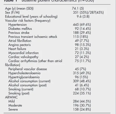
From the 639 patients in the LADIS cohort, one patient was excluded because of incomplete neuropsychological evaluation and hence 638 patients were included in the present study. Table 1 shows the patient characteristics and risk factors relevant to the present study.
Influence of ARWMC severity on neuropsychological evaluation
Analyses of variance (ANOVA) showed significant differences in performance on the neuropsychological tests according to the severity of ARWMC, most notably when comparing mild with severe ARWMC (table 2). Patients with severe ARWMC performed significantly worse on: (a) global measures of cognition (MMSE and ADAS total score), (b) compound measures of executive functions, speed and memory, (c) all individual tests of the same domains, (d) attention and (e) individual tests of memory, language and visuoconstructional praxis.
Influence of vascular risk factors on neuropsychological evaluation
T test results (data given in appendix 2; appendix 2 can be
viewed on the J Neurol Neurosurg Psychiatry website at http://
www.jnnp.com/supplemental) showed that patients with
hypertension performed significantly worse (p(0.001) on the
compound measure of executive functions, and on Stroop, TM and symbol–digit tests. Patients with diabetes had worse
performance (p(0.001) on TM, symbol–digit test and
immedi-ate word recall tests. Patients with previous stroke had worse
performance (p(0.001) on the digit cancellation, symbol digit
tests and on the compound measure of executive functions.
Table 1 Baseline patient characteristics (n = 638)
Age (y) (mean (SD)) 74.1 (5)
Sex (F/M) 351 (55%)/287(45%) Educational level (years of schooling) 9.6 (3.8) Vascular risk factors (frequency)
Hypertension 445 (69.6%) Diabetes mellitus 92 (14.4%) Previous stroke 188 (29.4%) Previous transient ischaemic attack 115 (18%) Atrial fibrillation 49 (7.7%) Angina pectoris 98 (15.3%) Heart failure 21 (3.3%) Myocardial infarction 72 (11.3%) Cardiac valvulopathy 27 (4.2%) Cardiac arrhythmias (other than atrial
fibrillation)
75 (11.7%)
Peripheral vascular disease 45 (7%) Hypercholesterolaemia 315 (49.3%) Hypertriglyceridaemia 96 (15%) Alcohol consumption (current) 309 (48.4%) Alcohol consumption (past) 41 (6.4%) Smoking (current) 68 (10.7%)
ARWMC, age related white matter changes.
Current alcohol intake was associated with better performance
(p(0.001) on MMSE comparing past with never drinkers.
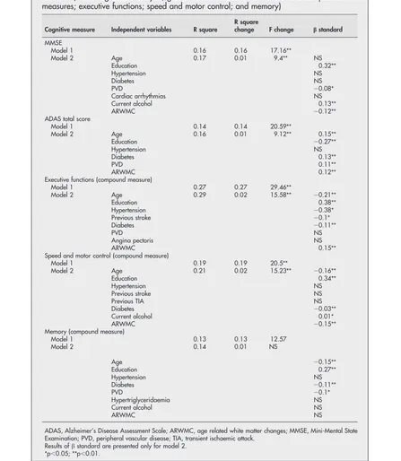
Linear regression analyses
Results of linear regression analyses to evaluate the impact of ARWMC and vascular risk factors on neuropsychological performance are shown in table 3. Results concerning global measures of cognition and compound measures are also shown in table 3. Results of regression analyses with individual tests
can be found in appendix 3 (appendix 3 can be viewed on theJ
Neurol Neurosurg Psychiatry website at http://www.jnnp.com/ supplemental) (positive results are described in the text). Linear regression analyses were repeated excluding stroke
patients (188 patients), and controlling for atrophy, lacunes22
and visual deficit (as potential confounders on the performance of neuropsychological tests) and produced similar results (data available on request).
Diabetes influenced the performance on the global measure of cognition (ADAS total score), compound measures of executive functions, speed and memory. Diabetes also influ-enced tests of attention, language, praxis and some tests of memory (word recall and delayed word recall). Arterial hypertension was associated with worse performance on the compound measure of executive functions, Trail B-A and symbol digit tests. Stroke was associated with lower perfor-mance on the compound measure of executive functions, on Stroop test 3–2, and tests of attention (digit cancellation) and memory (immediate word recognition). Patients with periph-eral vascular disease performed significantly worse on global measures (MMSE and ADAS total score), on the compound measure of memory and on word recall test. Patients with current alcohol intake had better performance on the MMSE, on mazes, word recall and commands tests and on the compound measure of speed, comparing never and past drinkers. Patients with current alcohol intake were not otherwise different on global status or other associated risk factors. Using one way ANOVA to evaluate the impact of alcohol quantity intake on neuropsychological performance,
moderate (1–3 drinks/day) and low drinkers (>1 drink/week
but,1 drink/day) performed significantly better on the MMSE
than never drinkers (27.95 vs 26.99 (p,0.05) and 27.88 vs
26.99 (p,0.01)). No differences were found for severe drinkers.
Introducing ARWMC into the model, we found that it was independently related to worse performance on global mea-sures of cognition (MMSE and ADAS total score), compound measures of executive functions and speed/motor control in all tests of executive functions, attention, language (naming) and visuoconstructional praxis. Education interfered with all
neuropsychological tests and the effect of age was retained.12
The presence of visual deficits and lacunes did not change the impact of vascular risk factors on neuropsychological perfor-mance.
DISCUSSION
This study confirmed that the severity of ARWMC is related to worse performance on global measures of cognition, executive functions, speed/motor control and tests of attention, naming and visuoconstructional praxis. In previous studies, ARWMC were associated with cognitive deficits in independent elderly subjects, mainly in executive functions, attention, speed and
motor control23 24
but also with global measures of
cogni-tion,1 4 23 25visuoconstructional1 23and memory tasks.3However,
the association was not consistent across all studies. We had the opportunity to study the impact of ARWMC and simulta-neously the impact of clinical vascular risk factors on neuropsychological performance, controlling for demographic variables and atrophy. We found that even controlling for the
severity of ARWMC and atrophy, vascular risk factors (such as diabetes, hypertension and previous stroke) emerged as relevant variables with an influence on neuropsychological tests. These results suggest that vascular risk factors impair cognition, independent of cerebral damage measurable, using current radiological methods. It is, however, conceivable that other imaging methods (eg, diffusion tensor MRI) might have demonstrated more extensive ARWMC and probably a less
independent effect of vascular risk factors.26
Our study has some limitations. The sample was selected based on minor complaints, and probably represents the first moment when non-disabled elderly people with cerebral white mater changes seek medical attention. However, this sample does not represent the global community. To increase consis-tency and homogeneity in the whole assessment, investigators
were provided with a specifically developed handbook with
guidelines for applying criteria and tolls.13
The methodology
and inclusion criteria were uniform in all centres.13
Sociocultural and nutritional differences between countries and consequent variation between vascular risk factors profile in the included population may limit the external validity of the results. However, the possible heterogeneity provided by linguistic, cultural and educational differences of the LADIS cohort makes the results more consistent and generalisable, as they are from a large sample with a wide variety of subjects. Moreover, validation and harmonisation of instruments and procedures among centres, including the neuropsychological battery, was done prior to the beginning of inclusion of
subjects, as discussed elsewhere.12 Differences across centres
were evaluated and could not explain the results.12
To exclude Table 3 Impact of vascular risk factors on neuropsychological performance: cognitive
domains (linear regression analysis; global tests MMSE and ADAS total and compound measures; executive functions; speed and motor control; and memory)
Cognitive measure Independent variables R square
R square
change F change bstandard
MMSE
Model 1 0.16 0.16 17.16**
Model 2 Age 0.17 0.01 9.4** NS
Education 0.32**
Hypertension NS
Diabetes NS
PVD 20.08*
Cardiac arrhythmias NS
Current alcohol 0.13**
ARWMC 20.12**
ADAS total score
Model 1 0.14 0.14 20.59**
Model 2 Age 0.16 0.01 9.12** 0.15**
Education 20.27**
Hypertension NS
Diabetes 0.13**
PVD 0.11**
ARWMC 0.12**
Executive functions (compound measure)
Model 1 0.27 0.27 29.46**
Model 2 Age 0.29 0.02 15.58** 20.21**
Education 0.38**
Hypertension 20.38*
Previous stroke 20.1*
Diabetes 20.11**
PVD NS
Angina pectoris NS
ARWMC 0.15**
Speed and motor control (compound measure)
Model 1 0.19 0.19 20.5**
Model 2 Age 0.21 0.02 15.23** 20.16**
Education 0.34**
Hypertension NS
Previous stroke NS
Previous TIA NS
Diabetes 20.03**
Current alcohol 0.01*
ARWMC 20.15**
Memory (compound measure)
Model 1 0.13 0.13 12.57
Model 2 0.14 0.01 NS
Age 20.15**
Education 0.27**
Hypertension NS
Diabetes 20.11**
PVD 20.1*
Hypertriglyceridaemia NS
Current alcohol NS
ARWMC NS
ADAS, Alzheimer’s Disease Assessment Scale; ARWMC, age related white matter changes; MMSE, Mini-Mental State Examination; PVD, peripheral vascular disease; TIA, transient ischaemic attack.
Results ofbstandard are presented only for model 2.
*p,0.05;**p,0.01.
possible interference on the performance of neuropsychological tests, visual deficits were considered in the regression analysis. Recent evidence found that diabetics have a higher risk of
dementia, including Alzheimer’s disease,10 27
and that non-demented diabetic patients performed worse on several
cognitive tasks compared with healthy controls.27 28However,
in these studies, ARWMC was not taken into account. A recent study found that patients with type 2 diabetes had a worse performance on attention, executive functions, processing
speed and memory, which was associated with ARWMC.29
In our study, diabetes was found to be the most important vascular risk factor interfering with global tests, compound measures of executive functions, speed/motor control and tests of attention, memory, praxis and language. This association was independent of the degree of ARWMC, atrophy, age, education and presence of visual deficits.
Cognitive changes and higher risk of dementia have been
reported in patients with hypertension,8 9 30 and recent results
of trials using antihypertensive medication suggest a beneficial
effect on cognition.31
Van Swieten and colleagues30
also found that patients with hypertension with confluent ARWMC performed worse on tests of executive functions (TM and Stroop), MMSE and the Wechsler Memory Scale visual subtest,
but not on other memory tests. Sierraet alfound that
middle-aged hypertensive patients with ARWMC performed signifi-cantly worse than hypertensive patients without ARWMC on
the digit span forward task.32
We found that hypertension was associated with a worse performance on tests of attention and mental flexibility, but not on other tests. One possible explanation for the relatively modest impact of hypertension may be related to the low proportion of non-hypertensive subjects (only 30%) in the LADIS sample.
In a previous study, ARWMC was related to speed and
attention in stroke survivors,33 and stroke was proposed as a
mediator between cerebral small vessel disease and cognitive
decline.34
We found previous stroke to be associated with worse performance on mental flexibility, attention and memory recognition tests, independent of the severity of ARWMC.
The influence of alcohol intake on brain structure and cognition is a controversial issue. Recent studies suggested that light to moderate alcohol consumption is associated with a
reduced risk of dementia.35 Conversely, brain atrophy is
associated with alcohol intake, even for low intake drinkers,36
and controversial effects on ARWMC and infarcts were reported. We found that mild and moderate drinkers performed better on global evaluation (MMSE) than non-drinkers, independent of education, age and ARWMC, but no other association was found between alcohol intake and neuropsy-chological testing, even when heavy drinkers were included. These results could possibly reflect a ‘‘survival’’ bias.
In conclusion, the neuropsychological performance of inde-pendent elderly subjects with ARWMC was influenced by biological and demographic variables: severity of ARWMC,
some vascular risk factors, but also age and education12. These
results emphasise the key role of risk factor control for the prevention of dementia and cognitive impairment.
ACKNOWLEDGEMENTS
The LADIS Study is supported by the European Union within the Vth European Framework Program ‘‘Quality of life and management of living resources’’ (1998–2002), contract No QLRT-2000-00446 as a concerted action.
Appendices 1–4 can be viewed on theJ Neurol
Neurosurg Psychiatrywebsite at http:// www.jnnp.com/supplemental.
Authors’ affiliations
. . . .
Ana Verdelho, Sofia Madureira, Jose´ M Ferro,Neurology Department, Centro de Estudos Egas Moniz, Santa Maria Hospital, Lisbon, Portugal
Anna-Maria Basile, Leonardo Pantoni, Emilia Salvadori, Domenico Inzitari,Department of Neurological and Psychiatric Sciences, University of Florence, Florence, Italy
Hugues Chabriat,Department of Neurology, Hoˆpital Lariboisie`re, Paris, France
Timo Erkinjuntti,Memory Research Unit, Department of Clinical Neurosciences, Helsinki University, Helsinki, Finland
Franz Fazekas,Department of Neurology and MRI Institute, Karl Franzens University Graz, Graz, Austria
Michael Hennerici,Department of Neurology, University of Heidelberg, Klinikum Mannheim, Mannheim, Germany
John O’Brien,Institute for Ageing and Health, University of Newcastle, Newcastle-upon-Tyne, UK
Philip Scheltens, Marieke C Visser,Department of Neurology, VU Medical Centre, Amsterdam, the Netherlands
Lars-Olof Wahlund,Karolinska Institute, Department of Clinical Neuroscience and Family Medicine, Huddinge University Hospital, Huddinge, Sweden
Gunhild Waldemar,Memory Disorders Research Unit, Department of Neurology, Copenhagen University Hospital, Copenhagen, Denmark
Anders Wallin,Institute of Clinical Neuroscience, Go¨teborg University, Go¨teborg, Sweden
Competing interests: None.
REFERENCES
1 Skoog I, Berg S, Johansson B,et al.The influence of white matter lesions on neuropsychological functioning in demented and non-demented 85-year-olds.
Acta Neurol Scand1996;93:142–8.
2 Leeuw FE, de Groot JC, Achten E,et al.Prevalence of cerebral white matter lesions in elderly people: a population based magnetic resonance imaging study. The Roterdam scan study.J Neurol Neurosurg Psychiatry2001;70:9–14. 3 Breteler MM, van Swieten JC, Bots ML,et al.Cerebral white matter lesions,
vascular risk factors, and cognitive function in a population-based study: The Rotterdam Study.Neurology1994;44:1246–52.
4 Longstreth WT Jr, Manolio TA, Arnold A,et al.Clinical correlates of white matter findings on cranial magnetic resonance imaging of 3301 elderly people. The Cardiovalscular Health Study.Stroke1996;27:1274–82.
5 Ylikoski A, Erkinjuntti T, Raininko R,et al.White matter hyperintensities on MRI in the neurologically nondiseased elderly. Analysis of cohorts of consecutive subjects aged 55 to 85 years living at home.Stroke1995;26:1171–7. 6 Basile AM, Pantoni L, Pracucci G,et al.Age, hypertension, and lacunar stroke
are the major determinants of the severity of age-related white matter changes. The LADIS Study.Cerebrovasc Dis2006;21:315–22.
7 Harrington F, Saxby BK, McKeith IG,et al.Cognitive performance in hypertensive and normotensive older subjects.Hypertension2000;36:1079–82. 8 Launer LJ, Ross GW, Petrovitch H,et al.Midlife blood pressure and dementia: the
Honolulu-Asia aging study.Neurobiol Aging2000;21:49–55.
9 Tzourio C, Dufouil C, Ducimetiere P,et al.Cognitive decline in individuals with high blood pressure: a longitudinal study in the elderly. EVA Study Group. Epidemiology of Vascular Aging.Neurology1999;53:1948–52. 10 Biessels GJ, Staekenborg S, Brunner E,et al.Risk of dementia in diabetes
mellitus: a systematic review.Lancet Neurol2006;5:64–74.
11 Leys D, Henon H, Pasquier F. White matter changes and poststroke dementia.
Dement Geriatr Cogn Disord1998;9:25–9.
12 Madureira S, Verdelho A, Ferro JM,et al.Development of a neuropsychological battery for a multinational study: the LADIS.Neuroepidemiology
2006;27:101–16.
13 Pantoni L, Basile AM, Pracucci G,et al.Impact of age-related cerebral white matter changes on the transition to disability—The LADIS study: rationale, design and methodology.Neuroepidemiology2005;24:51–62.
14 Fazekas F, Chawluk JB, Alavi A,et al.MR signal abnormalities at 1.5T in Alzheimer’s dementia and normal aging.AJNR Am J Neuroradiol
1987;8:421–6.
15 Lawton MP, Brody EM. Assessment of older people: self-maintaining and instrumental activities of daily living.Gerontologist1969;9:179–86. 16 Scheltens P, Leys D, Barkhof F,et al.Atrophy of the medial temporal lobes on
MRI in probable Alzheimer’s disease and normal aging: diagnostic value and neuropsychological correlates.J Neurol Neurosurg Psychiatry1992;55:967–72. 17 Folstein M, Folstein S, McHugh PJ. Mini-Mental State: a practical method for
grading the cognitive state of patients for clinicians.J Psychiatr Res
1975;12:189–98.
18 Ferris S. General measures of cognition.Int Psychogeriatr2003;15:215–17. 19 Stroop JR. Studies of interference in serial verbal reactions.J Exp Psychol
1935;18:643–62.
20 McLeod CM. Half a century of research on the Stroop effect: an integrative review.Psychol Bull1991;109:163–203.
22 van der Flier WM, van Straaten EC, Barkhof F,et al.Small vessel disease and general cognitive function in nondisabled elderly: the LADIS study.Stroke
2005;36:2116–20.
23 Ylikoski R, Ylikoski A, Raininko R,et al.Cardiovascular diseases, health status, brain imaging findings and neuropsychological functioning in neurologically healthy elderly individuals.Arch Gerontol Geriatr2000;30:115–30. 24 de Groot JC, de Leeuw FE, Oudkerk M,et al.Cerebral white matter lesions and
cognitive function: The Rotterdam scan study.Ann Neurol2000;47:145–51. 25 Garde E, Mortensen EL, Krabbe K,et al.Relation between age-related decline in
intelligence and cerebral white-matter hyperintensities in healthy octogenarians: a longitudinal study.Lancet2000;356:628–34.
26 Charlton RA, Barrick TR, McIntyre DJ,et al.White matter damage on diffusion tensor imaging correlates with age-related cognitive decline.Neurology2006;66:217–22. 27 Arvanitakis Z, Wilson RS, Bienias JL,et al.Diabetes mellitus and risk of Alzheimer
disease and decline in cognitive function.Arch Neurol2004;61:661–6. 28 Cukierman T, Gerstein HC, Williamson JD. Cognitive decline and dementia in
diabetes—systematic overview of prospective observational studies.Diabetologia
2005;48:2460–9.
29 Manschot SM, Brands A, Grond J,et al.Brain magnetic resonance imaging correlates of impaired cognition in patients with type 2 diabetes.Diabetes
2006;55:1106–13.
30 van Swieten JC, Geyskes GG, Derix MM,et al.Hypertension in the elderly is associated with white matter lesions and cognitive decline.Ann Neurol
1991;30:825–30.
31 Progress CG. Effects of blood pressure lowering with perindopril and indapamide therapy on dementia and cognitive decline in patients with cerebrovascular disease.Arch Intern Med2003;163:1069–75.
32 Sierra C, De La Sierra A, Salamero M,et al.Silent cerebral white matter lesions and cognitive function in middle-aged essential hypertensive patients.
Am J Hypertens2004;17:529–34.
33 Burton EJ, Kenny RA, O’Brien J,et al.White matter hyperintensities are associated with impairment of memory, attention, and global cognitive performance in older stroke patients.Stroke2004;35:1270–5.
34 Prins ND, Dijk EJ, Heijer T,et al.Cerebral small-vessel disease and decline in information processing speed, executive function and memory.Brain
2005;128:2034–41.
35 Ruitenberg A, van Swieten JC, Witteman JC,et al.Alcohol consumption and risk of dementia: the Rotterdam Study.Lancet2002;359:281–6.
36 Ding J, Eigenbrodt ML, Mosley TH Jr,et al.Alcohol intake and cerebral abnormalities on magnetic resonance imaging in a community-based population of middle-aged adults: the Atherosclerosis Risk in Communities (ARIC) study.
Stroke2004;35:16–21.
Keep up to date: sign up for our alerting services
Find out automatically when an article is published on a specific topic or by a particular author. We can also alert you when an article is cited or if an eLetter or correction is published. You can also choose to be alerted when a new issue is published online [and when we post articles Online First]. Check out the New Content Alerts and Citation tracker from the Online tools section on the home page.