Corresponding author: [email protected]
Correlation between alkaline phosphatase,
γ
-glutamyl transpeptidase, and bilirubin
with interleukin-1
β
level in dogs with
obstructive jaundice
Nurcahya Setyawan1* , Vicky S. Budipramana2
1Digestive Surgery Division, Department of Surgery, Faculty of Medicine, Gadjah Mada University/Dr. Sardjito Hospital, Yogyakarta, 2Digestive Surgery Division, Department of Surgery, Faculty of Medicine, Airlangga University/Dr. Soetomo Hospital, Surabaya
DOI: http://dx.doi.org/10.19106/JMedSci004801201602
ABSTRACT
Surgical management in obstructive jaundice still contributes to signiicant morbidity and mortality. One of complications following surgery in obstructive jaundice is sepsis. This complication is caused by the toxic effects of bilirubin and bile salts, endotoxins, bacterial translocation, modulation of the immune-inlammatory cascade, decreased cellular immunity and/or nutritional status. Many studies have shown the elevated inlammatory response indicator, interleukin-1 (IL-1β), in patients with obstructive jaundice. However, only a few report described the association between the indicators of obstructive jaundice (alkaline phosphatase [ALP], γ-glutamyl transpeptidase [GGT], and bilirubin) and the indicator of inlammatory response (interleukin-1β [IL-1β]). This study aimed to investigate the association between the indicator of obstructive jaundice (ALP, GGT, and bilirubin) and the level of interleukin-1β (IL-1β) in dogs as the animal model. We performed ligation on distal common bile ducts (CBD) to produce a model of obstructive jaundice. Every three days within a month, the blood samples from ten dogs were extracted to determine the ALP, GGT, direct and total bilirubin, and IL-1β levels. We found a signiicant correlation between the ALP and GGT with IL-1β level with p-value of 0.036 (r=0.626) and 0.003 (r=0.826). However, there was no association between the increased direct bilirubin with the IL-1β level (p=0.068; r=0.537). Moreover, the increased level of ALP and GGT had a strong correlation with the increased level of direct bilirubin with p-value of 0.004 (r=0.810) and p=0.011 (r=0.746). In conclusion, the increased level of GGT was the strongest indicator for inlammatory response in dogs with obstructive jaundice. Furthermore, the increased levels of GGT and ALP might imply the development of obstructive jaundice in dogs.
ABSTRAK
obstruksi (Alkaline Phosphatase, Gamma-Glutamyl Transaminase, dan bilirubin) dengan indikator respon inlamasi (IL-1β). Penelitian ini bertujuan untuk menilai hubungan antara indikator ikterus obstruksi (ALP, GGT, dan bilirubin) dengan indikator respon inlamasi (IL-1β) pada hewan coba anjing. Penelitian ini adalah penelitian eksperimental dengan hewan coba anjing. Sepuluh anjing dilakukan ligasi duktus koledokus distal sebagai model ikterus obstruksi. Setiap 3 hari selama 1 bulan, pada 10 hewan coba ini dilakukan pemeriksaan serum darah meliputi ALP, GGT, bilirubin direk dan total, serta kadar IL-1β setiap 3 hari selama 1 bulan. Didapatkan hubungan yang bermakna antara kenaikan kadar ALP dan GGT dengan IL-1βdengan nilai p masing-masing adalah 0,036 (r=0,626) dan 0,003 (r=0,826). Namun, tidak terdapat hubungan hubungan antara kenaikan kadar bilirubin direk dengan IL-1β (p=0,068; r=0,537). Selain itu, kenaikan kadar ALP memiliki korelasi yang sangat kuat dengan kenaikan kadar GGT(r=0,912; p=0,000) dan bilirubin direk (r=0,810, p=0,004). Demikian juga kenaikan kadar GGT berkorelasi yang sangat kuat dengan kenaikan kadar bilirubin direk (r=0,746; p=0,011). Kenaikan kadar GGT merupakan indikator paling kuat untuk menunjukkan respon inlamasi pada ikterus obstruksi pada hewan coba anjing. Selain itu, kenaikan kadar GGT dan ALP merupakan indikator terjadinya ikterus obstruksi pada hewan coba anjing.
Keywords: obstructive jaundice - inlammatory response - animal model - distal common
bile ducts
INTRODUCTION
Surgical management of cholestasis or obstructive jaundice, especially in malignancies is still highly correlated with
signiicant morbidity and mortality. Deinitive
surgical management is needed in cholestasis
in accordance with the cause. Some cases required prompt deinitive therapy, but the risk
of morbidity is still quite high, about 40-60%, such as sepsis, anastomotic failure, bleeding,
and failure of wound healing. The occurrence of surgical complication in the deinitive
surgical management of cholestasis is caused by the elevation of intraductal biliary tract
pressure. This effect is caused by the toxic effect of bilirubin and bile salt, endotoxins,
bacterial translocation, modulation of the
inlammatory immuno-inlammatory cascade,
decreased cellular immunity as well as
decreased in nutritional status. In some cases,
temporary surgical repair is done in a way of preoperative biliary drainage with can lower intraductal pressure thus improving or restoring liver function, which in turn
can reduce the risk of complications of liver failure and sepsis.1-3
Biochemical markers of cholestasis are the presence of elevated levels of Alkaline
Phosphatase (ALP), and Gamma-glutamyl
Transpeptidase (GGT). ALP and GGT are
the hepatocyte membrane and are released in
case of hepatocellular damage. In cholestasis,
the synthesis of these enzymes is induced
and made soluble. GGT is lifted because the leak out of the bile duct of the increased bile duct pressure. In the next stage of cholestasis; Aspartate Aminotransferase (AST), Alanine Aminotransferase (ALT) and bilirubin
will increase because of liver damage as a
secondary effect of cholestasis.4-8
The exposure of endotoxemia and
bacterial translocation due to obstructive jaundice cause uncontrolled induction of the
inlammatory cascade. In cholestasis, there is an increase of endotoxin concentration in the portal circulation, as a result of a lack
of bile salts in the intestinal lumen, which
lead to in an imbalance of microlora and
mucosal barrier, resulting in bacterial
translocation. The damage of the hepatocyte
and cholangiocyte cause activation and
release of proinlammatory cytokines. Tumor Necrosing Factor Alpha (TNFα) and interterleukin-1 (IL-1) and interleukin-6 (IL-6) are produced by macrophages,
endothelial cells, and Kupffer cells which
are the response of the body immune system. Animal experiments have shown increased concentrations of proinlammatory cytokines such as tumor necrosis factor (TNF), IL-1, IL-6, interleukin-8 (IL-8), and interleukin-10 (IL-10). Cytokine concentration increased contributes to increased risk of complications. This increased levels of cytokine is a manifestation of an inlammatory process, which induces SIRS and sepsis.3,9-14
The role of Preoperative Biliary Drainage (PBD) in obstructive jaundice is controversial.15 Study shows that PBD could increase the
surgical outcomes in patients with obstructive
jaundice. Several other studies in human have shown no beneit in PBD, so it is still being debated. Nevertheless, some experimental studies have shown PBD beneicial effects, such as reduction of systemic endotoxemia,
improvement of liver function and nutritional
status; reduction of cytokine release, and as
a result, enhance the body’s immune status
and signiicantly reduced mortality on animal model.14
Various studies have been conducted
and reported that the existence of elevated serum level of IL-1, IL-6, TNFα and IL-10 in patients with cholestasis.12-14 Few research
discusses the relationship between indicators
of cholestasis (ALP, GGT or bilirubin) on the occurrence of the inlammatory response, and
which are the most meaningful and sensitive
indicators.1,2 Based on above description, this
study was conducted to analyze the correlation
between increased level of ALP, GGT and
performed on 10 dogs to develop or mimic
a model of cholestasis or obstruction. Every three days during the irst month, blood serum was examined including Alkaline Phosphatase, Gamma-glutamyl Transpeptidase, direct and total bilirubin, and the level of Interleukin-1 beta. Examination of the inlammatory response (IL-1β) was measured from the irst
day and repeated every 3 days within a period
of 30 days.
The amount of sample was determined
by calculating the following formula: Large
Sample → n = 1+2C [s/d]² which C: the value selected in accordance with the level (ά) and power (1-ß) is 7.85; S: a mean estimate of surveyed → 0.3; D: represents the expected deviation → 0.4; ά: Conidence level → 12:05.
So we calculated the number of research
subjects with the formula: n = 1 + 2x7.85 [0.3 / 0.4]² is 9.8. We rounded the result so the total number of samples are 10. We
determined the inclusion criteria which were the animal subjects that had the same weight,
same sex and been given the same treatment. We determined the exclusion criteria which
were all animal subjects that were too small
or sick. Independent variables in this study were the clinical examination of obstructive jaundice and increased level of ALP, GGT, and bilirubin. Dependent variable in this study were the serum level of proinlammatory cytokine.
We deined jaundice or icterus as a yellowish pigmentation of the skin, the
conjunctival membranes over the sclerae (whites of the eyes), and other mucous
membranes. We deined obstructive jaundice
and might be due to blocked bile ducts caused by gallstones, or tumors of the bile duct which can block the area where the bile duct meets the duodenum. We deined the level of proinlammatory cytokine as an examination of serum level of IL-1β, using the ELISA technique.
This study was conducted in Digestive Surgery Department, Airlangga University/ Dr. Soetomo Hospital and in Department of Veterinary Medicine, Airlangga University within the period of April – June 2012. Bivariate correlation analysis was used to analyze the correlation between increased levels of alkaline phosphatase and GGT with the level of IL-1β in cholestasis.
RESULTS
A total of 10 samples (dogs) had an average age of 6 years. One dog year equals about 7 human years. So it was estimated the samples had an average 42 human years. The average weight was 10.8 kilograms consisted of 6 female dogs and 4 male dogs.
This study found a statistically signiicant correlation between ALP and IL-1β (r = 0.626, p = 0.036), between GGT and IL-1β (r = 0.826, p = 0.003). There were no statistically signiicant correlation between Bilirubin Direk and IL-1β (p> 0.05). Increased levels of GGT
and ALP occurred three days after ligation of
the CBD. Increased level of ALP had a strong correlation with increased level of GGT (r = 0.912; p = 0.000). Similarly, increased level of GGT was correlated very strongly with the increased of direct bilirubin levels (r = 0.746; p = 0.011). It was found a very strong positive
correlation between ALP and increased level
of bilirubin (r = 0.810, p = 0.004).
These results indicated that there was very strong and signiicant correlation between increased levels of GGT, ALP; and increased levels of IL-1β and direct bilirubin IL-1β, as a manifestation of the inlammatory process as well as damage of liver function. While the
increased level of direct bilirubin correlated
with IL-1β levels, had a moderate positive correlation but not statistically signiicant (r = 0.537, p = 0.068).
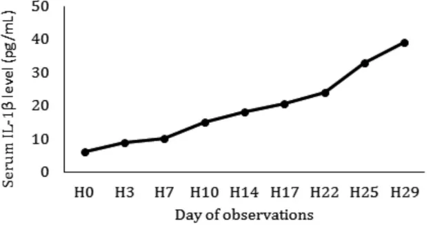
FIGURE 1. The Average Increased Level of IL-1β
The increased level of IL-1β occurred
since day 3 and continued to increase until
day 29. The increased level of IL-1β between
baseline and day 29, had a statistically
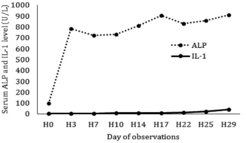
FIGURE 2. The Average Incerased Levels of ALP and IL-1
FIGURE 2 shows the high increased
levels of ALP on the third day after ligation
of the CBD, then luctuated until day 29.
FIGURE 3 shows a regular increased in GGT levels until day 29.
FIGURE 3. The Average Incerased Levels of GGT and IL-1β
Figure 4 shows the increased levels of
direct and total bilirubin until the 3rd day,
with a reduction on day 14, and rise again
until day 29.
DISCUSSION
At the initial examination of blood serum,
it was found two subjects had an increased
in white blood cell count (leukocytosis), but otherwise, they were clinically well. Clinically, we did not ind scleral icterus in the subjects. The evaluation began in day 3, we found increased serum level in almost all subjects.
20% of direct bilirubin was not increased, 10% of direct bilirubin had mild increased and 70% of direct bilirubin increased at
almost twice the normal value. However, we did not ind any signs of scleral icterus in all the subjects. It was found increased levels of alkaline phosphatase, GGT, leukocytes, and IL-1β that were quite high. The increased in leucocytes ranged between 17,100-59,800/uL, with the mean value of 29.620/uL, and 10% of the samples had no leukocytosis. There was a steep increased of the level og IL-1β (range from 0.897pg/dl to 9.274pg/dl). This showed that there were inlammatory or infection.
Biochemical markers of cholestasis are the presence of elevated levels of Alkaline
Phosphatase (ALP) and Gamma-Glutamyl
Transpeptidase (GGT). ALP and GGT is
a sensitive indicator of the occurrence of
cholestasis. ALP and GGT are the hepatocyte
membrane and released if there is a damage of
hepatocellular. In cholestasis, the synthesis of these enzymes is induced and made soluble. GGT is lifted because the leak out of the bile duct of the increased bile duct pressure. In the next stage of cholestasis; AST, ALT, and
bilirubin will increase because of liver damage
as a secondary effect of cholestasis.4-8
A study says that the level of ALP in a
dog is increased earlier than GGT, bilirubin, and clinical jaundice.16 High levels of ALP and GGT show the presence of biliary duct obstruction. High level of ALP (300-500U/L)
occurs in chronic liver disease and biliary
obstruction. Mild increased (<300U/L)
typically occurs in cirrhosis of the liver due
to viral, non-alcoholic fatty liver disease, cholestatic liver disease and hepatocellular
cancer. The level of ALP increased four
times higher than normal value in 1 or 2 days
after obstruction. The level of ALP can also
be increased in conditions apart from the
liver disorder and biliary obstruction; such
as malignancies (bronchogenic carcinoma,
Hodgkin’s lymphoma, renal cell carcinoma),
high fat diet, pregnancy, growing child and
chronic kidney disease.17
Cholestasis is associated with pro-inlammatory status, resulting from the portal and systemic endotoxemia. The concentration of endotoxin in the portal circulation increases, as a result of a lack of bile salts in the intestinal
lumen, with resulting in an imbalance of
microlora and increased the permeability
of the intestinal mucosal barrier, resulting in
bacterial translocation. Exposure endotoxemia
and bacterial translocation due to obstructive jaundice cause uncontrolled induction of
the inlammatory cascade. The damage
to the hepatocyte or cholangiocyte causes
activation and release of proinlammatory cytokines. The TNFα, IL-1 and IL-6 will
be produced by macrophages, endothelial cells, and Kupffer cells are still good that the
response of the systems of defense TNFα, IL-1) and IL-6 are produced by macrophages,
endothelial cells, and Kupffer cells which
are the response of the body immune system. Animal experiments have shown increased concentrations of proinlammatory cytokines such as TNFα, IL-1, IL-6 (IL-8), and IL-10. Cytokine concentration increased contributes to increased risk of complications. This increased levels of cytokine is a manifestation of an inlammatory process, which induces SIRS and sepsis.3,9-14 A study on the animal
model have shown increased concentrations
an increased incidence of complications. Increased level of this cytokine is a manifestation of an inlammatory process. The local Increased level of cytokines will cause the cardinal signs of inlammation,
such as heat (calor), swelling (tumor), redness (rubor), pain (dolor) and loss of function
(functio laesa). While a high level of cytokines contribute to SIRS and sepsis.3,9-14
It was found 2 dogs (2%) having anemia, with the hemoglobin values of 5.7g/dL and 3.6 g/dl on the day 7. The cause remained undetermined 100% leukocytosis obtained between 17,100-59,800/uL. There was a sharp increase of IL-1 level. Increased level of AST occurred in 90% of subjects as well as increased level of ALT occurred in 80% of subjects. Increased levels of SGOT and SGPT indicate the beginning of declining of liver function. Increased level of direct bilirubin occurred in 4 (40%) of the subjects. This suggests that an increased level of direct
bilirubin in obstruction, can last more than 7
days.18
On day 14, after blood sampling, 3 subjects found dead. Most likely it was due to the infection or sepsis. We found the hemoglobin level range from 4.3 to 4.6g/dl, the number of leukocyte 38,100-62,000u/L, ALP 623-1.233 in the dead subjects. Sepsis occurred with the level of SGOT/PT of 123-699U/L. It was found scleral icterus in the 3 dead subjects. There were no signifcant changes in valuation day 17-22. We obtained an increased level of ALP with the lowest level of 432U/L with the highest level of 1.676U/L on day 29. The highest level of direct bilirubin was 1.02mg/ dl. Until day 29, we did not ind clinically jaundice in the rest of the subjects. All the
subjects remain active and energetic without
showing signs of sepsis.
The occurrence of a strong and signiicant correlation between the increase in GGT and ALP with IL-1β, suggesting that the increased
levels of Gamma-glutamyl Transpeptidase and Alkaline Phosphatase will be followed by the occurrence of an inlammatory process. While the weak correlation between direct bilirubin and interleukin-1 β (IL-1β), indicating the
need of direct bilirubin to increase in order
to induce an inlammatory process. The inlammatory process will occur when there is a high enough level of direct bilirubin. In this study, inlammatory process was marked by a sharp elevated in the level of IL-1β and an increased white blood count (leukocytes) on the 3rd day after ligation of the CBD.
CONCLUSIONS
Increased levels of ALP and GGT are sensitive indicators of cholestasis. Increased levels of ALP and GGT had a strong positive and signiicant correlation to increased level of IL-1β as a pro-inlammatory cytokine. Increased level of GGT had the most powerful and signiicant positive correlation to increased level of IL-1β, compared to increased level f ALP. an increase ALP levels. Whereas,
increased levels of total and direct bilirubin had
a low and insigniicant correlation to increased level of IL-1β. Inlammatory process occurred on the third day after ligation of the CBD, so
that in the case of cholestasis or obstructive
jaundice, the risk of infection, SIRS, and
sepsis are considered very high, so it should be considered to perform a temporary surgical
repair, prior to deinitive or curative surgical management.
apprehension, encouragement, patient, and
digestive trainees for sharing their knowledge
and idea in helping the researchers in the
construction of the research.
REFERENCES
1. Aify M, Samy N, El Maksoud NA, Ragab HM, Yehia A. Biochemical alterations in
malignant obstructive jaundice: effect of
pre-operative drainage. New York Science Journal 2010; 3(2):80-9.
2. Sewnath ME, van der Poll T, van Noorden CJ, ten Kate FJ, Gouma DJ. Cholestatic interleukin-6–deicient mice succumb to endotoxin-induced liver injury and pulmonary inlammation, Am J Respir Crit Care Med 2004; 169(3):413-20.
3. Arrese M, Trauner M. Molecular aspects of bile formation and cholestasis. Trends Mol Med 2003; 9(12):558-64.
4. Anonim. EASL clinical practice guidelines:
management of cholestatic liver diseases, J
Hepatol 2009; 51(2):237-67. http://dx.doi. org/10.1016/j.jhep.2009.04.009.
5. Zajic S, Damnjanovic Z, Stojanovic M, Visnjic M, Dencic S, Ilic D, et al. Biochemical
markers in patients with extrahepatic cholestasis, Acta Medica Medianae 2008; 47(1):5-12.
6. Rege RV. Adverse effects of biliary
obstruction: implications for treatment of
patients with obstructive jaundice. AJR Am J Roentgenol 1995; 164(2):287-93.
7. Soares AF, Castro e Silva Jr O, Ceneviva R, Roselino JE, Zucoloto S. Biochemical
and morphological changes in the liver after hepatic artery ligation in the presence or
absence of extrahepatic cholestasis. Int J Exp Path 1993; 74(4):367-70.
8. Kinugasa T, Uchida K, Kadowaki M, Takase H, Nomura Y, Saito Y. Effect of bile duct ligation on bile acid metabolism in rats. J Lipid Res 1981; 22(2):201-7.
9. F Charles Brunicardi, MD, FACS, Schwartz’s, Principles of Surgery, Ninth Edition, 9th Eds,
2010.
10. Hirschield GM, Heathcote EJ, Gershwin ME. Pathogenesis of cholestatic liver disease and therapeutic approaches. Gastroenterology
2010; 139:1481-96. http://dx.doi.org/
10.1053/j.gastro.2010.09.004.
11. Chand N, Sanyal AJ. Sepsis-induced cholestasis. Hepatology 2007; 45(1):230-41.
12. Alpini G, Ueno Y, Tadlock L, Glaser SS, LeSage G, Francis H, et al. Increased
susceptibility of cholangiocytes to tumor
necrosis factor-α cytotoxicity after bile duct ligation. Am J Physiol Cell Physiol 2003; 285(1):C183-94.
13. Geier A1, Dietrich CG, Voigt S, Kim SK, Gerloff T, Kullak-Ublick GA, et al. Effects
of proinlammatory cytokines on ratorganic anion transporters during toxic liver injury and cholestasis. Hepatology 2003; 38(2):345-54.
14. Van der Gaag NA, Kloek JJ, De Castro SM, Busch OR, Van Gulik TM, Gouma DJ.
Preoperative biliary drainage in patients with obstructive jaundice: history and current
status. J Gastrointest Surg 2009; 13(4):814-20. http://dx.doi.org/ 10.1007/s11605-008-0618-4.
15. Bhandari M, Toouli J. Preoperative biliary
drainage (stenting) for treatment of obstructive
Jaundice. HPB (Oxford) 2006; 8(5):343-5. http:// 10.1080/13651820600804328.
16. Cornell University; Alkaline phosphatase (AP, ALP, SAP); from:http://ahdc.vet.cornell. edu/clinpath/modules/chem/alkphos.htm
17. Shaffer E. Laboratory test of the liver and gallbladder; The Merck Manual; last revision on Juni 2009.
18. Wikipedia; Alkaline Fosfatase; free
encyclopedia: from: http://en.wikipedia.org /