Regulation of proline accumulation in detached rice leaves
exposed to excess copper
Chien Teh Chen, Li-Men Chen, Chuan Chi Lin, Ching Huei Kao *
Department of Agronomy,National Taiwan Uni6ersity,Taipei,Taiwan,ROC Received 24 April 2000; received in revised form 18 July 2000; accepted 15 September 2000
Abstract
Accumulation of proline in response to excess Cu was studied in detached leaves of rice (Oryza sati6a). CuSO4was effective
in inducing proline accumulation in detached rice leaves under both light and dark conditions. CuSO4and CuCl2were equally
effective in inducing proline accumulation, indicating that proline accumulation is induced by Cu. Sulfate salts of Mg, Mn, and Fe were ineffective in inducing proline accumulation in detached rice leaves. Excess Cu had no effect on relative water content of detached rice leaves, suggesting that Cu-induced proline accumulation is unlikely due to water deficit. Proline accumulation induced by excess Cu was related to proteolysis and an increase in D1-pyrroline-5-carboxylate reductase or ornithine-d
-amino-transferase activity and could not be explained by proline utilization or stress-induced modifications in proline dehydrogenase or
D1-pyrroline-5-carboxylate dehydrogenase. The content of glutamic acid decreased by excess Cu. The increase in arginine but not
ornithine was found to be associated with the increase in proline content in Cu-stressed detached rice leaves. CuSO4treatment
resulted in an increase in abscisic acid content in detached rice leaves. The possibility that proline accumulation induced by excess Cu is mediated through abscisic acid is discussed. © 2001 Elsevier Science Ireland Ltd. All rights reserved.
Keywords:Abscisic acid; Copper;Oryza sati6a; Proline accumulation
www.elsevier.com/locate/plantsci
1. Introduction
Plants are exposed to various types of environ-mental stress. Among these stresses, osmotic stress, in particular that due to drought and salin-ity, is the most serious problem that limits plant growth and crop productivity in agriculture [1]. Proline accumulation in plant cells exposed to salt or water stress is a widespread phenomenon and is often considered to be involved in stress resistance
mechanisms, although its precise role continues to be controversial [2 – 4].
Copper is an essential micronutrient for plants. Excess Cu induces a wide range of biochemical effects and physiological processes, such as photo-synthesis, pigment photo-synthesis, protein metabolism, and membrane integrity [5]. Based on the assump-tion that excess Cu can operate as one stress factor and may deteriorate the plant water balance [6], it is logical to expect that excess Cu may cause proline accumulation in plant tissues. In fact, ex-cess Cu-induced accumulation of proline has been reported in wheat seedlings [7], Lemna minor [8],
Silene 6ulgaris [9],Anacystis [10] and Chlorella sp. [11,12].
Proline accumulation in plant tissues has been suggested to result from (a) a decrease in proline degradation; (b) an increase in proline biosynthe-Abbre6iations:ABA, abscisic acid; ELISA, enzyme-linked
immuno-sorbent assay; FW, fresh weight; GABA,g-aminobutyric acid; OAT, ornithine-d-aminotransferase; P5C, D1-pyrroline-5-carboxylate;
P5CDH,D1-pyrroline-5-carboxylate dehydrogenase; P5CR,D1
-pyrro-line-5-carboxylate reductase; PDH, proline dehydrogenase; RWC, relative water content.
* Corresponding author. Tel.: +886-2-23698159; fax: + 886-2-23620879.
E-mail address:[email protected] (C.H. Kao).
sis; (c) a decrease in protein synthesis or proline utilization; and (d) hydrolysis of proteins [13]. In plants, proline is synthesized from glutamic acid via D1-pyrroline-5-carboxylate (P5C) by two en-zymes, P5C synthetase and P5C reductase (P5CR). It has been shown from labelling experiments that ornithine can also serve as a precursor to proline biosynthesis in higher plants [14 – 16]. The isola-tion of cDNAs encoding ornithine-d -aminotrans-ferase (OAT) in higher plants [17,18] suggests that the formation of P5C from ornithine. Arginine can also contribute to proline biosynthesis, and the pathway from arginine proceeds via ornithine as a result of catalytic activity of arginase [14,19]. Pro-line is metabolized to glutamic acid via P5C by two enzymes, proline dehydrogenase (PDH) and P5C dehydrogenase (P5CDH) [3]. Studying the effect of excess Cu on enzyme activities involved in proline biosynthesis and degradation could provide valuable information on the physiological significance of its accumulation. However, to our knowledge, no such study has been undertaken. Neither do we know whether three amino acids (glutamic acid, ornithine and arginine) involved in the proline biosynthetic pathways are limiting fac-tors for proline accumulation induced by excess Cu. In this paper, we shall examine the regulation of proline accumulation in detached rice leaves exposed to excess Cu.
2. Materials and methods
2.1. Plant material
Rice (Oryza sati6a cv. Taichung Native 1) was cultured as previously described [20]. The apical 3-cm segments excised from the third leaves of 12-day-old seedlings were used. Briefly, rice seedlings were planted on a stainless net floating on half-strength Johnson’s modified nutrient solu-tion [21] in a 500 ml beaker. The nutrient solusolu-tion (pH 4.8) was replaced every 3 days. Rice plants were grown for 12 days in a greenhouse, where natural light was provided and the temperature was controlled at 30°C during the day and at 25°C at night. The apical 3 cm of the third leaf was used for the experiment. A group of ten segments was floated in a Petri dish containing 10 ml of test solution. Incubation was carried out at 27°C in the light (40 mmol m−2 s−1) or in the dark.
2.2. Determinations of proline, other amino acids,
protein and chlorophyll
Proline was extracted and its concentration de-termined by the method of Bates et al. [22]. Leaf segments were homogenized with 3% sulfosalicylic acid and the homogenate was centrifuged at 3000×g for 20 min. The supernatant was treated with acetic acid and acid ninhydrin, boiled for 1 h, and then absorbance at 520 nm was determined. Contents of proline are expressed as mmol g−1 FW. For determination of glutamic acid, glu-tamine, arginine, ornithine, g-aminobutyric acid (GABA), and total amino acids, leaf samples were extracted with 2% sulfosalicylic acid and the ho-mogenate was centrifuged at 15 000×g for 20 min. The supernatant was used directly for amino acid analysis. Amino acid analysis was carried out by an amino acid analyzer (Beckman 6300, CA, USA). Proline content reported in Figs. 3 and 5 was also determined by the amino acid analyzer. For protein determination, leaf segments were ho-mogenized in 50 mM sodium phosphate buffer (pH 6.8). The extracts were centrifuged at 17 600×g for 20 min, and the supernatants were used for determination of protein by the method of Bradford [23]. Chlorophyll was determined ac-cording to Wintermans and De Mots [24] after extraction in 96% (v/v) ethanol.
2.3. RWC and Cu measurements
RWC, defined as water content of leaf tissue as a percentage that of the fully turgid tissue, was determined by the method of Weatherly [25]. For determination of Cu, leaf segments were dried at 65°C for 48 h. Dried material was ashed at 550°C for 20 h. Ash residue was incubated with 31% HNO3 and 17.5% H2O2 at 72°C for 2 h, and dissolved in a mixture of 70% HNO3 and 60% HClO4 (9:1). Cu was then quantified using an atomic absorption spectrophotometer (Model AA-6800, Shimadzu, Kyoto).
2.4. ABA assay
in-terfere in the immunoassay, extracts were first passed through polyvinylprrrolidone column and C18 cartridges. The elutes were concentrated to dryness by vaccum-evaporation and resuspended in Tris-buffered saline before enzyme-linked im-munosorbent assay (ELISA). ABA was quanti-tated by ELISA [26]. ABA immunoassay detection kit (PGR-1) was purchased from Sigma Chemical Co. (St Louis, MO, USA), is specific for (+ )-ABA.
2.5. Enzyme assay
For enzyme extraction, leaf samples were ho-mogenized in prechilled mortar and pestle with extraction medium at 4°C. The extraction medium contained 100 mM potassium phosphate buffer (pH 7.4), 1 mM pyridoxal 5-phosphate, 1 mM
EDTA, 10 mM mercaptoethanol, 1%
polyvinylpyrrolidone, 5 mM MgCl2, and 0.6 M
KCl. The homogenate was centrifuged at
12 000×g for 20 min at 4°C. The resulting clean supernatant fractions were desalted by Sephadex G-25 column before the enzyme assay.
P5CR was assayed by a NADH dependent P5CR reaction [27]. The assay mixture contained 0.4 mM NADH, 1.5 mM P5C, 50 mM potassium phosphate buffer, 0.8 mM dithiothreitol, and the enzyme extract. The reaction was started by the addition of P5C and the decrease in absorbance was followed at 340 nm. One unit of P5CR was defined as 1 nmol NADH oxidized h−1. OAT activity was assayed according to Vogel and Ko-pac [28]. In the final volume of 1 ml, the reaction medium contained: 50 mM ornithine, 20 mM a -ketoglutarate, 1 mM pridoxal 5-phosphate, 100 mM potassium phosphate buffer, and the enzyme extract, the final pH was 8.0. The reaction medium was incubated at 37°C for 30 min. The reaction was stopped by adding 0.5 ml trichloroacetic acid (10%) and the color was developed by incubatng the reaction mixture with 0.5 mlo -aminobenzalde-hyde (0.5%) in ethanol (95%) for 1 h. After cen-trifugation at 12 000×g for 10 min, the clear supernatant fraction was taken to measure the absorbance at 440 nm. Enzyme activity was calcu-lated using the extinction coefficient (2.68 mM−1 cm−1 at 440 nm). The activity was expressed in units, where one unit of OAT was defined as 1 nmol P5C produced h−1. PDH was assayed by following the NAD reduction at 340 nm in a 100
mM Na2CO3-NaHCO3 buffer (pH 10.3) contain-ing 50 mM proline, 0.4 mM NAD, and the en-zyme extract [29]. One unit of PDH was defined as 1 nmol NAD reduced h−1. P5CDH was assayed by monitoring the production of NADH at 340 nm. The reaction mixture contained 50 mM potas-sium phosphate buffer (pH 8.0), 4 mM NAD, 1.5 mM P5C, and the enzyme extract [30]. One unit of P5CDH was defined as 1 nmol NAD reduced h−1.
2.6. Determination of proline utilization
For proline utilization, detached rice leaves were pretreated with 50 mM ornithine for 3 h (since addition of ornithine has been observed to be more effective than that of glutamic acid or arginine in increasing proline content in rice leaves [31]) to increase the endogenous proline content and then transferred to distilled water and 10 mM CuSO4 for 12 h in the light. Proline content was then determined. The decline in proline content will be considered as that proline is utilized [31].
3. Results
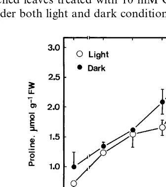
Proline content in detached rice leaves increased with the increase of CuSO4concentration (Fig. 1). Proline content increased about 2.5-fold in de-tached leaves treated with 10 mM CuSO4for 24 h under both light and dark conditions. It is obvious
Fig. 1. Effect of CuSO4 on proline content in detached rice
leaves under light and dark conditions. Detached rice leaves were incubated in solutions containing 0 – 10 mM CuSO4.
Fig. 2. Effect of various divalent metals on proline contents in detached rice leaves. Proline was determined 24 h after treat-ment of metals (10 mM) in the light. Vertical bars represent standard errors (n=4).
and control leaves. Unexpectedly, ABA content in CuSO4-treated rice leaves was observed to be much higher than that in water-treated controls throughout the entire duration of incubation.
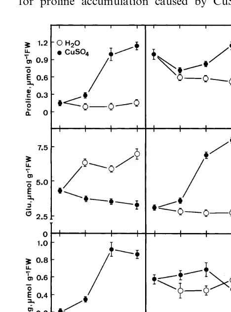
The results of Fig. 5 show that protein and chlorophyll a contents decreased and total amino acids increased with increasing duration of CuSO4 stress. Endogenous arginine, glutamine and GABA contents in detached rice leaves treated with CuSO4 were higher than those treated with water (Fig. 3). However, the content of glutamic acid in CuSO4-treated detached rice leaves was much lower than that in water-treated detached rice leaves in 12 h of incubation (Fig. 3). There was no clear differences in ornithine content be-tween CuSO4- and water-treated detached rice leaves in 12 h of incubation (Fig. 3).
To determine the role of biosynthetic pathways for proline accumulation caused by CuSO4, the
that CuSO4 in the light and CuSO4 in the dark were equally effective in inducing proline accumu-lation in detached rice leaves.
Fig. 2 shows the effect of various divalent metals on the proline content in detached rice leaves. It is clear that sulfate salt of Mg, Fe and Mn at 10 mM did not affect proline content in detached rice leaves. Fig. 2 also shows that CuSO4 and CuCl2 were equally effective in inducing pro-line accumulation. Chloride salt of Cd, Co, Zn, Ni and Al was also effective in increasing proline content in detached rice leaves (data not shown). Proline content in control leaves remained al-most unchanged during the first 12 h of incubation in the light (Fig. 3). It is clear from Fig. 3 that accumulation of proline induced by CuSO4 was evident at 4 h after treatment.
To be sure that the described proline accumula-tion was related to an increase in the leaf Cu content, Cu concentrations were determined in detached rice leaves treated with either water or 10 mM CuSO4 (Fig. 4). Cu content in control leaves remained unchanged during the first 12 h in the light. However, Cu content in CuSO4-treated de-tached rice leaves increased with increasing dura-tion of incubadura-tion. It is obvious that the increase in Cu content in CuSO4-treated detached rice leaves was evident 4 h after treatment. The effect of CuSO4on RWC and ABA content in detached rice leaves is shown in Fig. 4. No differences in RWC were observed between Cu-treated leaves
Fig. 3. Time course of the CuSO4effect on proline, glutamic
acid (Glu), glutamine (Gln), g-aminobutyric acid (GABA), arginine (Arg) and ornithine (Orn) contents in detached rice leaves. Detached rice leaves were incubated in water or 10 mM CuSO4 in the light. Amino acids were determined by
Fig. 4. Time course of the CuSO4 effect on Cu content,
relative water content (RWC), and ABA content in detached rice leaves. Detached rice leaves were incubated in water or 10 mM CuSO4 in the light. Vertical bars represent standard
errors (n=4).
4. Discussion
The present investigation shows that excess CuSO4 induces the accumulation of proline in detached rice leaves. This result is in agreement with those of other investigators using different plants as experimental materials [7 – 12]. The fact that CuSO4 and CuCl2 were equally effective in
Fig. 5. Time course of the CuSO4 effect on the contents of
chlorophyll, protein and total amino acids in detached rice leaves. Detached rice leaves were incubated in water or 10 mM CuSO4 in the light. Total amino acid contents were
determined by amino acid analyzer. Vertical bars represent standard errors (n=4).
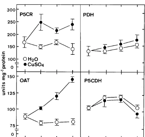
Fig. 6. Time course of the CuSO4effect on the activities of
P5CR, OAT, PDH and P5CDH in detached rice leaves. Detached rice leaves were incubated in water or 10 mM CuSO4 in the light. Vertical bars represent standard errors
(n=4).
such a difference except that it is possible that plant species (O.sati6a) we used in our experiment may somewhat have contributed to different re-sults. We also observed that promotion of senes-cence of detached rice leaves caused by CuSO4was not light dependent and that CuSO4-promoted senescence of detached rice leaves is linked to lipid peroxidation [38].
Exposure to heavy metals including Cu is known to deteriorate the plant water balance [6,39 – 41]. It has been suggested that proline accu-mulation induced by metals such as Cu, Cd and Zn depends on the development of a metal-in-duced water stress in leaves [9,40]. Since Cu had no effect on RWC of detached rice leaves (Fig. 4), it is unlikely that Cu-induced proline accumula-tion is attributed to the development of water stress. Kastori et al. [41] also observed that proline accumulation in Cu-exposed leaf discs and argued that this was due to Cu uptake per se, rather than to water stress.
The decrease in protein content was faster in CuSO4-stressed detached rice leaves than water-treated detached rice leaves (Fig. 5). Therefore, protein degradation might contribute to the CuSO4-induced proline accumulation in detached rice leaves. This conclusion is supported further by the observation that total amino acids are higher in detached rice leaves exposed to CuSO4 than in those treated with water (Fig. 5).
Proline is known to be synthesized from glu-tamic acid via P5C by two enzymes, P5C syn-thetase and P5CR [3,4]. In the present study, P5CR was found to be higher in CuSO4-treated detached rice leaves than in water-treated detached rice leaves (Fig. 6), suggesting that glutamic acid is converted to proline in rice leaves exposed to excess CuSO4. It was further supported by the decreased or low content of glutamic acid in CuSO4-stressed detached rice leaves (Fig. 3). Since GABA contents in CuSO4-treated detached rice leaves was observed to be higher than those in water-treated detached rice leaves, therefore the decreased content of glutamic acid also resulted from glutamic acid being metabolized to GABA [42]. The increase in glutamine content and the decrease in glutamic acid in CuSO4-treated de-tached rice leaves also suggest that CuSO4inhibits the conversion of glutamine to glutamic acid, a step catalyzed by glutamic acid synthase [42]. CuSO4 could also have an effect on the bifunc-inducing proline accumulation indicates that
pro-line accumulation induced by Cu is rather than by SO42− or Cl− (Fig. 2). Our results also show that proline content in CuSO4-treated detached rice leaves increased progressively with an increase in duration of the incubation time (Fig. 3) as well as in a concentration-dependent manner (Fig. 1).
There are many reports indicate that light pro-motes stress- and Cd-induced proline accumula-tion in plants [33 – 37]. Recently, Wu et al. [10] also demonstrated that Cu- or Cd-treated Chlorella sp. accumulated more proline under light condition than dark condition. However, our results indicate that there is no requirement for light in the accu-mulation of proline as a response to excess Cu (Fig. 1). We do not have a good explanation for
Table 1
Proline content in ornithine-pretreated detached rice leaves incubated in water and CuSO4a
Treatment Proline,mmol g−1FW
Ornithine, 3 h 25.490.5
Ornithine, 3 hH2O, 12 h 7.190.9
Ornithine, 3 hCuSO4, 12 h 7.990.7
aDetached rice leaves were pretreated with 50 mM
tional enzyme P5C synthetase involved in the syn-thesis of P5C from glutamic acid [4]. However, for unknown reasons, we were unable to detect any P5C synthetase activity in crude extracts. There-fore, this enzyme was not considered in the present work.
OAT catalyzes the first step in the pathway of conversion of ornithine to proline [4]. In the present investigation, OAT activity was found to increase significantly in detached rice leaves ex-posed to excess Cu (Fig. 6). The increase in OAT activity may have, to some extent, contributed to the elevated content of proline. Increase in OAT activity along with an increase in the content of proline has also reported in wheat under cold stress [13], and in Brassica juncea under salt stress [28]. Both ornithine and arginine can contribute to proline biosynthesis. It is interesting to note that the increase in arginine, but not ornithine content was found to be associated with the increase in proline content in CuSO4-stressed detached rice leaves (Fig. 3).
The enzymes, PDH and P5CDH, are reported to catalyze proline oxidation [3,4]. In the present investigation, no effect of CuSO4 has been ob-served on PDH and P5CDH activities in detached rice leaves (Fig. 6). These results suggest that proline oxidation (or degradation) contribute lit-tle, if any, to proline accumulation in detached rice leaves under CuSO4 stress condition.
Of particular interest is the finding that ABA accumulates in detached rice leaves exposed to excess Cu (Fig. 4). Since CuSO4 had no effect on RWC of detached rice leaves (Fig. 4), the accumu-lation of ABA caused by CuSO4is unlikely due to the development of water stress. Proline accumula-tion in detached rice leaves can be induced by ABA treatment [32]. It is not known whether Cu-induced proline accumulation in detached rice leaves is mediated through accumulation of ABA. Previously, we have demonstrated that ABA-in-duced proline accumulation was not due to less utilization of proline in detached rice leaves [32]. In the present investigation, we also observed that proline accumulation in detached rice leaves ex-posed to CuSO4was not caused by less utilization of proline (Table 1). However, further investiga-tion is required to establish the cause and effect link between ABA and proline accumulations in detached rice leaves exposed to CuSO4 stress.
Acknowledgements
This work was supported by the National Sci-ence Council of the Republic of China (NSC 88-2313-B-002-066).
References
[1] J.S. Boyer, Plant productivity and environment, Science 218 (1982) 443 – 448.
[2] D. Aspinall, L.G. Paleg, Proline accumulation: physio-logical aspects, in: L.G. Paleg, D. Aspinall (Eds.), The Physiology and Biochemistry of Drought Resistance in Plants, Academic Press, Sydney, 1981, pp. 205 – 241. [3] Y. Yoshiba, T. Kiyosue, K. Nakashima, K.
Yamaguchi-Shinozaki, K. Yamaguchi-Shinozaki, Regulation of levels of proline as an osmolyte in plants under water stress, Plant Cell Physiol. 38 (1997) 1095 – 1102.
[4] P.D. Hare, W.A. Cress, J. van Staden, Proline synthesis and degradation: a model system for elucidating stress-related signal transduction, J. Exp. Bot. 50 (1999) 413 – 434.
[5] J.C. Fernandes, F.S. Henriques, Biochemical, physiolog-ical and structural effect of excess copper in plants, Bot. Rev. 57 (1991) 246 – 273.
[6] J. Barcelo, V. Poschenrieder, Plant water relations as affected by heavy metal stress: a review, J. Plant Nutr. 13 (1990) 1 – 37.
[7] R. Bassi, S.S. Sharma, Proline accumulation in wheat seedlings exposed to zinc and copper, Phytochemistry 33 (1993) 1339 – 1342.
[8] R. Bassi, S.S. Sharma, Changes in proline content ac-companying the uptake of zinc and copper by Lemna minor, Ann. Bot 72 (1993) 151 – 154.
[9] H. Schat, S.S. Sharma, R. Vooijs, Heavy metal-induced accumulation of free proline in a metal-tolerant and a nontolerant ecotype of Silene 6ulgaris, Physiol. Plant.
101 (1997) 477 – 482.
[10] J.-T. Wu, S.-C. Chang, K.-S. Chen, Enhancement of intracellular proline level in cells of Anacystis nidulans
(Cyanobacteria) exposed to deleterious concentration of copper, J. Phycol. 31 (1995) 376 – 379.
[11] J.-T. Wu, M.-T. Hsieh, L.-C. Know, Role of proline accumulation in response to toxic copper inChlorellasp. (chlorophyceae) cells, J. Phycol. 34 (1998) 113 – 117. [12] J.-T. Wu, S.-J. Chang, T.-L. Chou, Intracellular proline
accumulation in some algae exposed to copper and cadmium, Bot. Bull. Acad. Sin. 36 (1995) 89 – 93. [13] C. Charest, C.T. Phan, Cold acclimation of wheat
(Triticum aest6um): properties of enzymes involved in
proline metabolism, Physiol. Plant. 80 (1990) 159 – 168. [14] D.H. Brown, L. Fowden, Metabolism of d
-acetylor-nithine in two Leguminous species, Phytochemistry 5 (1966) 887 – 892.
[16] R.G. Coleman, M.P. Hegarty, Metabolism of D, L-or-nithine-2 14C in normal and potassium-deficient barley,
Nature 179 (1957) 376 – 377.
[17] A.J. Delauney, C.A.A. Hu, P.B. Kavi Kishor, D.P.S. Verma, Cloning of ornithine d-aminotransferase cDNA fromVigna aconitifoliaby trans-complementation inEs
-cherichia coli and regulation of proline biosynthesis, J. Biol. Chem. 268 (1993) 18673 – 18678.
[18] N.H.C.J. Roosens, T.T. Thu, H.M. Iskandar, M. Ja-cobs, Isolation of the ornithine-d-aminotransferase cDNA and effect of salt stress on its expression in
Arabidopsis thaliana, Plant Physiol. 117 (1998) 263 – 271. [19] E.M. Lingnowski, W.E. Splittstoesser, The change in arginine levels and the metabolism of urea and ornithine in Cucubita moschatta seedlings, Physiol. Plant. 25 (1971) 225 – 229.
[20] J.-N. Lin, J.-W. Wang, C.H. Kao, Effect of abscisic acid and water stress on the senescence of detached rice leaves, Biol. Plant. 42 (1999) 313 – 316.
[21] C.M. Johnson, P.R. Stout, T.C. Broyer, A.B. Carlton, Comparative choline requirements of different plant spe-cies, Plant Soil 8 (1957) 337 – 353.
[22] L.S. Bates, S.P. Waldren, I.D. Teare, Rapid determina-tion of free proline for water-stress studies, Plant Soil 39 (1973) 205 – 207.
[23] M.M. Bradford, A rapid and sensitive method for the quantitation of microgram quantities for protein utiliz-ing the principles of protein-dye bindutiliz-ing, Anal. Biochem. 72 (1976) 248 – 254.
[24] J.F.G.M. Wintermans, A. De Mots, Spectrophotometric characteristics of chlorophyll a and b and their pheophytins in ethanol, Biochem. Biophys. Acta 109 (1965) 448 – 453.
[25] P.F. Weatherly, Studies in the water relation of cotton plant. I. The field measurement of water deficits in the leaves, New Phytol. 49 (1950) 81 – 97.
[26] M. Walker-Simmons, ABA levels and sensitivity in de-veloping wheat embryos of sprouting resistant and sus-ceptible cultivars, Plant Physiol. 84 (1987) 61 – 66. [27] S. Madan, H.S. Nainawatee, R.K. Jain, J.B.
Chowd-hury, Proline and proline metabolising enzymes in in-vitro selected NaCl-tolerantBrassia junceaL. under salt stress, Ann. Tot. 76 (1995) 51 – 57.
[28] R.H. Vogel, M.J. Kopac, Some properties of
ornithine-d-transaminase from Neurospore, Biochem. Biophys. Acta 37 (1960) 539 – 540.
[29] A.B. Rena, W.E. Splittstoesser, Proline dehydrogenase and pyrroline-5-carboxylate reductase from pumpkin cotyledons, Phytochemistry 14 (1975) 657 – 661.
[30] S.F. Boggess, L.G. Paleg, D. Aspinall, D1
-Pyrroline-5-carboxylic acid dehydrogenase in barley, a proline-accu-mulating species, Plant Physiol. 56 (1975) 259 – 262. [31] C.-W. Yang, C.-C. Lin, C.H. Kao, Endogenous
or-nithine and arginine contents and dark-induced proline accumulation in detached rice leaves, J. Plant Physiol. 155 (1999) 665 – 668.
[32] C.-W. Yang, J.W. Wang, C.H. Kao, The relation be-tween accumulation of abscisic acid and proline in de-tached rice leaves, Biol. Plant. 43 (2000) 301 – 314. [33] S. Arora, P. Pardha Saradhi, Light-induced
enhance-ment in proline levels in Vigna radiataexposed to envi-ronmental stress, Aust. J. Plant Physiol. 22 (1995) 383 – 386.
[34] A.D. Hanson, R.E. Tully, Light stimulation of proline synthesis in water-stressed barley leaves, Planta 145 (1979) 45 – 51.
[35] P.S. Joyce, L.G. Paleg, D. Aspinall, The requirement for low-intensity light in the accumulation of proline as a response to water deficit, J. Exp. Bot. 35 (1984) 209 – 218.
[36] C.R. Stewart, Role of carbohydrate in proline accumula-tion in wilted barley leaves, Plant Physiol. 61 (1978) 775 – 778.
[37] J.H. Venekamp, J.E.M. Lampe, J.T.M. Koot, Organic acids as sources of drought induced proline synthesis in field bean plants, Vicia faba L, J. Plant Physiol. 133 (1989) 654 – 659.
[38] L.-M. Chen, C.H. Kao, Effect of excess copper on rice leaves: evidence for involvement of lipid peroxidation, Bot. Bull. Acad. Sin. 40 (1999) 283 – 287.
[39] J.C. Barcelo, Poschenrieder, I. Andreu, B. Gunse, Cadmium induced decrease of water stress resistance in bush bean plants (Phaseolus6ulgarisL. cv. Conender).
I. Effect on water potential, relative water content and cell wall elasticity, J. Plant Physiol. 125 (1986) 17 – 25.
[40] G. Costa, J.-L. Morel, Water relations, gas exchange and amino acid content in Cd-treated lettuce, Plant Physiol. Biochem. 32 (1994) 561 – 570.
[41] R. Kastori, M. Petrovic, N. Petrovic, Effect of excess lead, cadmium, copper, and zinc of water relations in sunflower, J. Plant Nutr. 15 (1992) 2427 – 2439. [42] A.J. Ireland, P.J. Lea, The enzymes of glutamine,
gluta-mate, asparagine, and aspartate metabolism, in: B.K. Singh (Ed.), Plant Amino Acids, Mercel Dekker, New York, 1999, pp. 49 – 109.