Introduction
Fasciola spp. infection mainly by F. gigantica causing weight loss, lame growth and low endurance and damage the liver and bile as well (Mitchell, 2007). The prevalence of liver flukes by F. gigantica in Indonesia reached 90% (Abidin, 2002). With high prevalence of the disease, the loss in Indonesia reached $107 million/year (Spithill et al., 1999). In addition to material losses by zoonosis, an estimated 2.4 to 17 million people around the world suffer from fasciolosis including Asia (WHO, 2009).
According to Estuningsihet al.(2009), attempts to suppress economic loss and
morbidity due to worm infections only succeed through a control program that is based on early diagnosis. With early diagnosis, the presence of worms in the body which could cause pathological changes can be prevented. Diagnosis of fasciolosis generally done with conventional enforcement, which is based on the examination of worm eggs in feces. However, this method has the disadvantage of not being able to detect the infection F. gigantica during prepaten (8–10 weeks of infection), as well as the low sensitivity.
This shortage fosters the development of immune diagnosis methods of fasciolosis especiallyF. gigantica in order to get faster diagnosis results and more accurate among others by serological methods. Currently, serology inspection ofF. giganticais based on the detection of antibodies to the antigen contained in the liquid worm excretory secretory (ES). Anonymous (2011) reported that ES is a protective antigen that can trigger
Identification of Excretory Secretory (ES) Liquid Antigen Protein
Fasciola gigantica
with Polyethilen Glycol (PEG) Separation
Yendri Junaidi
1,*, Ima Malawati
1, Made Sriasih
21Postgraduate Program of the Faculty of Animal Science, Universitas Gadjah Mada,
Jalan Fauna No. 3 Bulaksumur, Yogyakarta 55281, Indonesia
2Laboratory of Microbiology and Biotechnology, Faculty of Animal Science,
Mataram University, Jalan Majapahit 62, Mataram, Nusa Tenggara Barat 83125, Indonesia
Abstract
Fasciola giganticadiagnosis usually performed by detection of worm eggs presence in the feces, but this conventional method has many disadvantages. Early diagnosis (early detection) cannot be performed in conventional methods because the worms in the host's body began to lay eggs at the age of 8–12 weeks of patency. The current detection method that is based on antibodyantigen reactions using excreted/secreted (ES) liquid by adult F. gigantica, is believed to be used for the early detection of fasciolosis. This study aimed to characterize the antigenic components ofF. giganticaextretory/secretory products that could be used as a vaccine candidate development for early fasciolosis diagnostics. ES products were separated by PEG4000 at various concentrations (8%, 16%, 24%), then precipitates (pellets) obtained were dialyzed and characterized using SDSPAGE and Western blotting. Results from SDS PAGE showed that there were 18 proteins bands with 7–70 kDa molecular weights. Western blotting on pellets derived from PEG separation at various concentrations affirmed that the proteins of 50, 25 and 20 kDa were antigenic at 8% PEG concentration, the 25 kDa and 50 kDa were antigenic at 16% PEG concentrations and the 25 kDa was antigenic at 25% PEG concentration.
Keywords: Antigenic, diagnostic,ES liquid,F. gigantica, fasciolosis, PEG4000
*Corresponding author:
Yendri Junaidi
Faculty of Animal Science, Universitas Gadjah Mada, Jalan Fauna No. 3 Bulaksumur, Yogyakarta 55281, Indonesia
a host immune response definitive. ES antigens have properties that could be recognized by the immune response system (Bird, 1991; Chowdhuly and Tada, 1994). Sahebabet al.
(1999) reported that the presence of liquid ES is an indication of alive and active worm infection. To produce a necessary protein, ES fluid separation process to other components that are excreted by the worm must be done. ES liquid protein separation could be done by using protein solvents like polyethilen glycol (PEG). Harris (1992) reported that PEG is a simple molecule with a linear or branched molecular structure. PEG with 700–900 molecular weights are semi solid and PEG with 900–1000 molecular weights or more are solid at room temperature. PEG with molecular weight <1000 are viscous, colorless liquid. While PEG with molecular weight> 1000 are candles and white solid form. The number of ethylene glycol determine molecular weight PEG produced. PEG is commonly used for trait selection agent resistance gene is a molecular weight PEG, 4000 and 6000 (Yuliana, 2010).
Xiet al.(2006) reported that the use of PEG has the advantage as a solvent because it can precipitate protein reagent of good enough for purification of proteins from a variety of sources. In addition, according to Dewiet al.(2012) protein precipitation with PEG does not affect the subsequent purification steps. Based on this study, experiment was conducted to detect the presence of proteins making up liquid separation ESF. giganticaresults with different concentrations of PEG, and selecting proteins with antigenic/immunogenic.
Materials and Methods
Isolation of fluid excretory secretory (ES)
Adult heartworms F. gigantica is collected and then put in a glass beaker containing 10 mL Phosphate Buffer Saline (PBS), and stand for 20 min. The regurgitant first discharged, then replaced with new PBS 10 mL, done three times. Adult worms were incubated for one night at 37°C. Fluid containing protein ES centrifuged 2,500 rpm at 4°C for 15 min and filtered with 0.22 lm
filter. Isolation of proteins were performed at 8%, 16%, and 24% PEG concentrations. Then 10 mL of fluid ES thawed in 5 mL falcon tube aside into another. PEG is added into the liquid gradually while shaked in the vortex. ES residual fluid is added until the volume reaches 10 mL or 800 mg/10 mL, slowly shaked in vortex for 30 min and centrifuged at 10,000 rpm in temperature of 40°C for 5 min. Pellet allowed to remain inside the tube (fraction I). The same process carried out three times for fractions II, and fractions III.
Dialysis of protein excretory secretory (ES)
Samples (supernatant 8%, 16%, 18% and pellets 8%, 6%, 24% and final supernatant) were dialyzed using PBS (87.2 g NaCl, Na2HPO412H2O 392.2 g, KH2PO452.0 g) and saline (174 g NaCl). Membrane tubing with a cut of 11,000 dalton prepared and dialyzed for 2–3 hours at 4°C for each sample.
Characterization of protein ES with SDS PAGE
Analysis of profile protein ES with Western blotting technique
Samples in the form of pellets, results of each fraction, were prepared. Once ready, SDSPAGE process began, followed by Western blotting results of SDSPAGE gel soaked in transfer buffer for 20 min. Then the nitrocellulose membrane (NC) was soaked in dH2O for 2 min. Membrane NC soaked back into the transfer buffer for 10 min. Western blotting stacking material on the tool with the sequence (filter paper, NC membranes, the results of SDSPAGE gel and the top of the filter paper). Power pac mounted on the machine Western blotting with a voltage of 5 V, 0:10 A, 1 W for 45 min. Removable tool and filter paper attached to the top of the gel removed. NC paper removed and transferred upwards board that has been coated with clear plastic. After the membrane cut to limit the formation of ribbon line desired. Blocking using TBS Tween been added with 5% skim milk and and stored overnight at 4°C. Samples were incubated using primary antibodies (1st Ab) in the form of skim milk with the addition of bovine serum positive and negative fasciolosis dilution of 1:200 and secondary antibody (2ndAb) prepared on skim milk with the addition of bovine IgGHRP dilution of 1:8000 and each incubated for 2 h. The last addition of the substrate, the detection and visualization of the results.
Results and Discussion
Profile protein of ES F. gigantica liquid results of the SDSPAGE
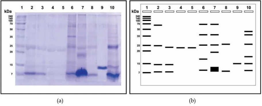
In this study, ES fluid isolated from wormsF. gigantica, then used as a candidate antigen for detecting fasciolosis on livestock, especially cattle. To get the protein constituent, the liquid is separated by using PEG4000 ES with several levels of concentration of 8% (fraction I), 16% (fraction II) and 24% (fraction III). ES result of mixing fluids with varying concentrations of PEG, showed no change in turbidity of the solution and the volume (fraction 1) 10 mL (fraction II) and 9.5 mL (fraction III) 8 mL. To know the profile protein molecules and molecular weight of each fraction of the process was then performed SDSPAGE. Garfin (2003) and Wibowo (2010) mentions that the SDSPAGE can be used to determine how the protein complex constituent purified sample in the sample. The results of the molecular characterization of proteins using acrylamide gel shown in Figure 1.
ES fluid protein profile of worm F. gigantica separation results with varying concentrations of PEG4000 (6%, 18% and 24%) gel electrophoresis using 10% in Figure 1 shows that the main constituent of protein molecular weight of each fraction ES fluid ranged between 7– 70 kDa. The results obtained from the count based on the value of RF
(a) (b)
(retardation factor). Results count of RF and BM Log in marker obtained linear regression line equation Y = 1,5248x + 1 9916 (R2 = 0.9964). Based on the results of a calculation and observation, band profiles of protein from the supernatant and pellet with a PEG concentration of 8%, 16%, and 24% varies from thick to thin. Lastutiet al., (2008) report that thin stained protein bands is a picture of many proteins and also due to genetic differences of protein obtained. Results of protein characterization using SDSPAGE in this study showed that there were 18 protein bands with molecular weight of 60.58, 58.71, 48.38, 40.59, 39.36, 27.20, 26.37, 25.57 , 24.80, 24.05, 19.99, 17.67, 13.39, 11.48, 10.79, 8.44, 8.18 and 7.93 kDa, with 7–70 kDa molecular weight range. This study was in line with Marcodo (1989), which states that there are 18 protein bands of liquid ESF. giganticawith the molecular weight range 14–96 kDa based on SDSPAGE. According Meshgiet al.(2007), the test protein characterization ES, there are three common molecular weight in the range of 24, 33 and 42 kDa, and in this study all three of these molecules are always visible, so it is said also in line with the results of the study (Mashagiet al., 2007).
Other researchers have reported differences in the number and size of the molecular weight of the liquid constituent ES. Kusnoto (2008) reported that based on the results of ES protein preparation techniques worm Fasciola spp. SDSPAGE and Western blotting were obtained a few specific proteins include 130, 108, 58, 45.40, 35, 26, 27, 25, 18, 15 and 8 kDa. Then Soulsby (1986) also added that the antigen F. giganticaof cows has 20 bands pattern with a molecular weight of 14–156 kDa. ElRahimy (2012) show that there were 13F. giganticaES liquid protein based on SDSPAGE with a molecular weight range of 9.1 to 35.7 kDa.
Differences SDSPAGE results are interpreted is influenced by several factors such as protein solvent used, topography where animals live, breed of cattle, the lifespan of worms that are used, as well as ribbons and long distance gel used. Kusnoto (2013) explains that the calculations using linear
regression probable cause relative differences and long distances protein bands and preliminary measurements of gel. Estuningsih and Widjajanti (1998) also mentions that at each level of different ages, wormsF. gigantica
also excrete proteins and different molecular weights. In the SDSPAGE result of this research, there are two dominant protein with an indication of the nature that is weighing 8 kDa and 25 kDa, because they always appear at each level of fractionation used, either in the pellet and supernatant.
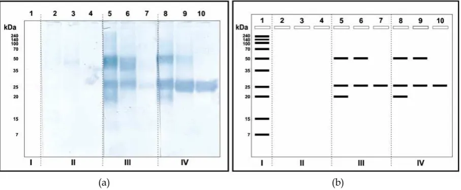
Profile protein antigenic worms F. gigantica result of western blotting
The process was done by comparing the Western bloting ES antigen in pellet results fractionation with antibodies from cows that positive (+) and negative () fasciolosis. Pellet used for Western blotting analysis was based on the results of SDSPAGE that the protein band pellet that looks more contrast and molecular weight of the protein is more diverse (Figure 2).
Visually lanes 2, 3 and 4 do not show the presence of protein bands due to the use of bovine serum negative () fasciolosis, with the sense that the antibodies and antigen binding does not occur. While the antigenic properties of the protein antigen ES treated with bovine serum positive (+) line 5 and 8 show three band of antigenic protein with a molecular weight of 50 kDa, 25 kDa and 20 kDa. Then line 6 and 9 show the antigenic protein band with a weight range of 50 kDa and 25 kDa. Line 7 as well as 10 showed antigenic protein band with a weight range of 25 kDa.
The overall data in Figure 2 shows it was found that one protein band weight range 25 kDa was always clearly visible on each lane both on the results of Western blotting and SDSPAGE, thus indicating that the separation with PEG does not affect the structure of antigenic proteins, it has proven to still recognized protein antigenic/immunogenic 25 kDa even pure on the concentration of PEG 24%.
the molecular weight antigenic ES liquid wormF. giganticaisolates is 7–25 kDa. Then according Farghally et al. (2009), a protein component of Fasciola with antigenic has a molecular weight of 29 kDa, followed by a 27 kDa and 25 kDa. Likewise, the results Mohamed et al. (2004) antigens that are immunoreactive against fasciolosis serum infected patients is 29 kDa, 25 kDa and 12 kDa. RiveraMarreroet al.(1988) also reported that the protein band with a weight of 25 kDa and 30 kDa of fluidF. giganticaES is a protein with specific properties for acute and chronic fasciolosis in rabbits, cows and sheep. Similar to the results of research Priago (2006) against worms F. gigantica and F. hepatica, the dominant protein profiles appear with a molecular weight of 29.3, 25, and 19 kDa.
However, some of the results reported by other researchers such as Estuningsih and Widjajanti (1998) reported that the antigenic properties of the fluid found in the ESweight proteins 46 kDa and 47 kDa. Then Naverrete
et al.(1993) reported that ESF. giganticaspecific molecule is a molecule with a weight of 23–37 kDa. In addition, the study SariMehmeoglu (2002) also explains the difference, which according to the research results showed that the infection fasciolosis three proteins with antigenic properties which is 36 kDa, 29 kDa and 17 kDa. The differences are likely
influenced by several things such as the influence of individual animals, feed and animal life as well as the geographic point solvent used to separate the protein components of ES. Xi et al. (2006) explains that the addition of PEG with various concentrations can purify many kinds of plant protein with a purity specification levels found in the concentration of PEG is higher (16% and 24%) based on the results of the analysis of 2D gel performed. Dewiet al.(2012) also confirmed that the precipitation of proteins with PEG does not affect the subsequent purification steps. The difference age of worms as research material also affects the pattern of antigenic protein bands (Estuningsih and Widjajati, 1998). MasComa et al. (2005) explains that the geographical location of a live worm can affect the antigen is excreted. Influence of host species against specific antigen immune response can vary (Sobhon
et al., 1996; Meshgi et al., 2008). Besides, Farghaly (2009) also showed that differences in the results of some research can be influenced by the method and course of study especially in making and sample preparation. Conclusions
The results of SDSPAGE for characterization of protein band profiles of constituent fluid ESF. gigantica showed 18
(a) (b)
protein band with a molecular weight ranging between 770 kDa. The Western blotting of proteins with weight range of 2050 kDa and one band with a weight of 25 kDa as an indication of the protein with antigenic properties were excellent. ES separation of proteins with PEG did not affect the subsequent purification step for the protein band with a weight of 25 kDa PEG is still seen at higher concentrations even more specific. References
Abidin, Z. (2002) Laporan Tahunan Dinas Peternakan Kabupaten Gowa. Dinas Peternakan Kabupaten Gowa. Jawa Barat. Anonymous. (2011) Pengembangan metode ELISA utuk mendeteksi keberadaan koproantigen Fasciola gigantica pada ruminansia: model uji diagnostik untuk
human fasciolosis. Bogor: Sekolah Pasca Sarjana IPB.
Astiti, L.G.S., Estuningsih, E., Hartiningrum, D.B., Muliyati, K., and Ichwan (2011) Pemberantasan dan pengendalian penularan fasciolosis pada sapi bali di Pulau Lombok. NTB: Balai Pengkajian Teknologi Pertanian Nusa Teggara Barat.
Chwohury, N., and Tada I. (1994) Helminthology. New Delhi: Springer VerlagNorasa Publising House. Dewi, N.C., Nurhidayati, T., Purwati, K.I.
(2012) Pengaruh penambahan PEG (Polyethylen Glicol) terhadap profil protein tembakau (Nicotiana tabacumL. var Prancak 95) pada media in vitro. Surabaya: FMIPA Institut Teknologi Sepuluh November.
ElRahimy, M. (2012) Evaluation of some
Fasciola giganticaantigens as vaccines against fasciolosis in goats. Giza: Department of Internal Medicine and Infectious Diseases, Faculty of Veterinary Medicine. Cairo University. Estuningsih, S.E., Spithill, T., Raadsma, H., Law, R., Adiwinata, G., Meusen, L., and Piedrafitall, D. 2009. Development and application of a fecal antigen diagnostic sandwich ELISA for estimating prevalence ofFasciola giganticain cattle
in Central Java, Indonesia. J. Parasitol., 95(2), 450–455.
Garfin, D.E. (2013) Gel electrophoresis of proteins. In: Davey J., and Lord, M. (Eds) Essential cell biology vol. 1, chapter 7: cell structure, a practical approach. Oxford: Oxford University Press.
Farghaly, A.M., Nada, S.M., Emam, W.A., Matter, M.A., Mohamed, S.M.A., Sharaf, E.M., and ElGamal, R.A. 2009. Role of FastELISA and Western Blot in diagnosis of human fasciolosis using
crude adult worm and
excretory/secretory Fasciola antigen. Int. J. Vet. Sci. Med., 1(2), 55–65. Guyton, A.C., and Hall, J.E. (2007) Fisiologi
kedokteran edisi 11 (Translation of the Textbook of Medical Physology 11th Edition). Jakarta: EGC.
Harris, M.J. (Ed.) (1992) Poly(ethylene glycol) chemistry: biotechnical and biomedical applications. New York: Plenum Press. Kim, K., Yang, H.J., and Chung, Y.B. 2003. Usefulness of 8 kDa protein ofFasciola hepaticain diagnosis of fasciolosis. The Korean J. Parasitol., 41(2), 121–123. Kumar, N, Ghosh, S., and Gupta, S.C. 2008.
Early detection of Fasciola gigantica
infections in buffaloes by enzim linked immunosorbent assay. Parasitol. Res., 103, 141–150.
Kusnoto, Subekti, S., Sudiana, I.K., and Soedarno. 2011. Karakterisasi dan isolasi protein spesifik dari material EkskretoriSekretori (ES)Toxocara cati
untuk pengembangan diaknostik
toxocariasisdengan teknik ELISA. JBP, 13(1), 56–65.
Lastuti, N.D.R., Pasila, A.R., and Nurhayati, T. 2008. Identifikasi profil protein eksresi sekresi cacing Haemonchus contortusdewasa dengan SDSPAGE. Veterinaria Medika, 1(1), 39–42. Laula, P.D., Koesdarto, S., and Atik, M,G.
Veterinary Medicine Airlangga University. Surabaya.
Marcodo, R. 1989. Detection of immunocreative peptides ofF. gigantica
with sera from infected subjects by enzyme immunoelectrotransfer. Bol. Chil. Parasitol., 44(3–4), 86–88.
MasComa, S., Valero, M.A., and Bargues, M.D. 2009. Fasciola, lymnaeids, and human fasciolosis with a global overview on disease transmission epidemiology, evolutionary genetics, molecular epidemiology and control. Adv. Parasitol., 69, 41–146.
Meshgi, B., Eslami, A., and Hemmatzadeh, F. 2008. Determination of somatic and excretorysecretory antigens ofFasciola hepatica and Fasciola gigantica using SDSPAGE. Iranian J. Vet. Res., 9(1), 77–80.
Mitchell, G.B.B. (2007) Liver fluke. In: Aitken, I.D. (Ed.). Disease of sheep 4th ed. London: Blackwell.
Mohamed, M.M., AlSherbiny, M.M., Sharaf, A.A., and Elmamlouk, T.H. 2004. Immunological identification ofFasciola hepatica antigens containing major human Tcell and Bcell epitopes. J. Egypt Soc. Parasitol., 34(3), 751–766. Neverrete, P. A., Yadav, S.C., and Raina, O.K.
1993. Vaccination of buffaloes with
Fasciola giganticarecombinant fatty acid binding protein. Parasitol. Res., 97, 129–135.
Priago, M.V., Valero, M.A., Panova, M., and MasComa, S. 2006. Phenotypic comparison of allopatric populations ofFasciola hepaticaandFasciola gigantica
from European and African bovines using a computer image analysis system (CIAS). Parasitol. Res., 99., 368–378.
Rivera, M.C.A., Santiago, N., and Hillyer, G.V. 1988. Evaluation of immunodiagnostic antigen in the ES production of Fasciola hepatica. J. Parasitol., 74(4), 646–652.
Shehab, A.Y., Hassan, E.M., Abou, B.L.M., Omar, E.A., Helmy, M.H., El Morshedy, H.N., and Farag, H.F. 1999.
Detection of circulating e/s antigen in the sera of patients with fascioliasis by ELISA: a tool of serodiagnosis and assessment of cure. Trop. Med. Int. Health, 4, 686–690.
Spithill, F.W. (1999) Fasciola gigantica: epidemiology, control, immunology and molecular biology. In: Fasciolosis. Wallingfort: CAB International Publishing.
Sriasih, M., Depamede, S.N., and Ali, M. (2013) Karakterisasi protein antigenik cairan eksretori sekretori cacing F. gigantica isolat lokal dengan teknik Western blotting. Research report. Mataram: Mataram University. Suryana, A. (2009). Dukungan teknologi
penyediaan produk pangan peternakan bermutu, aman dan halal. Jakarta: Agricultural Research and Development Agency.
WHO (Wold Health Organization). (2009) Position paper on foodborne Trematode infections and taeniasis/cysticercisis. Vientiane. Lao People’s Democratic Republic.
Wibowo, M.S. (2010) Elektriforesis. Bandung: School of Pharmacy ITB.
Yuliana. (2010) Pengaruh invigorasi menggunakan polyethylene glycol (PEG) 6000 terhadap viabilitas benih tembakau (Nicotiana tabacum). Thesis. Faculty of Science and Technology Islamic State University Maulana Malik Ibrahim. Malang.
Xi, J., Wang, X., Li, S., Zhou, X., Yue, L., Fan, J., and Hao, D. (2006) Polyethylene glycol fractionation improved detection of lowabundant proteins by two dimensional electrophoresis analysis of plant proteome. Phytochemistry, 67(21), 2341–2348.
Zhang, W., Peigne, F., Moreau, E., Chauvin, A., Huang, W. 2005. Comparison of modulation of sheep, mouse, and buffalo lympochocyte responses by
Fasciola hepatica and Fasciola gigantica