_______________________________________________________________________________________________________________________________
ID Design 2012/DOOEL Skopje, Republic of Macedonia
Open Access Macedonian Journal of Medical Sciences. 2017 Apr 15; 5(2):137-141. https://doi.org/10.3889/oamjms.2017.031
eISSN: 1857-9655 Basic Science
Serum VEGF Levels in
Helicobacter pylori Infection and
Correlation with Helicobacter pylori cagA and vacA Genes
Gontar Siregar1*, Dina Sari2, Taufik Sungkar1
1
University of Sumatera Utara, Gastroentero-Hepatology, Medan, Sumatera Utara 20122, Indonesia; 2University of Sumatera Utara, Nutrition, Medan, Sumatera Utara, Indonesia
Citation: Siregar G, Sari D, Sungkar T. Serum VEGF Levels in Helicobacter pylori Infection and Correlation with Helicobacter pylori cagA and vacA Genes. Open Access Maced J Med Sci. 2017 Apr 15; 5(2):137-141. https://doi.org/10.3889/oamjms.2017.031
Keywords: Helicobacter pylori; Vascular endothelial growth factor; cagA gene; vacA gene; Gastritis.
*Correspondence: Gontar Siregar. University of Sumatera Utara, Gastroentero-Hepatology, Medan, Sumatera Utara 20122, Indonesia. E-mail: [email protected]
Received: 25-Jan-2017; Revised: 09-Feb-2017;
Accepted: 10-Feb-2017; Online first: 19-Mar-2017
Copyright:© 2017 Gontar Siregar, Dina Sari, Taufik Sungkar. This is an open-access article distributed under the terms of the Creative Commons Attribution-NonCommercial 4.0 International License (CC BY-NC 4.0).
Funding: This research did not receive any financial support.
Competing Interests: The authors have declared that no competing interests exist.
Abstract
BACKGROUND: Helicobacter pylori vacA and cagA genes are associated with higher virulence. Vascular Endothelial Growth Factor (VEGF) is one important marker for neo-angiogenesis.
AIM: The purpose of this study was to investigate the relationship between VEGF serum levels with cagA and
vacA genes in H. pylori infection.
METHODS: A cross-sectional study was done on eighty patients that consecutive admitted to endoscopy unit. The diagnosis of H. pylori infection was based on rapid urease test. Serum samples were obtained to determine circulating VEGF level. Polymerase chain reaction was done to examine H. pylorivacA and cagA genes. Data analysis were carried-out using SPSS version 22.
RESULTS: A total of 80 patients were examined. There were 45 (56.3%) patients infected with Helicobacter pylori. There were 33 (73.3%) patients with H. pyloricagA positive. Serum VEGF levels in patients with the H. pylori positive were significantly higher compared to the patients that have no H. pylori. Serum levels of VEGF were significantly higher in cagA positive than negative.
CONCLUSION: Serum VEGF level is correlated with H. pylori infection and its virulence status. The more virulence of H. pylori, cagA gene, the higher serum VEGF levels were found.
Introduction
Helicobacter pylori (H. pylori) infection is estimated occurred in 50% of the population in the world where the majority of these infections occur in developing countries with a percentage between 70-90% while only 40-50% occur in industrialised
countries [1, 2]. The prevalence of H. pylori in
Western countries continues to decline due to the improvement of living standards, good hygiene, low population density, and the use of antibiotics, while in Asia including in Indonesia, H. pylori infection rate is very high [3, 4].
H. pylori infection is the most common cause of chronic gastritis in worldwide. H. pylori which colonize in the human stomach can cause chronic gastritis, peptic ulcer disease, gastric cancer, lymphoma mucosa related tissue (MALT). Status of vacA and cagA H. pylori most associated with higher
virulence of H. pylori. Individuals infected with H. pylori positive cagA / vacA status susceptible to severe gastritis that induce peptic ulcer and gastric malignancies [5].
_______________________________________________________________________________________________________________________________
_______________________________________________________________________________________________________________________________ 138 http://www.mjms.mk/ http://www.id-press.eu/mjms/ studies discussed the relationship serum levels of
VEGF with H. pylori virulence were limited. The purpose of this study was to investigate the
relationship between VEGF serum levels with cagA
and vacA gene in H. pylori infection.
Material and Methods
Patient Selection
This study was a cross-sectional study on eighty consecutive gastritis patients that were admitted to Endoscopy Unit at Adam Malik General Hospital and Permata Bunda Hospital, Medan, Indonesia between May and December 2016. Inclusion criteria are stated as followings: male or
female aged ≥ 18 years old, patients were diagnosed
with gastritis on endoscopy and histopathologic examination, willing to be recruited in the study and signed the patient consent forms. None of the patients had received antibiotics, a bismuth compound, H2 antagonists, proton pump inhibitors or immune modulating drugs within the last four weeks before endoscopy. Patients with evidence of malignancy,
immunosuppression, metabolic disorders, or
gastrointestinal haemorrhage, and patients who had a history of gastric surgery excluded. This study was approved by the local ethics committee. During endoscopy examination, gaster biopsy specimens were taken for rapid urease, histopathology and polymerase chain reaction tests.
Histological Assessment of Gastritis
A diagnosis of gastritis was made by a histopathologic examination. The following procedure was done by taking a biopsy from the gastric antrum and corpus, staining them using a Hematoxylin-Eosin stain, and analysing the pathology of the gastric mucosa referring to the visual analogue scale of the updated Sydney System [8]. All specimens were examined by the same professionals at the laboratory of anatomical pathology in the University of Sumatera Utara.
Helicobacter pylori detection
The rapid urease test (Pronto Dry®, Gastric,
France) was used to establish the diagnosis of H.
pylori infection. The results were read within 24 hours. The yellow colour is considered a negative result. A positive result was reported if the colour changed from amber to pink-red within 24 hours of incubation at room temperature [9].
Polymerase Chain Reaction
Antral gastric biopsy specimens were
collected during endoscopy. DNA was extracted from the biopsies by the QIAmp DNA Mini Kit (Qiagen,
Valencia, CA, USA) following the manufacturer’s
instructions. Extracted DNA was used for subsequent PCR experiments. Amplification was conducted in a
total volume of 25 µL. The reaction mixture contained
12.5 µL, 2X ready PCR mix (Thermo Scientific) and
consisted of 1.25 U Taq-Pol, 75 mM Tris-HCl (pH 8.8),
1.5 mM MgCl2, and 0.2 mM of each dNTP. The
reaction mixture contained 12.5 µL master mix, 1.0 µM of each forward and reversed primers, 1 µg DNA
template, and 8.5 µL RNase-free water to a total
volume of 25 µL. The amplification was carried out in
a C-1000 thermal cycler (Bio-Rad, USA) according to the following program: an initial denaturation step at
95oC for 10 min, followed by 35 cycles of denaturation
at 95oC for 30 s, annealing, primer specific for 1 min,
and a final extension step at 72oC for 5 min. Amplified
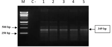
PCR products were resolved by agarose gel electrophoresis (5V/60 min) using 1.5% agarose in
Tris-Acetate-EDTA (TAE) buffer containing 0.5 µg/mL
of ethidium bromide. Molecular size ladder of 1 kb (Fermentans, Germany) was used to determine the size of the bands. The gel was observed and photographed on a Gel-Doc System (Bio-Rad, USA).
Figure 1: PCR Amplification product of cagA gene 349 bp of H. pylori. (Lane M: Ladder marker, Lane 1 to 5 biopsy samples, C- negative control)
Serum Levels of VEGF
Venous blood was drawn using a serum separator tube and allowed to clot for 30-45 minutes at room temperature before centrifugation for 15 minutes at approximately 1,000 g. Serum was immediately stored frozen in aliquots at -20oC until assay for VEGF was performed. Circulating VEGF levels were examined in serum using the Quantikine Human VEGF-ELISA (Quantikine, R&D Systems, Inc., Minneapolis).
Statistical Methods
Data analysis was performed through
univariate and bivariate analyses using the SPSS 22nd
______________________________________________________________________________________________________________________________
_______________________________________________________________________________________________________________________________
Results
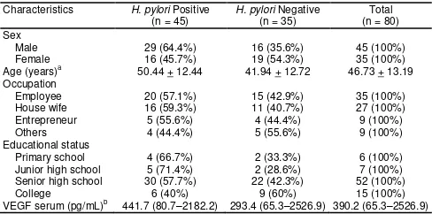
The mean age of the 80 subjects was 46.73 + 13.19 years, with a range between 19-68 years. There were 45 (56.25%) male patients and 35 (43.75%) female patients. Three major occupations of the patients were employees (43.7%), housewife (33.7%) and entrepreneurs (11.3%). There were 45 (56.3%) H. pylori-infected patients. The median of VEGF serum
was 390.2 pg/mL (65.3 – 2526.9 pg/mL) (Table 1).
Table 1: Basic characteristics of the subjects
Characteristics H. pylori Positive (n = 45) VEGF serum (pg/mL)b 441.7 (80.7–2182.2) 293.4 (65.3–2526.9) 390.2 (65.3–2526.9)
n: Total number of subjects; a mean + SD; b median (min – max).
From 45 patients infected with H. pylori, 33
(73.3%) patients had H. pylori with cagA gene
positive. All of them had H. pylori with vacA gene due
to the whole H. pylori strains carrying vacA gene
(Table 2).
Table 2: Distribution of H. pyloricagA and vacA gene status
H. pyloricagA & vacA Gene Status n = 45
n: Total number of subjects.
There was a significant difference in the mean
serum VEGF levels between patients with H. pylori
n: Total number of subjects; *p<0.05
There was a significant difference in the mean
serum VEGF levels between patients with H. pylori
cagA gene positive and negative (p = 0.017). Patients
with positive H. pylori cagA gene had serum levels of
VEGF significantly higher than H. pylori cagA gene
Logistic regression was performed to
ascertain the effect of cagA gene status on the
likelihood that subjects have a high level of serum VEGF. The logistic regression mode was statistically significant (p = 0.037). Patients with H. pylori cagA gene positive were 10.82 times more likely to have a
higher level of serum VEGF than H. pylori cagA gene
negative (Table 5).
Table 5: Logistic regression for the association between H. pyloricagA gene and serum VEGF levels (n=45)
Variable OR (95% CI) p
cagA gene 10.82 (1.14 – 101.93) 0.037*
n: Total number of subjects; an adjusted for age and sex; *p<0.05.
Discussion
The average age of H. pylori positive patients
was 50.44 ± 12.44 years old and 41.94 ± 12.72 years
old for H. pylori negative patients. Our results were comparable to a study conducted by Salimzadeh L et al., which reported that the average age of H. pylori
positive patients were 46.74 ± 16.79 years old and
48.56 ± 19.82 years old for H. pylori negative [10].
Meanwhile, in Laos, the mean age of H. pylori positive patients was 46 years old [11].
The prevalence of H. pylori in this study
(56.5%) was higher than other studies. Salimzadeh L et al. reported that the prevalence of H. pylori among Iran was 44.4% [10]. While Myint T et al. revealed that
the prevalence of H. pylori in Myanmar was 48.0%
[12]. In Indonesia, Syam AF et al. reported the prevalence of H. pylori was 22.1% [13]. The difference occurred as this study was not population-based study.
_______________________________________________________________________________________________________________________________
The vacuolating cytotoxin A (vacA) is also one
of major virulence factors released by H. pylori. VacA
causes the formation of large vacuoles and the induction of apoptosis in gastric epithelial cells [14].
Almost all H. pylori contain the vacA gene that
encodes a vacuolating cytotoxin [15, 18]. In this study,
vacA gene was found in all H. pylori, positive
patients.
Vascular endothelial growth factor (VEGF) is
a central regulator of angiogenesis and
vasculogenesis. There are evidence showing that VEGF expression is closely associated with poor prognosis and adverse clinical characteristics of gastric cancer such as tumour invasion and lymph
node metastasis and H. pylori upregulates VEGF
expression in gastric epithelial cells. Several
mechanisms such as NF-ĸB, cyclooxygenase-2
(COX-2), and epidermal growth factor receptor (EGFR) signalling are considered to mediate H. pylori-induced VEGF production in gastric epithelial cells [14]. This study also found that serum VEGF level in the infected group significantly higher compared to H. pylori negative (p < 0.05). The previous study also
suggested that H. pylori can upregulate the VEGF
serum levels [19].
Cytotoxin-associated genes pathogenicity
island (cagPAI) expresses a needle-like structure, type IV secretion system (T4SS) that is required for the injection of the protein of cytotoxin-associated gene A (cagA) or peptidoglycan into the cytosol of host cells. H. pylori peptidoglycan is recognised by a cytosolic receptor, nucleotide-binding oligomerization
domain (NOD) 1, which leads to NF- ĸB activation and
IL-8 production. A study conducted by Kang et al.
reported that H. pylori could induce VEGF production
in gastric epithelial cells via both T4SS-dependent and T4SS-independent pathways [14]. In this study, there was a significant difference in VEGF serum levels between cagA positive and cagA negative [719.27 + 525.60 vs. 402.80 + 442.67 pg/ml; p = 0.002]. CagA-expressing H. pylori are associated with an enhanced
host inflammatory response [15]. Subjects with H.
pylori cagA gene positive were 10.82 times more likely to have a higher level of serum VEGF than H. pylori cagA gene negative.
The limitation of this study was that the diagnosis of H. pylori only used one method (rapid urease test) whiles other methods may give different results. Also, the sample size was small.
In conclusion, serum VEGF level is correlated with H. pylori infection and its virulence status. The more virulence of H. pylori, cagA gene, the higher serum VEGF levels were found.
References
1. Sipponen P, Maaroos HI. Chronic gastritis. Scandinavian Journal of Gastroenterology. 2015;50:657-67. PMid:25901896 PMCid:PMC4673514
2. Varbanova M, Frauenschläger K, Malfertheiner P. Chronic
gastritis – An update. Best Practice & Research Clinical Gastroenterology. 2014;28:1031-42.
https://doi.org/10.1016/j.bpg.2014.10.005 PMid:25439069
3. Rehnberg-Laiho L, Rautelin H, Koskela P, Sarna S, Pukkala E, Aromaa A, et al. Decreasing prevalence of helicobacter antibodies in Finland, with reference to the decreasing incidence of gastric cancer. Epidemiol Infect. 2001;126:37-42. PMid:11293681 PMCid:PMC2869672
4. Matsukura N, Yamada S, Kato S, Tomtitchong P, Tajiri T, Miki M, et al. Genetic differences in interleukin-1 beta polymorphisms among four Asian populations: an analysis of the Asian paradox between H. pylori infection and gastric cancer incidence. J Exp Clin Cancer Res. 2003; 22: 47-55. PMid:12725322
5. Rugge M, Genta RM. Staging and grading of chronic gastritis. Human pathology. 2005;36:228-33.
https://doi.org/10.1016/j.humpath.2004.12.008 PMid:15791566 6. Tuccillo C, Cuorno A, Rocco A, Martinelli E, Staibano S, Mascolo M, et al. Vascular endothelial growth factor and neo-angiogenesis in H. pylori gastritis in humans. J Pathol. 2005;207: 277–84. https://doi.org/10.1002/path.1844 PMid:16184519 7. Caputo R, Tuccillo C, Manzo BA, Zarrilli R, Tortora G, Blanco CDV, et al. Helicobacter pylori VacA Toxin Up-Regulates Vascular Endothelial Growth Factor Expression in MKN 28 Gastric Cells through an Epidermal Growth Factor Receptor-, Cyclooxygenase-2-dependent Mechanism. Clin Cancer Res. 2003;9:2015-21. PMid:12796363
8. Rugge M, Pennelli G, Pilozzi E, et al. Gastritis: the histology report. Dig Liver Dis. 2011;43S:S373–84.
https://doi.org/10.1016/S1590-8658(11)60593-8
9. Rojborwonwitaya J, Vijitjunykul N. Comparison of the Accuracy
of Two Commercial Rapid Urase Tests, CLOtest® and Pronto Dry®, in detecting Helicobacter pylori Infection. Thai J
Gastroenterol. 2005:6(2):55-60.
10. Salimzadeh L, Bagheri N, Zamanzad B, et al. Frequency of virulence factors in Helicobacter pylori-infected patients with gastritis. Microbiol Pathogenesis. 2015;30:1-6.
https://doi.org/10.1016/j.micpath.2015.01.008
11. Vannarath S, Vilaichone RK, Rasachak B, et al. Virulence genes of Helicobacter pylori in gastritis, peptic ulcer and gastric cancer in Laos. Asian Pac J Cancer Prev. 2014;15(20):9027-31. https://doi.org/10.7314/APJCP.2014.15.20.9027 PMid:25374247
12. Myint T, Shiota S, Vilaichone RK, et al. Prevalence of Helicobacter pylori infection and atrophic gastritis in patients with dyspeptic symptoms in Myanmar. World J Gastroenterol. 2015;21(2):629-36. https://doi.org/10.3748/wjg.v21.i2.629 PMid:25605987 PMCid:PMC4296025
13. Syam AF, Miftahussur M, Makmun D, et al. Risk factors and prevalence of Helicobacter pylori in five largest islands of Indonesia: A preliminary study. PLoS ONE. 2015: 1-14. https://doi.org/10.1371/journal.pone.0140186
14. Kang MJ, Song EJ, Kim BY, et al. Helicobacter pylori induces vascular endothelial growth factor production in gastric epithelial cells through hypoxia-inducible factor-1α-dependent pathway. Helicobacter. 2014;19:476-83. https://doi.org/10.1111/hel.12169 PMid:25231285
15. Yamaoka Y, Graham DY. Helicobacter pylori virulence and cancer pathogenesis. Future Oncol. 2014;10(8):1487-500. https://doi.org/10.2217/fon.14.29 PMid:25052757 PMCid:PMC4197059
16. Yakut M, Örmenci N, Erdal H, et al. The association between
______________________________________________________________________________________________________________________________
_______________________________________________________________________________________________________________________________ a status. Clinics and Research in Hepatology and
Gastroenterology. 2014;37:302-11.
https://doi.org/10.1016/j.clinre.2012.09.013 PMid:23137754
17. Trang TTH, shiota S, Matsuda M, et al. The prevalence of Helicobacter pylori virulence factors in Bhutan, Vietnam, and Myanmar is related to Gastric Cancer Incidence. BioMed Research International. 2015;2015:1-8. https://doi.org/10.1155/2015/830813 PMid:26090448 PMCid:PMC4450262
18. Atherton JC, Cover TL, Papini E, Telford JL. Vacuolating Cytotoxin. In: Mobley HLT, Mendz GL, Hazell SL eds. Helicobacter
pylori. Physiology and Genetics, 2001: 97–109. https://doi.org/10.1128/9781555818005.ch9