83 Copyright © 2018 WJC | ISSN 2621-5985 (online) | ISSN 2549-385X (print)
Volume 2, Nomor 2, Oktober 2018
Ag-ZSM-11 ZEOLITE SYNTHESIS USING SILICA FROM ELEPHANT GRASS
FOR LED APPLICATION
Citra Deliana Dewi Sundari
1*, Soni Setiadji
2, Atthar Luqman Ivansyah
3, Dede
Abdurrahman
2and Denia Febby Nurbaeti
21Department of Chemistry Education, UIN Sunan Gunung Djati Bandung, Jl. Cimencrang, Cimenerang, Panyileukan, Bandung, West Java, 40292, Indonesia
2Department of Chemistry, Faculty of Science and Technology, UIN Sunan Gunung Djati Bandung, Jl. A.H. Nasution No.105 Bandung, West Java, 40614, Indonesia
3Master Program in Computational Science, Faculty of Mathematic and Natural Science, Institut Teknologi Bandung, Jl.Ganesha No. 10, Bandung, West Java, 40132, Indonesia
*Email:
[email protected]
Abstracts
Silica (SiO2) has been successfully isolated from elephant grass by the sol gel method through pH regulation using NaOH and HCl. The resulting silica has an amorphous character with crystalline phase impurities. The isolated silica was then used as a source of silica for the synthesis of ZSM-11 zeolite using the hydrothermal method with a ratio of SiO2 : TBAOH : H2O = 1 : 0.35 : 25. XRD characterization results confirmed the formation of ZSM-11 zeolite. The SEM results show that the ZSM-11 zeolite crystal morphology is oval with crystal growth in all directions. Immersion of the synthesized ZSM-11 zeolite into AgNO3 solution and followed by calcination resulted in zeolite Ag-ZSM-11 which has different optical properties compared to the initial ZSM-11 zeolite. Zeolite ZSM-11 gives purple luminescence while zeolite Ag-ZSM-11 gives pink luminescence when illuminated under UV lights.
Keywords: silica isolation, elephant grass, zeolite, ZSM-11, LED
Introduction
The use of sub-nanometer-sized precious metal clusters as luminescent materials has been reported by several researchers in the past few decades (Bhandari et al., 2016). Interest in this materials is caused by intense light absorption, efficient emissions, and bright color emissions. However, controlled
84
Copyright © 2018 WJC | ISSN 2621-5985 (online) | ISSN 2549-385X (print) Volume 2, Nomor 2, Oktober 2018
(Kunwar et al., 2014), and peptides (Yu et al., 2007), or by inorganic supporting materials such as glass (Royon et al., 2010). A different approach involves the synthesis of "ship-in-bottle" where the growth of metal clusters is limited by the pore size of supporting materials such as zeolites (Coutino-Gonzalez et al., 2014).
Zeolite is a material that can be used as a host for silver clusters because Ag+ ions can easily occupy zeolite cavities through cation exchange into zeolite pores, and also because of well-defined crystal porosity (Van Oers et al., 2014). Several studies that have been reported, have shown that emissions from Ag-zeolite are in the visible light spectrum (De Cremer et al., 2009) with external quantum efficiency approaching one (Fenwick et al., 2016). Therefore, its use as a next generation phosphor material in fluorescence lamps (Vosch et al., 2009) and as a wavelength converter in solar cells has been recommended by researchers (Vosch et al., 2009b). The main disadvantage of light emitting diode (LED) material that is commonly used today, both organic and inorganic, is its susceptibility to oxidation and aggregation, which results in the loss of character of the material emission (Schwabegger et al., 2014). The presence of Ag clusters in zeolite pores can prevent Ag aggregation, so that the Ag-zeolite material has the potential to be used as a light-emitting diode. Kennes et al reported for the first time, the first OLED with a silver cluster that was LED materials, still using commercial silica so
85
Copyright © 2018 WJC | ISSN 2621-5985 (online) | ISSN 2549-385X (print) Volume 2, Nomor 2, Oktober 2018 Methodology
Chemicals used in this research was HCl (37%, Merck®), NaOH (Merck®), TBAOH (Merck®), AgNO3 (Merck®), and Aqua DM. Silica Isolation from Elephant Grass
Elephant grass leaves are washed thoroughly and dried in an oven at 110 oC. The dried elephant grass is then burned by using the furnace at a temperature of 550 oC for 5 hours to remove organic substances. The ash obtained is dissolved with 1 M NaOH solution which is accompanied by heating at a temperature of 150 oC and stirring for 1 hour followed by the filtration. Then 3 M HCl solution is added dropwise to the filtrate solution until the pH of the filtrate solution becomes neutral, then left to stand overnight. The gel formed is then washed with hot aqua DM and dried in an oven at a temperature of 110 oC for 24 hours.
Zeolite ZSM-11 Synthesis
TBAOH is pipetted and put into a polypropylene (pp) container and stirred using a magnetic stirrer, then silica and water are added in succession and done slowly. After stirring for 3 hours at a constant speed, the solution is left overnight for the aging process. After the aging process, the mixture is put into an autoclave and put in the oven for 2 days at 170 °C. The residue is filtered and washed using aqua until the pH of the filtrate is neutral. The residue is then dried in an oven for 12 hours at 100 ᵒC and then calcined at 600 °C for 4 hours. Crystallinity and morphology of zeolites were measured by X-ray diffractometer (XRD) and Scanning Electron Microscope (SEM). Silica content
from the leaves of elephant grass was measured by X-Ray Fluorence (XRF).
Synthesis and characterization of Zeolite Ag-ZSM-11 as a ZEOLED material
The principle of synthesizing Ag-ZSM-11 zeolite is by inserting Ag+ ions into the ZSM-11 zeolite pores. The strategy is to mix Zeolite ZSM-11 into a certain concentration of AgNO3 and stir it for several hours. Then filtered using a vacuum pump accompanied by washing with aqua DM several times, and the residue was separated. The residue obtained in the form of Ag-ZSM-11 zeolite was then dried in an oven at a temperature of 110 oC for 24 hours. Furthermore, to eliminate the possible organic impurities in zeolite, calcination was carried out using a
Silica Isolation from Elephant Grass Leaves (Pennisetum purpureum)
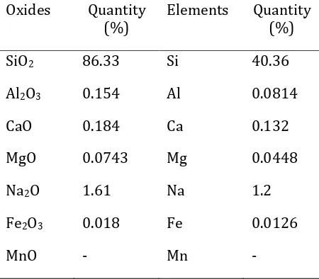
Silica in elephant grass leaves (Pennisetum purpureum) was isolated using alkali precipitation method. The isolated silica was characterized by XRF, XRD, and FT-IR. Table 1 shows the results of XRF which gives information about the content of the oxide minerals in silica isolated from elephant grass.
86
Copyright © 2018 WJC | ISSN 2621-5985 (online) | ISSN 2549-385X (print) Volume 2, Nomor 2, Oktober 2018
21.7°. This shows that the silica from the isolation has an amorphous phase formed due to the presence of silanol (Si-OH) groups on its surface (Shim et al., 2015). However, the presence of peaks at 2θ = 30.38°, 31.45° and 44.31° indicates that crystalline SiO2 phase was also formed as an impurity (Shim et al., 2015).
Table 1. Results of characterization of silica from elephant grass leaves using XRF
Oxides Quantity (%)
Elements Quantity (%)
SiO2 86.33 Si 40.36
Al2O3 0.154 Al 0.0814
CaO 0.184 Ca 0.132
MgO 0.0743 Mg 0.0448
Na2O 1.61 Na 1.2
Fe2O3 0.018 Fe 0.0126
MnO - Mn -
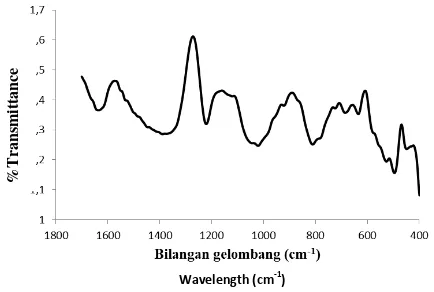
To find out the bond vibrations of the isolated silica, FT-IR analysis was performed. The results of silica analysis with FT-IR shown in Figure 2 indicate a peak at wave number 514 cm-1, 663 cm-1, 805 cm-1, 984 cm -1, 1195 cm-1 and 1641 cm-1. Vibration at wave number 1641 cm-1 shows the vibration of H2O or Si-OH molecules which are reactive groups on the surface of amorphous silica (Mozgawa et al., 2011).
The silanol group is a reactive group located on the surface of amorphous silica. In addition to indicating the presence of silanol groups, the wave number also indicates that there is still a water content in the isolated
silica. The wave number 984 cm-1 indicates the vibration of the Si-O-Si bond (Lu et al., 2014).
Figure 1 Difffractogram of Silica isolated from Elephant Grass
Figure 2 FT-IR spectrum of silica isolated from elephant grass
Synthesis and Characterization of ZSM-11 Zeolite
87
Copyright © 2018 WJC | ISSN 2621-5985 (online) | ISSN 2549-385X (print) Volume 2, Nomor 2, Oktober 2018 elephant grass leaves. TBAOH will direct the
filling of spaces in the structure of zeolites by Si and O so that later a ring will form which consists of SiO2 tetrahedra and forms a crystal lattice in the form of D5R (consisting of 10 rings). After zeolite is formed, then it is characterized using XRD.
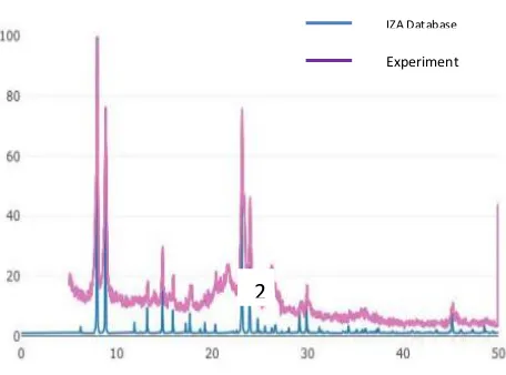
Figure 3 is a diffractogram of ZSM-11 zeolite synthesized in this study which is compared with the IZA (International Zeolite Association) database. In addition, the diffractogram of the experimental results was also compared with the journal written by Lu et al., where the specific diffractogram peak that indicates a ZSM-11 Zeolite has been
formed at 2θ = 7.92ᵒ, 8.78ᵒ, 23.14ᵒ, 23.98ᵒ, and 45.21ᵒ (Lu et al., 2014), Based on the diffractogram shown in Figure 3, there is a
peak at 2θ = 7.93˚, 8.85˚, 23.11˚, 23.96˚, and
45.05˚. Thus it can be concluded that ZSM-11 Zeolite was formed in this study. The
presence of two peaks at 2θ between 23-25˚
and one peak at 45.4˚ indicates that in the
synthesized ZSM-11 zeolite there is no ZSM-5 zeolite phase.
SEM image the synthesized ZSM-11 synthesis can be seen in Figure 4. From Figure 4 it can be seen that the synthesized ZSM-11 zeolite has an oval shape with a fairly pointed tip and crystal growth in various directions. It can be seen in figure (a) that the overall shape of zeolite has been evenly distributed with the same size. This morphology is different when compared to the ZSM-11 zeolite synthesized by Lu et al. (Lu et al., 2014). In ZSM-11 zeolites synthesized by Lu, et al. showing morphology with an oval shape that is not too sharp at the edges. This difference occurs because of differences in the source of silica used, the mole ratio, and the temperature at stirring
and incubation, as well as the length of incubation time.
Figure 3 Diffractogram of ZSM-11 Zeolite
Figure 4 ZSM-11 Zeolite SEM Test Results (a) 500x; (b) 1000x; (c) 2000x; (d) 5000x
magnification.
Figure 5 shows the results of zeolite analysis of ZSM-11 using FTIR. FTIR analysis was carried out at wavenumber 1700-400 cm -1, the wavenumber range is the fingerprint
IZA Database
Experiment
2
88
Copyright © 2018 WJC | ISSN 2621-5985 (online) | ISSN 2549-385X (print) Volume 2, Nomor 2, Oktober 2018
region to show what functional groups it contains. At wavenumber of 495.687 cm-1 is a bending vibration TO4 (T = Si). At wavenumber of 526.743 and 1220.926 cm-1 it is a double 5-ring (D5R) bending vibration in the ZSM-11 zeolite structure. This double 5-ring is an external link between one zeolite layer and another. At wavenumber of 811.090 and 1021.146 cm-1 it is stretching vibration of Si-O-Si (Dey et al., 2012 & Zhang et al., 2012). Synthesis and Characterization of Ag-ZSM-11 Zeolite
ZSM-11 zeolite was immersed into AgNO3 solution. This will cause Ag+ ions to enter the pores of ZSM-11 zeolite, forming the Ag-ZSM-11 zeolite. The optical properties related to the application of LEDs possessed by ZSM-11 and Ag-ZSM-11 have differences in luminescence when illuminated by UV lamps as shown in Figure 6.
Figure 5 FT-IR spectrum of the synthesized ZSM-11 Zeolite
When the ZSM-11 zeolite was illuminated by a UV lamp, it produced a purple glow. However, when Ag-ZSM-11 zeolite is illuminated by UV light, pink luminescence is produced. ZSM-11 zeolite is
89
Copyright © 2018 WJC | ISSN 2621-5985 (online) | ISSN 2549-385X (print) Volume 2, Nomor 2, Oktober 2018
(a) (b)
Figure 6 (a) ZSM-11 Zeolite luminescence when illuminated by UV lights; (b) Zeolite Ag-ZSM-11 luminescence when illuminated by UV lamps
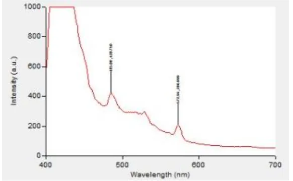
Figure 7 Photoluminescence spectrum of Ag-ZSM-11 zeolite
Conclusion
Silica can be isolated from elephant grass with SiO2 content of 86.33% w/w. Silica isolated from elephant grass has amorphous properties accompanied by the presence of other crystalline silica impurities. From the
Ag-90
Copyright © 2018 WJC | ISSN 2621-5985 (online) | ISSN 2549-385X (print) Volume 2, Nomor 2, Oktober 2018
zeolites have different optical properties related to LEDs when illuminated by UV lights, ZSM-11 zeolites produce purple luminescence while Ag-ZSM-11 zeolite produces pink luminescence.
References
Bhandari, S., Pramanik, S., Khandelia, R., Chattopadhyay, A.. 2016. Gold Nanocluster and Quantum Dot Complex in Protein for Biofriendly White-Light-Emitting Material. ACS Appl. Mater. Interfaces. 8, 1600.
Zheng, J., Dickson, R. M.. 2002. Individual Water-Soluble Dendrimer-Encapsulated Silver Nanodot Fluorescence. J. Am. Chem. Soc. 124, 13982.
Temboury, M. R. C., Paolucci, V., Hooley, E. N., Latterini, L., Vosch, T.. 2016. Probing DNA-stabilized fluorescent silver nanocluster spectral heterogeneity by time-correlated single photon counting. Analyst. 141(1), 123.
Kunwar, P., Hassinen, J., Bautista, G., Ras, R. H. A., Toivonen, J. 2014. Direct Laser Writing of Photostable Fluorescent Silver Nanoclusters in Polymer Films. ACS Nano. 8 (11), 11165-71.
Yu, J., Patel, S. A., Dickson, R. M. 2007. In vitro and intracellular production of peptide-encapsulated fluorescent silver nanoclusters. Angew. Chem. Int. Ed. 46 (12), 2028-30.
Royon, A., Bourhis, K., Bellec, M., Papon, G., Bousquet, B., Deshayes, Y., Cardinal, T., Canioni, L. 2010. Silver clusters embedded in glass as a perennial high capacity optical recording medium. Adv. Mater. 22 (46), 5282-6.
M., Kvashnina, K., Fron, E., Dieu, B., De Cremer, G., Lievens,P., Sels, B., Hofkens, J. 2014. X-ray irradiation-induced formation of luminescent silver clusters in nanoporous matrices. Chem. Commun. 50 (11), 1350-2.
Van Oers, C. J., Kurttepeli, M., Mertens, M., Bals, S., Meynen, V., Cool, P. 2014. Zeolite β nanoparticles based bimodal structures: Mechanism and tuning of the porosity and zeolitic properties. Microporous Mesoporous Mater. 185, 204-212.
De Cremer, G., Coutiño-Gonzalez, E., Roeffaers, M. B. J., Moens, B., Ollevier, J., Van der Auweraer, M., Schoonheydt, R., Jacobs, P. a., De Schryver, F. C., Hofkens, J., De Vos, D. E., Sels, B. F., Vosch, T. 2009. Characterization of fluorescence in heat-treated silver-exchanged zeolites. J. Am. Chem. Soc. 131 energetics and tailoring the optical properties of silver clusters confined in zeolites. Nat. Mater. 15(9), 1017-22.
91
Copyright © 2018 WJC | ISSN 2621-5985 (online) | ISSN 2549-385X (print) Volume 2, Nomor 2, Oktober 2018 Baekelant, W., Roeffaers, M.B. J., Van der
Auweraer, M. 2017. Silver Zeolite Composites-Based LEDs: A Novel Solid-State Lighting Approach. Adv. Funct. Mater. 27 (14), 1606411.
Vaibhav, V., Vijayalakshmi, U., Roopan, S. M. 2015. Agricultural waste as a source for the production of silica nanoparticles. Spectrochimica Acta Part A: Molecular and Biomolecular Spectroscopy. 139, 515. Nakanishi, E.Y., Frías, M., Martínez-Ramírez,
S., Santos, S. F., Rodrigues, M. S., Rodríguez, O., Savastano Jr., H. 2014. Characterization and properties of elephant grass ashes as supplementary cementing material in pozzolan/Ca(OH)2 pastes. Construction and Building Materials. 73, 391.
Nakanishi, E. Y., Frías, M., Santos, S. F., Rodrigues, M. S., de la Villa, R. V., Rodriguez, O., Junior, H. S. 2016. Investigating the possible usage of elephant grass ash to manufacture the eco-friendly binary cements. J. Clean. Prod. 116, 236-43.
Strezov V., Evans T. J., Hayman C. 2008. Thermal conversion of elephant grass (Pennisetum Purpureum Schum) to bio-gas, bio-oil and charcoal. Bioresource Technology. 99, 8394–8399.
Srisittipokakun, N., Sangwaranatee, N. 2014. Comparative Study of Physical and Optical Properties of Glasses from SiO2 and Napier Ash. Adv. Mat. Res. 979, 401.
Shim, J., Velmurugan, P., Oh, B-T. 2015. Extraction and physical characterization of amorphous silica made from corn cob ash at variable pH conditions via sol gel processing. J. Indust. Eng. Chem. 30, 249-53.
FT-IR Studies of Zeolites from Different Structural Groups. Chemik, 65 (7), 667-674.
Lu, M., Cheng, Y., Pan, S.-l., Wei, G.-y. 2014. Des. Wat. Treat. 57, 1.
Dey, K. P., Gosh, S., Naskar, M. K. 2012. A facile synthesis of ZSM-11 zeolite particles using rice husk ash as silica source. Mat. Lett. 87, 87-89.
92