10.1128/CMR.00008-08.
2008, 21(4):583. DOI:
Clin. Microbiol. Rev.
Shyam Daya and Kenneth I. Berns
Virus Vectors
http://cmr.asm.org/content/21/4/583
Updated information and services can be found at:
These include:
REFERENCES
http://cmr.asm.org/content/21/4/583#ref-list-1
at:
This article cites 80 articles, 40 of which can be accessed free
CONTENT ALERTS
more»
articles cite this article),
Receive: RSS Feeds, eTOCs, free email alerts (when new
http://journals.asm.org/site/misc/reprints.xhtml
Information about commercial reprint orders:
http://journals.asm.org/site/subscriptions/
To subscribe to to another ASM Journal go to:
on November 13, 2013 by guest
http://cmr.asm.org/
Downloaded from
on November 13, 2013 by guest
http://cmr.asm.org/
0893-8512/08/$08.00⫹0 doi:10.1128/CMR.00008-08
Copyright © 2008, American Society for Microbiology. All Rights Reserved.
Gene Therapy Using Adeno-Associated Virus Vectors
Shyam Daya
1and Kenneth I. Berns
1,2*
Department of Molecular Genetics and Microbiology, College of Medicine,1and University of
Florida Genetics Institute,2University of Florida, Gainesville, Florida
INTRODUCTION ...583
BIOLOGY OF AAV ...583
Virus Classification...583
AAV-2 Genome ...583
AAV Life Cycle ...584
AAV-2 Site-Specific Integration ...584
AAV Infection ...585
AAV AS A GENE THERAPY VECTOR...586
Rational Design of AAV Capsids ...587
Immune Response to AAV ...587
AAV CLINICAL TRIALS...588
FUTURE PROSPECTS...590
DISCUSSION ...591
FINAL COMMENTS...591
ACKNOWLEDGMENTS ...592
REFERENCES ...592
INTRODUCTION
Adeno-associated virus (AAV) vectors are currently among the most frequently used viral vectors for gene therapy. At recent meetings of the American Society for Gene Therapy, nearly half of the presentations involved the use of AAV. This represents a significant turnaround. Historically, AAV has not been of great medical interest, because it has not been identi-fied as a pathogen; thus, the lack of widespread knowledge of the virus initially inhibited its broad use as a vector. Twelve human serotypes of AAV (AAV serotype 1 [1] to AAV-12) and more than 100 serotypes from nonhuman primates have been discovered to date. The lack of pathogenicity of the virus, the persistence of the virus, and the many available serotypes have increased AAV’s potential as a delivery vehicle for gene therapy applications. This review will focus on the biology of AAV and its use as a vector for gene therapy.
BIOLOGY OF AAV
Virus Classification
AAV is a small (25-nm), nonenveloped virus that packages a linear single-stranded DNA genome. It belongs to the family
Parvoviridaeand is placed in the genusDependovirus, because productive infection by AAV occurs only in the presence of a helper virus, either adenovirus or herpesvirus. In the absence of helper virus, AAV (serotype 2) can set up latency by inte-grating into chromosome 19q13.4, establishing itself as the only mammalian DNA virus known to be capable of site-specific integration.
AAV-2 Genome
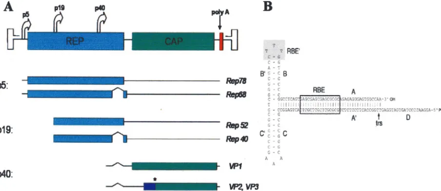
The AAV-2 genome is a linear, single-stranded DNA of 4.7 kb (Fig. 1A) (60). Both sense and antisense strands of AAV DNA are packaged into AAV capsids with equal frequency. The genome is structurally characterized by 145-bp inverted terminal repeats (ITRs) that flank two open reading frames (ORFs) (Fig. 1B).
The first 125 nucleotides of the ITR constitute a palindrome, which folds upon itself to maximize base pairing and forms a T-shaped hairpin structure. The other 20 bases, called the D sequence, remain unpaired. The ITRs are importantcis-active sequences in the biology of AAV. A key role of the ITRs is in AAV DNA replication. In the current model of AAV replica-tion, the ITR is the origin of replication and serves as a primer for second-strand synthesis by DNA polymerase. The double-stranded DNA formed during the synthesis, called replicating-form monomer, is used for a second round of self-priming replication and forms a replicating-form dimer. These double-stranded DNA intermediates (replicating-form monomer and replicating-form dimer) are processed via a strand displace-ment mechanism, resulting in single-stranded DNA used for packaging and double-stranded DNA used for transcription. Critical to the replication process are the Rep binding ele-ments (RBEs) (RBE and RBE⬘) and a terminal resolution site (TRS), which is located within the ITR (Fig. 1B). These fea-tures are used by the viral regulatory protein Rep during AAV replication to process the double-stranded intermediates. In addition to their role in AAV replication, the ITR is also essential for AAV genome packaging, transcription, negative regulation under nonpermissive conditions, and site-specific integration.
The left ORF contains the Rep gene, which produces four Rep proteins, Rep78, Rep68, Rep52, and Rep40. The larger Rep proteins (Rep78 and Rep68) are produced from
tran-* Corresponding author. Mailing address: P.O. Box 103610, Univer-sity of Florida Genetics Institute, 1376 Mowry Road, Gainesville, FL 32610-3610. Phone: (352) 273-8072. Fax: (352) 273-8284. E-mail: [email protected].
583
on November 13, 2013 by guest
http://cmr.asm.org/
scripts using the P5 promoter, whereas the smaller Rep pro-teins (Rep52 and Rep40) are produced from transcripts using the P19 promoter. Rep78 and Rep68 are produced from un-spliced and un-spliced transcripts, respectively, and are important regulatory proteins that act intransin all phases of the AAV life cycle. Specifically, they positively and negatively regulate AAV gene expression in the presence or absence of helper virus, respectively, and are required for DNA replication (48). The smaller Rep proteins, Rep52 and Rep40, produced from unspliced and spliced transcripts, respectively, are involved in the accumulation of single-stranded viral DNA used for pack-aging within AAV capsids. All four Rep proteins possess he-licase and ATPase activity. In addition, the larger Rep proteins possess strand- and site-specific endonuclease activity (nicking at the TRS) and site-specific DNA binding activity (binding at the RBE).
The right ORF contains the Cap gene, which produces three viral capsid proteins (VP1, VP2, and VP3) using the P40 pro-moter. Alternative splicing of the P40 transcript is used to produce the three viral proteins from two transcripts. The unspliced transcript produces VP1 (87 kDa), the biggest of the capsid proteins. The spliced transcript produces VP2 (72 kDa) and VP3 (62 kDa). VP2 is produced using a nonconventional ACG start codon, whereas VP3 is produced using a down-stream conventional AUG codon. The AAV-2 capsid com-prises 60 viral capsid proteins arranged into an icosahedral structure with symmetry equivalent to a triangulation number of 1. The capsid proteins (VP1, VP2, and VP3) are present in a 1:1:10 molar ratio.
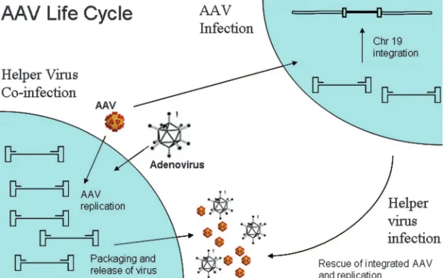
AAV Life Cycle
There are two stages to the AAV life cycle (Fig. 2) after successful infection, a lytic stage and a lysogenic stage. In the presence of helper virus (adenovirus or herpesvirus), the lytic stage ensues. During this period, AAV undergoes productive infection characterized by genome replication, viral gene
ex-pression, and virion production. The adenoviral genes that provide helper functions regarding AAV gene expression have been identified and include E1a, E1b, E2a, E4, and VA RNA. Herpesvirus aids in AAV gene expression by providing viral DNA polymerase and helicase as well as the early functions necessary for HSV transcription. Although adenovirus and herpesvirus provide different sets of genes for helper function, they both regulate cellular gene expression, providing a per-missive intracellular milieu for AAV productive infection.
In the absence of adenovirus or herpesvirus, there is limited AAV replication, viral gene expression is repressed, and the AAV genome can establish latency by integrating into a 4-kb region on chromosome 19 (q13.4), termed AAVS1 (36, 37). The AAVS1 locus is near several muscle-specific genes, TNNT1 and TNNI3 (16). The AAVS1 region itself is an up-stream part of a recently described gene, MBS85. The exact function of this gene is not clear, but its product has been shown to be involved in actin organization (64). Whether AAV integration into this site is suitable for human gene therapy applications remains to be evaluated. Tissue culture experi-ments suggest that the AAVS1 locus is a safe integration site.
AAV-2 Site-Specific Integration
One of the features of AAV is its ability to specifically integrate to establish latent infection. Current AAV vectors do not have this ability, and the development of such a vector would ensure long-term transgene expression in tissues with-out problems associated with insertional mutagenesis.
Some of the viral and cellular requirements for targeted integration have been elucidated. The AAV components that are required have been identified. These include the ITRs (in
cis), Rep78 or Rep68 (intrans), and a 138-bp sequence termed the integration efficiency element (IEE), located within the P5 promoter incis(49). It is unclear if the entire 138-bp IEE in P5 is required, since a recent study showed that a 16-bp RBE in P5 is sufficient (18). Latent infection with wild-type AAV-2
FIG. 1. Map of the wild-type AAV-2 genome. (A) Rep and Cap genes flanked by ITRs. The different Rep and Cap transcripts are produced from their respective promoters (P5, P19, and P40). The star indicates the alternative ACG codon used to produce VP3. (B) Secondary structure of an AAV-2 ITR showing the RBEs (RBE, GAGCGAGCGAGCGCGC; RBE⬘, CTTG) and the TRS (GTTGG).
on November 13, 2013 by guest
http://cmr.asm.org/
appears to be nonpathogenic in tissue culture, when Rep is expressed under its own promoter. Such expression is regu-lated by negative feedback. Excess Rep expression has been shown to arrest cell division (75) and induce cellular apoptosis (58).
A 33-bp minimum AAVS1 sequence, which contains an RBE-like and a TRS-like sequence separated by 8 nucleotides, is necessary and sufficient to target AAV integration (26). The intervening sequence may be varied, but a central 5⬘ CTC is required. The actual integration site is somewhat downstream from the target sequence and can be variable. Many RBEs have been identified in the human genome, with AAVS1 being the only site that has an RBE and a TRS in close proximity to one other. Interestingly, the AAV genome and AAVS1 can be tethered to each other via Rep68 in vitro (68). These observa-tions provide a molecular explanation for why AAVS1 is tar-geted, even though the exact mechanism remains unknown.
The process of site-specific integration is not completely specific even under ideal conditions of Rep78 and Rep68 ex-pression, with approximately 40 to 70% of integrants occurring in AAVS1. Moreover, the mechanism is imprecise, as judged by there not being reproducible breakpoints for vector-AAVS1 junctions; however, clusters of integrants appear within a 2-kb fragment of AAVS1. While thecis- andtrans-acting viral fac-tors required for site-specific integration have been identified, much less is known about cellular factors that are required or may be involved. Only recently has a study provided evidence that a cellular protein, human immunodeficiency virus trans-acting response element-RNA loop binding protein 185 (TRP-185), can promote AAV integration into AAVS1 further downstream from the RBE via interactions with both Rep and AAVS1 (73). Moreover, site-specific integration has been demonstrated in mice and rats transgenic for AAVS1, suggest-ing that the AAVS1 open-chromatin structure is maintained in
vivo and that the cellular factors that mediate site-specific integration are present in nondividing cells (56).
AAV Infection
AAV-2 gains entry into target cells by using the cellular receptor heparan sulfate proteoglycan (62). Internalization is enhanced by interactions with one or more of at least six known coreceptors including␣V5integrins (63), fibroblast growth factor receptor 1 (53), hepatocyte growth factor re-ceptor (35),␣v1integrin (7), and laminin receptor (3). The cellular events that mediate AAV trafficking postentry are not completely characterized. Cells defective for dynamin significantly hindered AAV-2 infection, suggesting that AAV is endocytosed into clathrin-coated vesicles (15). For successful AAV infection, AAV particles need to escape these endocytic vesicles. Infection experiments with bafilo-mycin A1 (a drug that inhibits the proton pump for endo-somes) suggested that the low pH in the endosomes is es-sential for virus escape and successful infection (9). Moreover, cellular signaling involving the activation of the Rac1 protein and the phosphatidylinositol 3-kinase pathway is necessary for intracellular trafficking of AAV particles using microtubules (57). Interestingly, a conserved phospho-lipase A2 motif identified in the N terminus of the VP1 protein was reported to be important for successful infec-tion (27). Specifically, the phospholipase A2motif seemed to be playing a crucial role during AAV trafficking, possibly helping AAV escape the late endosome. Mutational analysis of the AAV capsid structure indicated that the fivefold pore structure may also serve as the site for phospholipase do-main presentation during viral infection (10). Moreover, endosomal cysteine proteases, cathepsins B and L, have implied roles in AAV trafficking and capsid disassembly (4).
FIG. 2. AAV life cycle. AAV undergoes productive infection in the presence of adenovirus coinfection. This is characterized by genome replication, viral gene expression, and virion production. In the absence of adenovirus, AAV can establish latency by integrating into chromosome 19 (AAVS1). The latent AAV genome can be rescued and replicated upon superinfection by adenovirus. Both stages of AAV’s life cycle are regulated by complex interactions between the AAV genome and AAV, adenoviral, and host proteins.
on November 13, 2013 by guest
http://cmr.asm.org/
Exactly how AAV enters the nucleus after escaping the endosome is not known and is currently an active area of research. Although AAV is theoretically small enough to enter the nucleus via the nuclear pore complex, an early study suggested that AAV entry may be nuclear pore com-plex independent (29).
AAV AS A GENE THERAPY VECTOR
There are several considerations for any viral vector. These include the ability to attach to and enter the target cell, suc-cessful transfer to the nucleus, the ability to be expressed in the nucleus for a sustained period of time, and a general lack of toxicity. AAV vectors have been highly successful in fulfilling all of these criteria. Moreover, a variety of modifications have served to enhance their utility. Several considerations have guided the development of current AAV vectors, especially the lack of pathogenicity of the wild-type virus and its persistence. The small size of the AAV genome and concerns about potential effects of Rep on the expression of cellular genes led to the construction of AAV vectors that do not encode Rep and that lack thecis-active IEE, which is required for frequent site-specific integration. The ITRs are kept because they are thecissignals required for packaging. Thus, current recombi-nant AAV (rAAV) vectors persist primarily as extrachromo-somal elements (1, 59).
rAAV vectors for gene therapy have been based mostly on AAV-2. AAV-2-based rAAV vectors can transduce muscle, liver, brain, retina, and lungs, requiring several weeks for op-timal expression. The efficiency of rAAV transduction is de-pendent on the efficiency at each step of AAV infection: bind-ing, entry, viral traffickbind-ing, nuclear entry, uncoatbind-ing, and second-strand synthesis. Inefficient AAV trafficking (30) and second-strand synthesis (19) have been identified as being rate-limiting factors in AAV gene expression. Interestingly, the binding of cellular protein FKBP52 to the AAV ITR inhibits second-strand synthesis, and this inhibition is dependent on the
phosphorylation state of FKBP52 (51, 52, 80). Moreover, epi-dermal growth factor receptor kinase signaling has been im-plicated in regulating both AAV trafficking and second-strand synthesis (80).
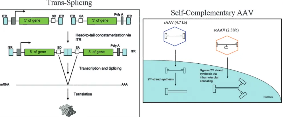
Several novel AAV vector technologies have been devel-oped to either increase the genome capacity for AAV or en-hance gene expression (Fig. 3). The idea oftrans-splicing AAV vectors has been used to increase AAV vector capacity (74). This system takes advantage of AAV’s ability to form head-to-tail concatemers via recombination in the ITRs. In this ap-proach, the transgene cassette is split between two rAAV vec-tors containing adequately placed splice donor and acceptor sites. Transcription from recombined AAV molecules, fol-lowed by the correct splicing of the mRNA transcript, results in a functional gene product. This application becomes useful for using AAV to deliver therapeutic genes up to 9 kb in size.trans
splicing has been successfully used for gene expression in the retina (55), the lung (39), and, more recently, muscle (25).
trans-Splicing vectors are less efficient than rAAV vectors. The design and use of self-complementary AAV (scAAV) vectors to bypass the limiting aspects of second-strand synthe-sis have been described (44). The rationale underlying the scAAV vector is to shorten the lag time before transgene expression and potentially to increase the biological efficiency of the vector. scAAV vectors can fold upon themselves, imme-diately forming transcriptionally competent double-stranded DNA. One consequence of the use of scAAV is that the max-imal size of the transgene is reduced by⬃50% (2.4-kb capac-ity), but up to 3.3 kb of DNA can be encapsidated (71). Rapid transduction has been observed using scAAV in both tissue culture and in vivo experiments.
Many clinically relevant tissues are not susceptible to infec-tion by AAV-2. Greater gene expression was seen in muscle, retina, liver, and heart using AAV serotypes 1, 5, 8, and 9, respectively. The cell surface receptors have been identified for only some of the many AAV serotypes: AAV-3 (heparan
sul-FIG. 3. (Left)trans-Splicing approach. The head-to-tail formation of two different AAV vector results in functional product after splicing. (Right) Comparison of scAAV and rAAV vectors.
on November 13, 2013 by guest
http://cmr.asm.org/
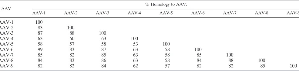
fate proteoglycan), AAV-4 (O-linked sialic acid), and AAV-5 (platelet-derived growth factor receptor). In addition, a 37-kDa/67-kDa laminin receptor has been identified as being a receptor for AAV serotypes 2, 3, 8, and 9 (3). The attachment receptors for the other serotypes have not yet been identified. All of these serotypes are potential candidates for testing as vectors for gene therapy. To date, most of the testing has involved serotypes 1 to 9, which have considerable differences at the capsid amino acid sequence level, except for AAV-1 and AAV-6 (Table 1), and has succeeded in identifying vectors with widely divergent tissue specificities.
The use of the different AAV serotypes in a pseudotyping approach (the genome of one ITR serotype being packaged into a different serotype capsid) has allowed broad tissue tro-pisms. However, some tissues remain refractory to transduc-tion using available serotypes. This presents a major challenge for AAV-based gene therapy for clinically relevant tissues.
Rational Design of AAV Capsids
A deeper understanding of the AAV capsid properties has made the rational design of AAV vectors that display selective tissue/organ targeting possible, thus broadening the possible applications for AAV as a gene therapy vector. Two ap-proaches have been used for AAV vector retargeting: (i) direct targeting and (ii) indirect targeting. In direct targeting, vector targeting is mediated by small peptides or ligands that have been directly inserted into the viral capsid sequence. This ap-proach has been used successfully to target endothelial cells (61, 69). Direct targeting requires extensive knowledge of the capsid structure. Important aspects involve the following: pep-tides or ligands must be positioned at sites that are exposed to the capsid surface, the insertion must not significantly affect capsid structure and assembly, and it is important that the native tropism be ablated to maximize targeting.
In indirect targeting, vector targeting is mediated by an as-sociating molecule that interacts with both the viral surface and the specific cell surface receptor. The use of bispecific antibod-ies (8) and biotin (6, 50) has been described for AAV vectors. The advantages of this approach are that different adaptors can be coupled to the capsid without significant changes in capsid structure, and the native tropism can be easily ablated. One disadvantage of using adaptors for targeting may involve the decreased stability of the capsid-adaptor complex in vivo. The development of efficient AAV targeting vectors will re-quire a better understanding of all aspects of the AAV
infec-tion process: binding and entry, viral processing, and nuclear entry and expression. Significant progress has been made in all these categories, and the development of efficient AAV tar-geting vectors will expand AAV’s use as a vector for many clinical applications.
Immune Response to AAV
One of the biggest challenges facing AAV gene delivery is the host immune response. The host defense mechanism at the adaptive level is made up of cell-mediated and humoral im-munity. The cell-mediated response functions at the cellular level, eliminating the transduced cells using cytotoxic T cells, whereas the humoral response produces neutralizing antibod-ies (Nab), preventing the readministration of vector. Almost no innate response is seen in AAV infection (76).
Immune response to AAV is primarily a humoral response (72). Preexisting Nab in patients, because of prior infection, account for the humoral response seen toward AAV. In a study by Chirmule et al. (13), antibodies to AAV were seen in 96% of the subjects (patients with cystic fibrosis [CF] and healthy subjects), and 32% showed neutralizing ability in an in vitro assay. Nab to AAV have been to show limit AAV transduction in liver (47) and lung (28); however, no such effect was seen in muscle (21), brain (43), and retina (5). Interestingly, the hu-moral response to AAV may be T-cell dependent; the inhibi-tion of T-cell funcinhibi-tion using anti-CD4 antibodies prevents Nab formation and allows vector readministration (14, 28, 40).
Cell-mediated responses to AAV vectors have been docu-mented, but this response may be dependent on the route of administration (11) and AAV serotype (67). A potent immune response to AAV-ovalbumin was observed when AAV was administered intraperitoneally, intravenously, or subcutane-ously but not when administered intramuscularly. Moreover, AAV-2 has been shown to induce a weak cell-mediated im-mune response. This may be attributed to AAV inefficiently infecting mature dendritic cells (DC); however, a recent study demonstrated an efficient infection of immature DC and gen-erated a cytotoxic-T-lymphocyte (CTL) response when used in adoptive transfer experiments (77). The extent to which ma-ture and immama-ture DC are transduced by AAV in vivo and the mechanism of how AAV induces a cellular immune response are not known.
In a recent clinical trial for hemophilia B, an unexpected liver toxicity was observed and was attributed to a CTL re-sponse to AAV-2-transduced hepatocytes (42). Subsequently,
TABLE 1. Capsid homology among AAV serotypes 1 to 9
AAV % Homology to AAV:
AAV-1 AAV-2 AAV-3 AAV-4 AAV-5 AAV-6 AAV-7 AAV-8 AAV-9
AAV-1 100
AAV-2 83 100
AAV-3 87 88 100
AAV-4 63 60 63 100
AAV-5 58 57 58 53 100
AAV-6 99 83 87 63 58 100
AAV-7 85 82 85 63 58 85 100
AAV-8 84 83 86 63 58 84 88 100
AAV-9 82 82 84 62 57 82 82 85 100
on November 13, 2013 by guest
http://cmr.asm.org/
it was discovered that the AAV-2 capsid heparin binding motif was responsible for T-cell activation (65). This correlated well with a study in mice that showed that AAV-2 infection can activate a CTL response, whereas AAV-7 and AAV-8 do not (67). Moreover, Wang et al. (67) suggested that the cross-presentation of input AAV capsids via major histocompatibil-ity complex class I presentation may be playing a role in the observed activation of cytotoxic T cells; however, this response does not diminish transgene expression via the targeted de-struction of transduced hepatocytes, a finding confirmed by another group (38). Taken together, these studies suggest that immune responses are a major hurdle and that a deeper un-derstanding of AAV-host interactions in humans is required for the efficient use of AAV as a gene transfer vector.
AAV CLINICAL TRIALS
AAV has become increasingly common as a vector for use in human clinical trials; as of now, 38 protocols have been ap-proved by the Recombinant DNA Advisory Committee and the Food and Drug Administration (FDA). The increased pop-ularity of AAV vectors reflects the appreciation of the long-term transgene expression observed in animal models and the relative lack of immune response and other toxicities in the models. Other factors that have played a role in encouraging the use of AAV vectors include the discovery of new serotypes and the appreciation that matching the tissue specificity of the serotype with the presumptive target tissue can greatly en-hance the potential effectiveness of therapy. In general, the goal of gene therapy can be classified into one of two catego-ries, the correction of an intracellular defect or the synthesis of a secreted protein, which is active at an extracellular level. In the latter case, the site of protein synthesis would not seem to be critical as long as it has no deleterious intracellular effects and is successfully secreted into blood. This assumption has been tested in clinical trials in which proteins normally synthe-sized in the liver are now induced to be produced in skeletal muscle. Whether the assumption is correct is still not certain, in part because different vector target sites may induce differ-ent host immune responses (41, 42).
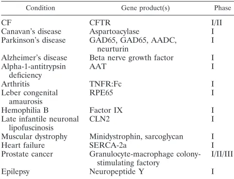
Despite the small packaging capacity of AAV vectors, clever investigators have devised ways of engineering transgenes and associated regulatory sequences so that their sizes can be re-duced sufficiently to allow packaging into AAV capsids. In general, the expectations with regard to minimal toxicity have been met, although there have been two notable exceptions to this, which will be discussed below. To date, no clinical cures have been effected, although there have been anecdotal data that have kept hopes up. Trials that have been concluded or are in progress are listed in Table 2. Several of these will be discussed below in some detail to illustrate specific points of interest and concern.
Initial targets for gene therapy included monogenic diseases in which the gene product either was altered to become non-functional or was missing. First among these was CF, a lethal, autosomal recessive disease in which the CF transmembrane regulator (CFTR) is inactivated by mutation. CFTR is a com-ponent of the Cl⫺
channel and the lack of functional CFTR affects the transmembrane electrical potential. This leads to the accumulation of thick secretions in the lung coupled with a
loss of the normal respiratory epithelial ciliary activity. The primary difficulty is pulmonary, with an increased incidence of pulmonary infection, especially by Pseudomonas aeruginosa. Additional difficulty occurs with pancreatic secretion, but the loss of the pancreatic enzymes can be treated with supple-ments. Thirteen protocols have been approved for phase I and phase II clinical trials using an AAV vector (2, 22, 23, 46, 66). Delivery of the vector was achieved by bronchoscope or by aerosol into the lung and in several cases by delivery to the maxillary sinus (to make measurement of the transmembrane potential, which is affected in CF, possible). The primary and most important observation in early trials was the lack of mea-surable toxicity and a very modest immune response evoked by the route of pulmonary delivery. Serum antibody was evoked but did not affect the subsequent administration of the vector. The measurement of efficacy in the lung is pretty much re-stricted to measures of pulmonary function; any improvement that was noted in this manner was not statistically significant. However, in those patients who had vector instilled into the maxillary sinus, it was possible to make a somewhat more direct measurement. The most notable effect was an increase in levels of interleukin-10, a cytokine that is anti-inflammatory, and a concomitant decrease in levels of interleukin-8, which has the opposite effect. Major challenges with vector delivery to the lung through the airway included rapid, regular shedding of the respiratory epithelium, which means that cells that have taken up the vector are fairly quickly lost and that the uptake of the AAV-2 vector in cell culture was mostly through the basolateral surface, which is not very accessible via the airway. Thus, consideration must be given to alternative routes of delivery and the possibility of vectors with alternative sero-types.
A second monogenic disease that could be amenable to gene therapy is hemophilia. Although this disease can be lethal, it is functionally chronic with current modes of therapy. The two common forms are hemophilia A and hemophilia B. Clotting requires a complex series of enzymatic reactions. Two of the required enzymes are factors VIII and IX; a lack of the former results in hemophilia A, and a lack of the latter results in hemophilia B. Initial efforts concentrated on the replacement
TABLE 2. Clinical trials involving AAV vectors
Condition Gene product(s) Phase
CF CFTR I/II
Canavan’s disease Aspartoacylase I
Parkinson’s disease GAD65, GAD65, AADC, neurturin
I
Alzheimer’s disease Beta nerve growth factor I Alpha-1-antitrypsin
Hemophilia B Factor IX I
Late infantile neuronal lipofuscinosis
CLN2 I
Muscular dystrophy Minidystrophin, sarcoglycan I
Heart failure SERCA-2a I
Prostate cancer Granulocyte-macrophage colony-stimulating factory
I/II/III
Epilepsy Neuropeptide Y I
on November 13, 2013 by guest
http://cmr.asm.org/
of factor IX, because the coding region and regulatory se-quences could readily be encapsidated in the AAV vector. A factor IX AAV vector could be used to “cure” mice with hemophilia B (31) and, more excitingly, also performed well in a canine model of hemophilia (32). Initial phase I studies were performed by the intramuscular injection of an AAV-2 vector (36, 41). Disappointingly, although no vector toxicity was noted, no transgenic factor IX could be detected in serum. The notion had been that although factor IX is normally expressed in hepatocytes, the expression of factor IX, which could be excreted, in muscle cells could raise serum factor IX concen-tration to a therapeutic level (⬃10% of normal). The conse-quence of the failure to see a rise in the factor IX serum level was to alter the vector target to the liver, the normal site of synthesis, with administration via the hepatic artery (42). Vec-tor was administered to the patients in increasing amounts. At the first two doses, there was no detectable toxicity nor any detectable transgenic factor IX in the serum. At the highest dose (2⫻1012
vector genomes/kg), there was detectable trans-genic factor IX in the serum for 4 to 9 weeks in the two subjects. However, in contrast to what was observed in animal models, the serum concentration went back to baseline levels. More troublesome was a rise in liver transaminases in the serum, a sign of liver inflammation. Subsequently, the inflam-matory response was shown to be caused by the induction of a CTL response (45). The first question was whether the im-mune response was due to the transgene product or the vector. It turned out to be due to the AAV capsid. While an antibody response to capsid had been anticipated, the CTL response to AAV proteins had not been anticipated, because all AAV genes had been deleted from the vector. The working hypoth-esis is that at the highest dose, where the inflammatory re-sponse had occurred, the multiplicity of infection (MOI) was sufficiently high that degradation products of the capsid were displayed on the surface of the transduced hepatocytes in suf-ficient quantity to induce the CTL response. Thus, there is a conundrum: with the vectors used, the dose required to pro-duce a detectable level of factor IX was also sufficient to inpro-duce a CTL response, which destroyed the cells expressing factor IX. Possible solutions to this problem include being able to induce tolerance to the AAV capsid fragments displayed on the surface of the hepatocytes or developing a more efficient vector, which would enable a much lower MOI or dose so that a CTL response would not be evoked. The latter may be able to be achieved by use of alternative AAV serotypes such as AAV-8 or by modification of the surface of the AAV capsid to render trafficking of the ingested AAV particle to the cell nucleus, with ensuing expression of the transgene being much more efficient. An example of the latter approach will be de-scribed below in the section on future prospects for AAV vectors.
A much more serious problem arose in a clinical trial in-volving rheumatoid arthritis (33, 70). Rheumatoid arthritis is a disabling inflammatory disease in which the immune system reacts against the body’s joint tissue. Current therapy involves blocking the host response against itself. One way of achieving this inhibition is to counteract the effects of the cytokine tumor necrosis factor alpha (TNF-␣) by use of the drug adalimumab (Humira). Repeated use of the drug is required whenever there is an exacerbation of the disease in a particular joint. An
alternative approach would be to design an AAV vector that could express a TNF inhibitor for an extended period of time, with expression located primarily in the joint that had the vector injected. Promising data were achieved in the animal model of disease. Unfortunately, in the phase I clinical trial, one patient became extremely ill the day after the administra-tion of the AAV vector and died within 4 days. Subsequent investigation established that the patient had died of an over-whelmingHistoplasmosis capsulatumfungal infection. The pa-tient had also been treated with adalimumab, one of whose side effects is known to be sepsis. Thus, the question was what role, if any, that the AAV vector played in the demise of the patient. While this was studied, the clinical trial was put on hold by the FDA. The study showed that the patient had already had a systemic histoplasmosis infection before the in-jection of the vector and that this infection was not controlled, most probably because of adalimumab, which is a systemic drug. Among the conclusions of the investigation was that the AAV vector carrying the transgene had not contributed to any toxicity. The possibility remained that the TNF-␣ inhibitor expressed from the transgene might have contributed to a reduction in the ability of the host immune system to combat the infection; this was deemed to be highly unlikely because little, if any, of the vector was able to be detected outside of the joint that had been injected. Thus, in a relatively short period of time, the FDA essentially exonerated the AAV vector and permitted the clinical trial to resume.
Parkinson’s disease, a chronic neurodegenerative disease, has also been an area in which there has been an AAV clinical trial. In Parkinson’s disease, a loss of dopaminergic neurons leads to the loss of inhibitory gamma aminobutyric acid-sensi-tive input to the subthalamic nucleus. Kaplitt et al. (34) and Feigin et al. (17) described a study in which 12 patients with advanced Parkinson’s disease had an AAV vector carrying a transgene encoding glutamic acid decarboxylase injected into the subthalamic nucleus on one side. The therapy was well tolerated, with no adverse effects attributable to gene therapy noted for any of the patients, who had been divided into three groups that received low, moderate, or high doses of the vec-tor. The clinical impression was that motor activity on the treated side was improved significantly relative to the un-treated side regardless of dose. No change in cognition was noted. The clinical impression was supported by position emis-sion tomography scan data, which measured the reduced met-abolic activity on the treated side, consistent with enhanced inhibition. Of particular interest was that motor improvement was not noted until 3 months postinjection so that it did not seem directly related to trauma associated with the injection. Also very encouraging was that the observed improvement in motor activity persisted for at least 1 year. Although the trial involved an open surgical procedure, the dramatic improve-ments noted, if consistent and reproducible, suggest that AAV gene therapy for chronic, degenerative neurological diseases has great promise. Additional clinical trials for Parkinson’s disease, Alzheimer’s disease, and Batten disease have been approved.
From these three examples of the 38 clinical trials that have been approved, two approaches can be noted. On one hand, the original notion of replacing a defective gene in a mono-genic disease is exemplified by the trials involving patients with
on November 13, 2013 by guest
http://cmr.asm.org/
CF or hemophilia B. The second approach is demonstrated in the Parkinson’s disease trial, in which the intention was to block the consequences of a chronic disease characterized more by a metabolic defect caused by the lack of dopaminergic neurons rather than a “cure” of the primary lesion.
Two inferences can be drawn from those clinical trials that have already been done. The first is that there has been rela-tively little toxicity that can be directly attributed to the AAV vector platform. The one area of potential toxicity appears to arise from an inflammatory response involving cytotoxic T cells responding to fragments of the coat proteins from input vector that are presented on the cell surface as major histocompati-bility complexes. This has been observed when very high doses of vector were given via the hepatic artery or by intramuscular injection. It is a particularly complex reaction, because dosage, location of injection, and the possibility of induction of toler-ance all have to be taken into consideration. Humoral immu-nity seems to play a role in some instances when the subse-quent administration of a vector may be blocked, but toxicity per se has not been a significant observation. Again, the route of administration seems to be important; little humoral immu-nity has been noted when the pulmonary route is used. The ability of humoral antibody to block vector activity is significant because the seropositivity of the population to AAV is high (⬃80 to 90% for AAV-2). However, the discovery of many new AAV serotypes and the ability to package the AAV-2-based vector DNA into many, if not most, of them suggest that preexisting humoral immunity will not pose a significant bar-rier to therapy.
FUTURE PROSPECTS
Although significant progress has been made in the use of AAV vectors for human gene therapy, several developments are likely to enhance the potential utility of the system. The host immune response remains of concern so that approaches to mitigate the response would constitute a definite advance. One such approach would be to reduce the vector dose re-quired for a therapeutic response. The discovery of additional AAV serotypes is one possibility (24). Another is to modify the surface of the vector capsid to include specific ligands for attachment to target tissues (see “Rational Design of AAV Capsids”). Recently, an alternative approach was described by Srivastava et al. (79). The particle-to-infectivity ratio of AAV vector preparations usually ranges from 10:1 to 100:1. These ratios reflect, in part, incomplete or empty vector particles. However, an additional reason for the high ratios includes trafficking from the endocytoplasmic vesicle to the nucleus. In the course of trafficking, the vector particle may become ubiq-uitinated and thus directed to a proteasome for degradation rather than to the nucleus, where the transgene may be ex-pressed. Srivastava’s group found that ubiquitinylation and direction to the proteasome require the phosphorylation of tyrosine residues on the surface of the vector capsid. There are seven tyrosines on the surface of the AAV-2 capsid, and Sri-vastava et al. (A. SriSri-vastava et al., unpublished data) system-atically replaced each of these tyrosine residues with phenyl-alanine. The consequence of these modifications is that the MOI required for the detection of transgene expression has been greatly reduced, both in cell culture and in several mouse
models of transduction of cells in the liver and eye. This inno-vation is likely to greatly enhance the ability to increase trans-gene expression in several diseases to therapeutic levels.
One of the most attractive features of current AAV vectors is the continued expression of the transgene for prolonged periods of time. This is in spite of the extrachromosomal lo-cation of the vector. However, the infrequent integration of the vector means that transduction must occur in cells that either do not turn over or do so very slowly. Additionally, the rarity of integration reduces the likelihood of insertional mutagenesis, but the possibility does remain. Recent experience with the induction of leukemia in patients in two clinical trials who were successfully treated for severe combined immunodeficiency disease with retroviral vectors has heightened awareness of the problem (20). Although AAV vectors seem to be highly un-likely to cause such problems in postmitotic tissues, the issue remains of some concern. In contrast to the wild-type AAV genomes, recombinant AAV vector genomes do not integrate site specifically into chromosome 19 in human cells in vitro and have been shown to remain episomal in animal models in vivo. However, all previous studies were carried out with cells and tissues that are postmitotic. In hematopoietic stem cells, which must proliferate and differentiate to give rise to progenitor cells, recombinant AAV genomes would be lost in the absence of stable integration into chromosomal DNA. Srivastava and colleagues, using a murine bone marrow serial transplant model in vivo, documented the stable integration of the pro-viral genomes, and integration sites were localized to different mouse chromosomes (A. Srivastava et al., unpublished data). None of the integration sites was found to be in a transcribed gene or near a cellular oncogene. All animals monitored for up to 1 year exhibited no pathological abnormalities. Thus, an AAV proviral integration-induced risk of oncogenesis was not found in these studies.
One of the features of AAV is its ability to specifically integrate into chromosome 19q13.4 to establish latent infec-tion. Current AAV vectors do not have this ability because they lack both thecis-active signal in P5 (IEE) and thetrans -active proteins (Rep68 and Rep78) required for site-specific integration. The development of such a vector would enable the transduction of germ or progenitor cells and thus help to ensure long-term transgene expression in tissues where cell turnover is a consideration. If transduction were done ex vivo, it would theoretically be possible to clone cells in which site-specific integration had occurred in the absence of significant, additional random integration. Appropriate vector design would allow therepgene to be expressed during transduction but not itself be incorporated. Two such vectors were de-scribed: one is an AAV/adenovirus hybrid (54), and the other is a bipartite AAV vector (78). In the latter case, Rep is expressed from one component, and the second component contains thecis-active IEE. Both systems are promising but in early stages of development.
Another area with great potential for improvement is the route of administration. This is particularly true for the use of AAV vectors in the central nervous system (CNS). Currently, vector administration requires an open neurosurgical proce-dure. This is true because of the blood-brain barrier and be-cause of the desire to target specific areas of the CNS. The development of vectors that could achieve the required
on November 13, 2013 by guest
http://cmr.asm.org/
ing specificity and the ability to penetrate the blood-brain bar-rier would greatly facilitate CNS gene therapy. A number of different methods have been suggested and tried with limited success to date.
Other possible advances include a better understanding of the host response and the requirements for the induction of tolerance and the development of more efficient systems for the production of AAV vectors (12).
DISCUSSION
Gene therapy requires three things: the identification of the defect at the molecular level, a correcting gene, and a way to introduce the gene into appropriate host cells (i.e., a vector). We now have a sophisticated understanding of the basic mech-anisms of many genetic diseases, and many corresponding genes have been cloned and can be produced at high levels. The major hurdle to be surmounted is the development of adequate vectors. The wide variety of approaches that have been tried, many of which are still being studied, points to the challenge of developing effective vectors. The delivery methods that have been tried include purified DNA under hydrody-namic pressure, the shotgun approach using DNA adhering to gold particles, lipid-DNA complexes, and, finally, virus-based vectors. Although the first three methods have an inherent simplicity that is attractive, in practice, the efficiency of gene delivery and expression has been lower than what is required for therapeutic efficacy. Viruses, on the other hand, represent nature’s vectors for the delivery and expression of exogenous genes in host cells. Here, the challenge is to maintain the efficiency of delivery and expression while minimizing any pathogenicity of the virus from which the vector was derived. In practice, the challenge has been significant. In the only clearly documented instance of therapeutic correction of an inborn error, the inherent oncogenic properties of the original virus (Moloney murine leukemia virus) were retained; 4 of 12 patients with X-linked severe combined immunodeficiency dis-ease developed leukemia. In this experiment, bone marrow precursor cells were transduced and allowed to differentiate. Under these conditions, a vector that would integrate was needed.
Among current viral vectors, only those derived from retro-viruses have the ability to integrate at a reasonable frequency; retroviruses require cell division for integration to occur, whereas lentiviruses and foamy viruses can enter the nucleus and integrate in nondividing cells. Lentiviral vectors carry the psychological burden of being derived from significant patho-gens, but foamy viruses infect a high percentage of humans without having been implicated as the cause of disease. Al-though there are production challenges, very promising results have been obtained in a canine model of congenital granulo-matosis. The overriding theoretical consideration is that retro-viruses integrate at many sites in the human genome, so there is always the concern of insertional mutagenesis possibly caus-ing oncogenesis.
AAV-2, and presumably other serotypes, has been reported to integrate at a specific site in the q arm of chromosome 19 (AAVS1). The frequency with which integration occurs in AAVS1 has been reported to be from 60 to 90%. This exceeds the frequency that has been observed, with the most successful
vectors being derived from bacteriophage systems. However, current AAV vectors do not have this ability (they lack the sequences required both intrans and incis), and integration, which has been observed to occur at a low frequency (10⫺7
), is random. As discussed above (Future Prospects), it is possible to design AAV vectors that can integrate in a site-specific manner; therefore, a DNA virus vector is possible.
AAV was initially considered as a vector by only a few laboratories. This undoubtedly reflected the lack of familiarity with the virus, since it is nonpathogenic and, thus, of interest only to those inherently interested in its distinctive biology. However, as noted above, with time, it has become among the most commonly used viral vectors. This is likely the conse-quence of several factors. First, almost all other viral vectors lead to an initial burst of transgene expression that commonly disappears after a relatively short time, measured in weeks. AAV transgene expression, on the other hand, frequently per-sists for years or the life time of the animal model. Second, other viral vectors have a greater capacity with which to insert the transgene(s). However, with time and clever engineering, it has been possible to insert originally very large transgenes into AAV vectors. Interestingly, it has also proved to be feasible to have split vectors in which one construct has slight sequence overlap with a second construct so that recombination after vector nuclear entry leads to the intact transgene product being expressed. Thus, the consequences of the small size of the AAV genome have been overcome to a large extent.
Another significant positive feature of AAV vectors is that they frequently do not elicit a deleterious immune response. This feature is dependent on the site of administration and the effective MOI of the vector used. Another factor is that AAV appears to be taken up poorly by dendritic cells. Finally, the small capacity of the genome has meant that no viral genes remain. In a parallel manner, the latest version of adenovirus vectors is the gutless vectors from which all viral genes have also been removed. Interestingly, the gutless adenovirus vec-tors still do not perform as well as AAV vecvec-tors in terms of expression persistence. It is tempting to speculate that the difference reflects the special structure of the AAV ITR, which could serve both as an insulator and to protect against cellular exonucleases.
Thus, AAV has become appreciated as a good vector for the transduction of postmitotic cells. At this time, retroviral vec-tors remain the vector of choice for the transduction of stem or progenitor cells despite the inherent concern of possible on-cogenesis. These considerations apply for situations in which long-term transgene expression is desired. In cases such as immunization or vector-induced oncolysis, where expression at higher levels for relatively short periods of time is desirable, other viral vectors such as those derived from adenovirus and herpesvirus have more useful characteristics. What has become apparent is that different vectors have characteristics that are advantageous in specific cases. Thus, the notion of best vector depends on the question of what is best for what purpose.
FINAL COMMENTS
AAV remains a promising delivery system for the realization of the dream of gene therapy. It compares favorably to other viral vectors, especially when sustained transgene expression is
on November 13, 2013 by guest
http://cmr.asm.org/
desired. Although nonviral vector systems such as lipid-medi-ated vectors, hydrodynamic delivery, and the gene gun have been advocated and tried, to date, none have approached the efficacy of the viral delivery systems. Whether such develop-ment will occur remains unknown. AAV vectors have achieved some success, and it seems likely that some of the advances described above and others not yet envisioned will enable AAV to become an effective therapeutic agent.
ACKNOWLEDGMENTS
We thank A. Srivastava for his helpful suggestions.
The work was supported in part by a grant from the USPHS, grant DK58327.
REFERENCES
1.Afione, S. A., C. K. Conrad, W. G. Kearns, S. Chunduru, R. Adams, T. C. Reynolds, W. B. Guggino, G. R. Cutting, B. J. Carter, and T. R. Flotte.1996. In vivo model of adeno-associated virus vector persistence and rescue. J. Vi-rol.70:3235–3241.
2.Aitken, M. L., R. B. Moss, D. A. Waltz, M. E. Dovey, M. R. Tonelli, S. C. McNamara, R. L. Gibson, B. W. Ramsey, B. J. Carter, and T. C. Reynolds.
2001. A phase I study of aerosolized administration of tgAAVCF to cystic fibrosis subjects with mild lung disease. Hum. Gene Ther.12:1907–1916. 3.Akache, B., D. Grimm, K. Pandy, S. R. Yant, Y. Xu, and M. A. Kay.2006. The
37/67-kilodalton laminin receptor is a receptor for adeno-associated virus serotypes 8, 2, 3, and 9. J. Virol.80:9831–9836.
4.Akache, B., D. Grimm, X. Shen, S. Fuess, S. R. Yant, D. S. Glazer, J. Park, and M. A. Kay.2007. A two-hybrid screen identifies cathepsin B and L as uncoating factors for adeno-associated virus 2 and 8. Mol. Ther.15:330–339. 5.Anand, V., B. Duffy, Z. Yang, N. S. Dejneka, A. M. Maguire, and J. Bennett.
2002. A deviant immune response to viral proteins and transgene product is generated on subretinal administration of adenovirus and adeno-associated virus. Mol. Ther.5:125–132.
6.Arnold, G. S., A. K. Sasser, M. D. Stachler, and J. S. Bartlett.2006. Meta-bolic biotinylation provides a unique platform for the purification and tar-geting of multiple AAV vectors serotypes. Mol. Ther.14:97–106. 7.Asokan, A., J. B. Hamra, L. Govindasamy, M. Agbandje-McKenna, and R. J.
Samulski.2006. Adeno-associated virus type 2 contains an integrin␣51 binding domain essential for viral cell entry. J. Virol.80:8961–8969. 8.Bartlett, J. S., J. Kleinschmidt, R. C. Boucher, and R. J. Samulski.1999.
Targeted adeno-associated virus vector transduction of nonpermissive cells mediated by a bispecific F(ab⬘gamma)2 antibody. Nat. Biotechnol.17:181– 186.
9.Bartlett, J. S., R. Wilcher, and R. J. Samulski.2000. Infectious entry path-ways of adeno-associated virus and adeno-associated virus vectors. J. Virol.
74:2777–2785.
10.Bleker, S., F. Sonntage, and J. A. Kleinschmidt.2005. Mutational analysis of narrow pores at the fivefold symmetry axes of adeno-associated virus type 2 capsids reveal a dual role in genome packaging and activation of phospho-lipase A2 activity. J. Virol.79:2528–2540.
11.Brockstedt, D. G., G. M. Podsakoff, L. Fong, G. Kurtzman, W. Mueller-Ruchholtz, and E. G. Engleman.1999. Induction of immunity to antigens expressed by recombinant adeno-associated virus depends on route of ad-ministration. Clin. Immunol.92:67–75.
12.Cao, O., C. Furlan-Frequia, V. R. Arruda, and R. W. Herzong.2007. Emerg-ing role of regulatory T cells in gene transfer. Curr. Gene Ther.7:381–390. 13.Chirmule, N., K. J. Propert, S. A. Magosin, Y. Qian, R. Qian, and J. M. Wilson.1999. Immune response to adenovirus and adeno-associated virus in humans. Gene Ther.6:1574–1583.
14.Chirmule, N., W. Xiao, A. Truneh, M. A. Schnell, J. V. Hughes, P. Zoltick, and J. M. Wilson.2000. Humoral immunity to adeno-associated virus type 2 vectors following administration to murine and non-human primate muscle. J. Virol.74:2420–2425.
15.Duan, D., Q. Li, A. W. Kao, Y. Yue, J. E. Pessin, and J. F. Engelhardt.1999. Dynamin is required for recombinant adeno-associated virus type 2 infec-tion. J. Virol.73:10371–10376.
16.Dutheil, N., F. Shi, T. Dupressoir, and R. M. Linden.2000. Adeno-associ-ated virus site-specifically integrates into a muscle-specific DNA region. Proc. Natl. Acad. Sci. USA97:4862–4866.
17.Feigin, A., M. G. Kaplitt, C. Tang, T. Lin, P. Mattis, V. Dhawan, M. J. During, and D. Eidelberg.2007. Modulation of metabolic brain networks after subthalamic gene therapy for Parkinson’s disease. Proc. Natl. Acad. Sci. USA104:19559–19564.
18.Feng, D., J. Chen, Y. Yue, H. Zhu, J. Xue, and W. W. Jia.2006. A 16bp rep binding element is sufficient for mediating rep-dependent integration into AAVS1. J. Mol. Biol.358:38–45.
19.Ferrari, F. K., T. Samulski, T. Shenk, and R. J. Samulski.1996.
Second-strand synthesis is a rate-limiting step for efficient transduction by recombi-nant adeno-associated virus vectors. J. Virol.70:3227–3234.
20.Fischer, A., S. Hacein-Bey-Abina, C. Lagresle, A. Garrigue, and M. Cavazana-Calvo.2005. Gene therapy of severe combined immunodeficiency disease: proof of efficiency and safety issues. Bull. Acad. Natl. Med.189:
779–785. (In French.)
21.Fisher, K. J., K. Jooss, J. Alston, Y. Yang, S. E. Haecker, K. High, R. Pathak, S. E. Raper, and J. M. Wilson.1997. Recombinant adeno-associated virus for muscle directed gene therapy. Nat. Med.3:306–312.
22.Flotte, T. R., B. Carter, C. Conrad, W. Guggino, T. Reynolds, B. Rosenstein, G. Taylor, S. Walden, and R. Wetzel.1996. A phase I study of an adeno-associated virus-CFTR gene vector in adult CF patients with mild lung disease. Hum. Gene Ther.7:1145–1149.
23.Flotte, T. R., P. L. Zeitlin, T. C. Reynolds, A. E. Heald, P. Pedersen, S. Beck, C. K. Conrad, L. Brass-Ernst, M. Humphries, K. Sullivan, R. Wetzel, G. Taylor, B. J. Carter, and W. B. Guggino.2003. Phase I trial of intranasal and endobronchial administration of a recombinant adeno-associated virus sero-type 2 (rAAV2)-CFTR vector in adult cystic fibrosis patients: a two-part clinical study. Hum. Gene Ther.14:1079–1088.
24.Gao, G., L. H. Vandenberge, and J. M. Wilson.2005. New recombinant serotypes of AAV vectors. Curr. Gene Ther.5:285–297.
25.Ghosh, A., Y. Yue, and D. Dongsheng.2006. Viral serotype and transgene sequence overlapping adeno-associated virus (AAV) vector-mediated gene transfer in skeletal muscle. J. Gen. Med.8:298–305.
26.Giraud, C., E. Winocour, and K. I. Berns.1994. Site-specific integration by adeno-associated virus is directed by a cellular DNA sequence. Proc. Natl. Acad. Sci. USA91:10039–10043.
27.Girod, A., C. E. Wobus, Z. Zadori, M. Ried, K. Leike, P. Tijssen, J. A. Kleinschmidt, and M. Hallek. 2002. The VP1 capsid protein of adeno-associated virus type 2 is carrying a phospholipase A2 domain required for virus infectivity. J. Gen. Virol.83:973–978.
28.Halbert, C. L., T. A. Standaert, C. B. Wilson, and A. D. Miller.1998. Successful readministration of adeno-associated virus vectors to the mouse lung requires transient immunosuppression during the initial exposure. J. Vi-rol.72:9795–9805.
29.Hansen, J., K. Qing, and A. Srivastava.2001. Infection of purified nuclei by adeno-associated virus 2. Mol. Ther.4:289–296.
30.Hauck, B., W. Zhao, K. High, and W. Xiao.2004. Intracellular viral process-ing, not single-stranded DNA accumulation, is crucial for recombinant adeno-associated virus transduction. J. Virol.78:13678–13686.
31.Herzog, R. W., J. N. Hagstrom, S. H. Kung, S. J. Tai, J. M. Wilson, K. J. Fisher, and K. A. High.1997. Stable gene transfer and expression of human blood coagulation factor IX after intramuscular injection of recombinant adeno-associated virus. Proc. Natl. Acad. Sci. USA94:5804–5809. 32.Herzog, R. W., E. Y. Yang, L. B. Couto, J. N. Hagstrom, D. Elwell, P. A.
Fields, M. Burton, D. A. Bellinger, M. S. Read, K. M. Brinkhous, G. M. Podsakoff, T. C. Nichols, G. J. Kurtzman, and K. A. High.1999. Long-term correction of hemophilia B by gene transfer of blood coagulation factor IX mediated by adeno-associated viral vector. Nat. Med.5:56–63.
33.Kaiser, J.2007. Gene transfer an unlikely contributor to patient’s death. Science318:1535.
34.Kaplitt, M. G., A. Feigin, C. Tang, H. L. Fitzsimons, P. Mattis, P. A. Lawlor, R. J. Bland, D. Young, K. Strybind, D. Eidelberg, and M. J. During.2007. Safety and tolerability of gene therapy with an adeno-associated virus (AAV) borne GAD gene for Parkinson’s disease: an open label, phase I trial. Lancet
369:2097–2105.
35.Kashiwakura, Y., K. Tamayose, K. Iwabuchi, Y. Hirai, T. Shimada, K. Matsumoto, T. Nakamura, K. Oshimi, and H. Daida.2005. Hepatocyte growth factor receptor is a co-receptor for adeno-associated virus type 2 infection. J. Virol.79:609–614.
36.Kay, M. S., C. S. Manno, M. V. Ragni, P. J. Larson, L. B. Couto, A. McClelland, B. Glader, A. J. Chew, S. J. Tai, R. W. Herzog, V. Arruda, F. Johnson, C. Scallan, E. Skarsgard, A. W. Flake, and K. A. High.2000. Evidence for gene transfer and expression of factor IX in hemophilia B patients treated with an AAV vector. Nat. Genet.24:257–261.
37.Kotin, R. M., M. Siniscalco, R. J. Samuslki, X. D. Zhu, L. Hunter, C. A. Laughlin, S. McLaughlin, N. Muzyczka, M. Rocchi, and K. I. Berns.1990. Site-specific integration by adeno-associated virus. Proc. Natl. Acad. Sci. USA87:2211–2215.
38.Li, H., S. L. Murphy, W. Giles-Davis, S. Edmonson, Z. Xiang, Li, Y., M. O. Lasaro, K. A. High, and H. C. Ertl.2007. Pre-existing AAV capsid specific CD8⫹T cells are unable to eliminate AAV-transduced hepatocytes. Mol. Ther.15:792–800.
39.Liu, X., M. Luo, L. N. Zhang, Z. Yan, R. Zak, W. Ding, S. G. Mansfield, L. G. Mitchell, and J. F. Engelhardt.2005. Spliceosome-mediated RNA trans-splicing with recombinant adeno-associated virus partially restores cystic fibrosis transmembrane conductance regulator function to polarized human cystic fibrosis airway epithelial cells. Hum. Gene Ther.16:1116–1123. 40.Manning, W. C., S. Zhou, M. P. Bland, J. A. Escobedo, and V. Dwarki.1998.
Transient immunosuppression allows transgene expression following read-ministration of adeno-associated viral vectors. Hum. Gene Ther.9:477–485. 41.Manno, C. S., A. J. Chew, S. Hutchison, P. J. Larson, R. W. Herzog, V. R.
on November 13, 2013 by guest
http://cmr.asm.org/
Arruda, S. J. Tai, M. V. Ragni, A. Thompson, M. Ozelo, L. B. Couto, D. G. Leonard, F. A. Johnson, A. McClelland, C. Scallan, E. Skarsgard, A. W. Flake, M. A. Kay, K. A. High, and B. Glader.2003. AAV-mediated factor IX gene transfer to skeletal muscle in patients with severe hemophilia B. Blood
101:2963–2972.
42.Manno, C. S., G. F. Pierce, V. R. Arruda, B. Glader, M. Ragni, J. J. Rasko, M. C. Ozelo, K. Hoots, P. Blatt, B. Konkle, M. Dake, R. Kaye, M. Razavi, A. Zajko, J. Zehnder, P. K. Rustagi, H. Nakai, A. Chew, D. Leonard, J. F. Wright, R. R. Lessard, J. M. Sommer, M. Tigges, B. Sabatino, A. Luk, H. Jiang, F. Mingossi, L. Couto, H. C. Ertl, K. A. High, and M. A. Kay.2006. Successful transduction of liver in hemophilia by AAV-factor IX and limi-tations imposed by the host immune response. Nat. Med.12:342–347. 43.Mastakov, M. Y., K. Baer, C. W. Symes, C. B. Leichtlein, R. M. Kotin, and
M. J. During.2002. Immunological aspects of recombinant adeno-associated virus delivery to the mammalian brain. J. Virol.76:8446–8454.
44.McCarty, D. M., P. E. Monahan, and R. J. Samulski.2001. Self comple-mentary recombinant adeno-associated virus (scAAV) vectors promote ef-ficient transduction independently of DNA synthesis. Gene Ther.16:1248– 1254.
45.Mingozzi, F., M. V. Maus, D. J. Hui, D. E. Sabatino, S. L. Murphy, J. E. Rasko, M. V. Ragni, C. S. Manno, J. Sommer, H. Jiang, G. F. Perce, H. C. Ertl, and K. A. High.2007. CD8(⫹) T-cell responses to adeno-associated virus capsid in humans. Nat. Med.13:419–422.
46.Moss, R. B., D. Rodman, L. T. Spencer, M. L. Aitken, P. L. Zeitlin, D. Waltz, C. Milla, A. S. Brody, J. P. Clancy, B. Ramsey, N. Hamblett, and D. Milla.
2004. Repeated adeno-associated virus serotype 2 aerosol-mediated cystic fibrosis transmembrane regulator gene transfer to the lungs of patients with cystic fibrosis; a multicenter, double-blind placebo-controlled trial. Chest
125:509–521.
47.Murphy, S. L., H. Li, S. Zhou, A. Schlachterman, and K. High.2008. Prolonged susceptibility to antibody-mediated neutralization for adeno-as-sociated vectors targeted to the liver. Mol. Ther.16:138–145.
48.Pereira, D. J., D. M. McCarty, and N. Muzyczka.1997. The adeno-associated virus (AAV) Rep protein acts as both a repressor and an activator to regulate AAV transcription during a productive infection. J. Virol.71:1079– 1088.
49.Philpott, N. J., J. Gomos, K. I. Berns, and E. Falck-Pedersen.2002. A p5 integration efficiency element mediates rep-dependent integration into AAVS1 at chromosome 19. Proc. Natl. Acad. Sci. USA99:12381–12385. 50.Ponnazhagen, S., G. Mahendra, S. Kumar, J. A. Thompson, and M.
Castil-las, Jr.2002. Conjugate-based targeting of recombinant adeno-associated virus type 2 vector using avidin-linked ligands. J. Virol.76:12900–12907. 51.Qing, K., J. Hansen, K. A. Weigel-Kelley, M. Tan, S. Zhou, and A.
Srivas-tava.2001. Adeno-associated virus type 2-mediated gene transfer: role of cellular FKBP52 protein in transgene expression. J. Virol.75:8968–8976. 52.Qing, K., W. Li, L. Zhong, M. Tan, J. Hansen, K. A. Wiegel-Kelly, L. Chen,
M. C. Yoder, and A. Srivastava.2003. Adeno-associated virus type 2-medi-ated gene transfer: role of cellular T-cell protein tyrosine phosphatase in transgene expression in established cell lines in vitro and transgenic mice in vivo. J. Virol.77:2741–2746.
53.Qing, K., C. Mah, J. Hansen, S. Zhou, V. Dwarki, and A. Srivastava.1999. Human fibroblast growth factor 1 is a co-receptor for infection by adeno-associated virus 2. Nat. Med.5:71–77.
54.Recchia, A., L. Perani, D. Sartori, C. Olgiati, and F. Malvilio.2004. Site-specific integration of functional transgenes into the human genome by adeno/AAV hybrid vectors. Mol. Ther.10:660–670.
55.Reich, S. J., A. Auricchio, M. Hildinger, E. Glover, A. M. Maguire, J. M. Wilson, and J. Bennett.2003. Efficient trans-splicing in the retina expands the utility of adeno-associated virus as a vector for gene therapy. Hum. Gene Ther.14:37–44.
56.Rizzuto, G., B. Gorgoni, M. Cappelletti, D. Lazzaro, G. Ciliberto, I. Gloag-uen, V. Poli, A. Sgura, D. Cimini, R. Cortese, E. Fattori, and N. La Monica.
1999. Development of animal models for adeno-associated virus site-specific integration. J. Virol.73:2517–2526.
57.Sanlioglu, S., P. K. Benson, J. Yang, E. M. Atkinson, T. Reynolds, and J. F. Engelhardt.2000. Endocytosis and nuclear trafficking of adeno-associated virus type 2 are controlled by Rac1 and phophatidylinositol-3 kinase activa-tion. J. Virol.74:9184–9196.
58.Schmidt, M., S. Afione, and R. M. Kotin.2000. Adeno-associated virus type 2 Rep78 induces apoptosis through caspase activation independently of p53. J. Virol.74:9441–9450.
59.Schnepp, B. C., R. L. Jensen, C. L. Chen, P. R. Johnson, and K. R. Clark.
2005. Characterization of adeno-associated virus genomes isolated from human tissues. J. Virol.79:4793–4803.
60.Srivastava, A., E. W. Lusby, and K. I. Berns.1983. Nucleotide sequence and organization of the adeno-associated virus 2 genome. J. Virol.45:555–564. 61.Stachler, M. D., and J. S. Bartlett.2006. Mosaic vectors comprised of modified AAV 1 capsid proteins for efficient vector purification and target-ing to vascular endothelial cells. Gene Ther.13:926–931.
62.Summerford, C., and R. J. Samulski.1998. Membrane-associated heparin sulfate proteoglycan is a receptor for adeno-associated virus type 2 virions. J. Virol.72:1438–1445.
63.Summerford, C., and J. S. Bartlett, and R. J. Samulski.1999.␣V5integrin:
a co-receptor for adeno-associated virus type 2 infection. Nat. Med.5:78–82. 64.Tan, I., C. H. Ng, L. Lim, and T. Leung.2001. Phosphorylation of a novel myosin binding subunit of protein phosphatase 1 reveals a conserved mech-anism in the regulation of actin cytoskeleton. J. Biol. Chem.276:21209– 21216.
65.Vandenberghe, L. H., L. Wang, S. Somanathan, Y. Zhi, J. Figueredo, R. Calcedo, J. Sanmiguel, R. A. Desai, C. S. Chen, J. Johnston, R. L. Grant, G. P. Gao, and J. M. Wilson.2006. Heparin binding directs activation of T-cells against adeno-associated virus serotype 2 capsid. Nat. Med.12:967– 971.
66.Wagner, J. A., T. Reynolds, M. L. Moran, R. B. Moss, J. J. Wine, T. R. Flotte, and P. Gardner.1998. Efficient and persistent gene transfer of AAV-CFTR in maxillary sinus. Lancet351:1702–1703.
67.Wang, L., J. Figueredo, R. Calcedo, J. Lin, and J. M. Wilson.2007. Cross-presentation of adeno-associated virus serotype 2 capsids activated cytotoxic T cells but does not render hepatocytes effective cytolytic targets. Hum. Gene Ther.18:185–194.
68.Weitzman, M. D., S. Kyostio, R. M. Kotin, and R. A. Owens.1994. Adeno-associated virus (AAV) rep proteins mediate complex formation between AAV DNA and its integration site in human DNA. Proc. Natl. Acad. Sci. USA91:5808–5812.
69.White, S. J., S. A. Nicklin, H. Buning, M. J. Brosnan, K. Leike, E. D. Papadakis, M. Hallek, and A. H. Baker.2004. Targeted gene delivery to vascular tissue in vivo by tropism-modified adeno-associated virus vectors. Circulation109:513–519.
70.Williams, D.2007. A RAC reviews serious adverse event associated with AAV therapy trial. Mol. Ther.15:2053.
71.Wu, J., W. Zhao, L. Zhong, Z. Han, B. Li, W. Ma, K. A. Weigel-Kelley, K. H. Warrington, and A. Srivastava. 2007. Self-complementary recombinant adeno-associated viral vectors: packaging capacity and the role of the rep proteins in vectors purity. Hum. Gene Ther.18:171–182.
72.Xiao, X., J. Li, and R. J. Samulski.1996. Efficient long-term gene transfer into muscle of immunocompetent mice by adeno-associated virus vectors. J. Virol.70:8098–8108.
73.Yamamoto, N., M. Suzuki, M. A. Kawano, T. Inoue, R. U. Takahashi, H. Tsukamoto, T. Enomoto, Y. Yamaguchi, T. Wada, and H. Handa.2007. Adeno-associated virus site-specific integration is regulated by TRP-185. J. Virol.81:1990–2001.
74.Yan, Z., Y. Zhang, D. Duan, and J. F. Engelhardt. 2000.trans-Splicing vectors expand utility of adeno-associated virus for gene therapy. Proc. Natl. Acad. Sci. USA97:6716–6721.
75.Yang, Q., F. Chen, and J. P. Trempe.1994. Characterization of cell lines that inducibly express the adeno-associated virus Rep proteins. J. Virol.68:4847– 4856.
76.Zaiss, A. K., Q. Liu, G. P. Bowen, N. C. Wong, J. S. Bartlett, and D. A. Muruve.2002. Differential activation of innate immune response by adeno-virus and adeno-associated adeno-virus vectors. J. Virol.76:4580–4590. 77.Zhang, C., N. Cortez, and K. I. Berns.2007. Characterization of a bipartite
recombinant adeno-associated virus vector for site-specific integration. Hum. Gene Ther.18:787–797.
78.Zhang, Y. I., N. Chirmule, G. P. Gao, and J. M. Wilson. 2000. CD40 ligand-dependent activation of cytotoxic T lymphocytes by adeno-associated virus vector in vivo: role of immature dendritic cells. J. Virol.74:8003–8010. 79.Zhao, W., J. Wu, L. Zhong, and A. Srivastava.2007. Adeno-associated virus 2-mediated gene transfer: role of a cellular serine/threonine protein phos-phatase in augmenting transduction efficiency. Gene Ther.14:545–550. 80.Zhong, L., W. Zhao, J. Wu, B. Li, S. Zolotukhin, L. Govindasamy, M.
Agbandje-McKenna, and A. Srivastava.2007. A dual-role of EGFR protein kinase signaling in ubiquitination of AAV 2 capsids and viral second strand synthesis. Mol. Ther.17:1323–1330.