31
CHAPTER 5
RESULTS AND DISCUSSION
5.1. Results
5.1.1. Primordial follicle counting
Hematoxilline-eosine staining of paraffin sections of ovaries from P6, P25, ~20wk and ~40wk mice was performed to study the growth and development of follicles in premutation mice.
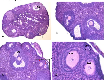
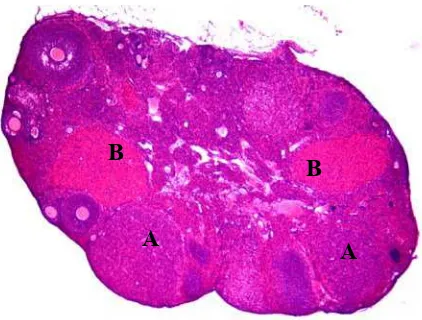
Figure 5.1. Photomicrograph of ovarian histology of ~20wk premutation mice using Hematoxyllin-Esoine (HE) staining. Magnification 40x for A, 100x for B, C and 400x for D. The pictures are showing all of stage of ovarian follicles. (A) showing graafian follicle (GF) determined by size with transparent vesicle (anthrum), cumulus oophorus and its position which protrudes from the surface of the ovary. (B) showing anthral follicle (AF) determined by
GF
AF
PAF
PAF PF
A
B
diameter <310 m with anthrum and its position near cortex of ovary. (C) Showing preovulatory follicle (PF) determined by diameter >370 m with anthrum and its position near cortex of ovary and preanthral follicles (red arrow). (D) Showing Primordial follicle (black arrow and circle) recognized by the mean diameter of the follicle (< 20 m), partially or completely encapsulated by one layer of flattened squamous pregranulosa cells and consist of oocyte, and preanthral follicle (PAF) recognized by its diameter 100-220 m without transparent vesicle (anthrum).
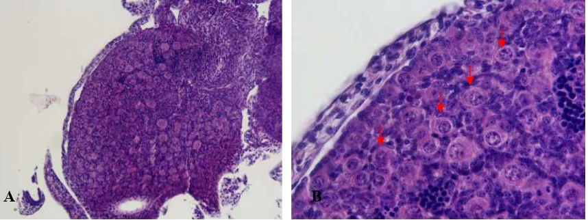
Ovarian histology of premutation mice were compared to wt mice. All stages of follicle were showed in ovaries of adult (~20wk) premutation mice, showing all stages of follicles (See Figure 5.1), and from P6 ovaries only primordial follicles is showed (See Figure 5.2).
Figure 5.2. Photomicrograph of ovarian histology of P6 premutation mice. (A) showing only primordial follicles in ovary of P6 mice. (B) showing primordial follicle (red arrow) recognized by the mean diameter of the follicle (< 20 m) and morphological criteria (primordial follicles are in rest and consist of an oocyte partially or completely encapsulated by flattened squamous pregranulosa cells). Magnification x100 (A) and x200 (B). Hematoxylline-Eosine Staining.
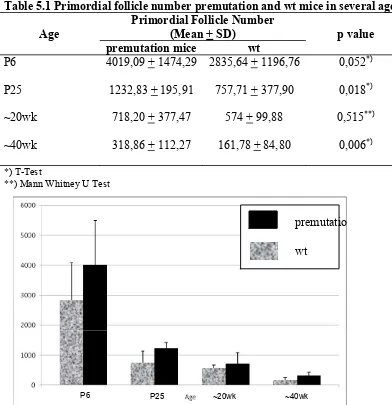
The number of primordial follicles were quantified in ovaries from wt and premutation mice at different ages to study the effect of expanded CGG repeat on the folliculogenesis. The means of number of primordial follicles from P6, P25, ~20wk, ~40wk mice are depicted in table 5.1. There is no significant difference was detected in the number of primordial follicles between wt and premutation mice in P6 and ~20wk (p=0.052 for P6 mice, p=0.515 for ~20wk mice). However
at P25 and ~40wk premutation mice the number of primordial follicles was significantly higher compared to wt mice (p=0.018 for P25 mice, p=0.006 for ~40wk mice).
Table 5.1 Primordial follicle number premutation and wt mice in several ages
Age
Primordial Follicle Number
(Mean + SD) p value
premutation mice wt
P6 4019,09 + 1474,29 2835,64 + 1196,76 0,052*)
P25 1232,83 + 195,91 757,71 + 377,90 0,018*)
~20wk 718,20 + 377,47 574 + 99,88 0,515**)
~40wk 318,86 + 112,27 161,78 + 84,80 0,006*)
*) T-Test
**) Mann Whitney U Test
Figure 5.3. Primordial follicle population in P6, P25, ~20wk and ~40wk premutation, and wt mice. The difference of primordial follicle number was statistically significant in P25 and 40wk premutation mice compared to wt. but it was not in P6 and 20wk premutation mice.
P6 P25 ~20wk ~40wk
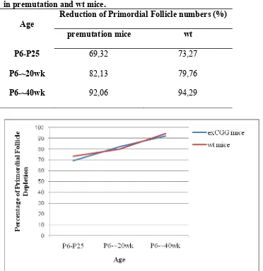
Following this results, Primordial follicle depletion in several age were counted to determine the progressivity of primordial follicle depletion in premutation mice and wt. The percentage of primordial follicle depletion are depicted in Table 5.2 and Figure 5.4. Primordial follicle depletion in premutation mice from P6 to varying age are lower than wt mice.
Table 5.2. Percentage of primordial follicle depletion from P6 to varying age in premutation and wt mice.
Age
Reduction of Primordial Follicle numbers (%)
premutation mice wt
P6-P25 69,32 73,27
P6-~20wk 82,13 79,76
P6-~40wk 92,06 94,29
5.1.2. Recent corpora lutea identification in premutation mice
The presence of recent ovulations in ovaries from premutation mice were compared to wt mice using recent corpora lutea identification to identify the early cessation of ovulation cycle. Recent corpora lutea were detected by its appearance as a large rounded or irregularly shaped structure consists of lutein cells without oocyte in the middle.
Figure 5.5. Photomicrograph of ovarian histology of premutation mice. (A) showing recent corpora lutea determined by its appearance as a large rounded shaped structure without oocyte in the middle and stained homogeneously bright blue because it is contain of lutein cells. (B) showing old corpora lutea determined by its appearance as a large irregularly shaped structure stained homogeneously bright red because of its cellular elements degenerate and undergo autolysis and change with fibrous tissue. Magnification x 40.
There is no corpora lutea from P25 ovaries. Recent ovulation were showed only in 60% of 20-week-old premutation mice (n=10), whereas in wt mice recent ovulation were detected in all ovaries studied (n=6; 100%). There is no significance difference of these features between premutation mice and wt mice (p=0.234). (See table 5.3)
A total lack of recent corpora lutea was found in 40-week-old premutation mice (n=7), whereas in wt mice 78% (n=9) of ovaries contained recent corpora
B
A
B
lutea. Using Fischer exact test there is significance difference of these features between premutation mice and wt mice (p=0.003). (See table 5.4)
Table 5.3. Proportion of presence recent corpora lutea from ~20wk premutation mice compared to wt mice
Mice group Recent corpus lutea p value*)
Yes No
Mice group Recent corpus lutea p value*)
Yes No
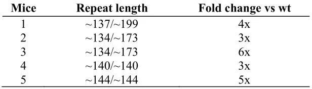
Fmr1 mRNA levels were analyzed in P25 ovaries from premutation mice
using Reverse transcriptase and Quantitative PCR (RT and Q PCR) to support the hypothesis of RNA gain toxicity in POI. Using specific primer sets for Fmr1
Ct. 2- Ct results in fold change. The fold change of Fmr1 mRNA levels are depicted in Table 5.5.
Table 5.5. Fold change of Fmr1 mRNA levels in P25 ovaries from
premutation mice compared to wt mice at the same age.
Mice Repeat length Fold change vs wt
1 ~137/~199 4x
2 ~134/~173 3x
3 ~134/~173 6x
4 ~140/~140 3x
5 ~144/~144 5x
Fmr1 mRNA levels were elevated 3-6 fold with an average of four-fold
compared to age-matched wt mice.
5.2. Discussion
POI can occur at various stages of follicular development and could be related to 1) an initial decrease in the primordial follicle pool, 2) altered recruitment of the dormant follicle, 3) interrupted maturation of the follicle or 4) increased follicular atresia. FMRP, the protein product of the FMR1 gene, is
expressed in several tissues that may influence any of these development stages: in the brain, in the region that is critical for hormonal regulation, in the primordial germ cells in the fetus, and in the granulosa cells and oocyte of developing follicles in adults (2). Thus, histopathological studies in ovarium are important to understand POI.
Bachner et al (1993) demonstrated that FMR1 serves a special function
Drosophila showed that dFMR1 null ovaries contain egg chambers with both
fewer and supranumerary germ cells (42). FMRP also controls the level of Cbl
mRNA in the ovary by reducing Cbl gene dosage (42). These results suggest that
FMRP controls germline proliferation during oogenesis by regulating the expression of Cbl in the developing ovary. Cbl gene is responsible for germ cell
proliferation. In Drosophila, dFMR1 is also known to be involved in germ cells
and oocyte specification (43).
Immunohistochemistry study demonstrated that in murine ovaries, Fmrp is predominantly expressed in granulosa cells and oocytes of developing follicles (39). Willemsen et al (2009) demonstrated that Fmrp expression in ovary of premutation mice is enhanced compared to wt mice.
Primordial follicles are formed just after birth in mice, and this pool of primordial follicles constitutes the complete supply of ovarian follicles during reproductive life (48). Initiation of primordial follicle growth occurs as soon as they are formed, but not all primordial follicles start to grow at the same time and some will remain dormant for months in rodents and even for years in the human . The factors that regulate initiation of primordial follicles growth are still largely unknown (18).
The absence of a reduced number of primordial follicles in P6 premutation mice suggests that the fragile X premutation has no effect on the initial pool of primordial follicles from birth. In fact, this results show a trend in the opposite direction, that is a higher number of primordial follicles in premutation mice. This finding seems to contradict with hypothesis that there is a reduction in the number of oocyte at birth in female carrying premutation allele (36).
An explanation for a trend showing higher number of primordial follicles in premutation mice is that Willemsen et al (unpublished, reviewed in Dept of Clinical Genetics ErasmusMC Rotterdam 2009) demonstrated the expression of Fmrp in ovaries of premutation mice is increased than wt mice. The increase expression of Fmrp in ovary of premutation mice might cause the Cbl mRNA
levels lower than normal. Cbl gene is responsible for inhibition of germ cell
proliferation (42). The reduction of Cbl levels more than normal results in germ
cells over proliferation.
The underlying mechanism of this alteration in these mice has not yet been defined. A possible cause might be a RNA-gain of function-toxicity. Recently, a similar mechanism has been postulated for the elevated levels of FMR1 mRNA in
the development of an adult-onset neurological disorder (FXTAS) in males who carry the fragile X premutation.
Previous studies demonstrated that at the molecular level, all individuals with premutation alleles show elevated FMR1 mRNA levels, 2-10 times higher
than normal, in peripheral blood leukocytes, but normal or reduced FMRP levels (45). Using Q and RT PCR analysis using RNA isolated from mice brain, Willemsen et al (2003) showed a two fold increase of Fmr1 mRNA levels at
different ages in premutation mice, when compared with age-matched wt mice (20). Four-fold elevated Fmr1 mRNA in ovaries of 25-day-old premutation mice
in comparison with age-matched wt mice is reported here. Thus, these results confirm the earlier findings in brain tissue from the premutation mice (20). This finding does not mean that increase levels of Fmr1 mRNA is always causing an
altered initial recruitment of primordial follicles, because Tejada et al (2008) demonstrated that increased levels of FMR1 mRNA was found in lymphocytes
from a female carrying a premutation without POI. Actually, POI occurs only in 30% of female premutation carriers (66).
The unlinear relationship between FMR1 repeat size and POI in female
premutation repeat size between 80-100 have the highest risk to develop POI (13). Premutation mice that have CGG repeat sizes between 100-199 repeats were used, which does not fully reflect the highest risk group in the human situation. On the other hand, female carriers of the premutation are always heterozygous for the premutation allele, whereas our premutation mice were homozygous.