Vol 5, No 3, July - September 1996 Case
Report
149Large Solitary
Non Paracytic Cyst of
Liver:
Management
by External
Drainage
Vivek Agrawal*, Kumar Krishnan**
Abstrak
Seiak Michel nelaporkan kasus kista hati pertama pada 1856, banyak laporan tentang kista hati nonparasitik tanpa kotnplilcasi diiumpai dalatn lccpustalcaan. Penyakit ini ununtnyaasinûomatilç kecuali jika adnknmplikasi ataumembesar. Pielografi intravena perlu dilakukan untukmenyingkirknn adanya patologi pada ginjal. Hasil uemuasknn diperoleh dengan memasang drainase dengan binbingan radiologik. Tidak diperlukan anestesi u,nuû, atau laparotomi. Kateter dipertahankan selama 2-3 bulan.
Abstracts
Ever since in 1856 when Michel reported the first case cf hepatic cyst, much literature has been adlcd on the solitary uncomplicated nonparacytic cystic disease of the liver. This distinct disease entity renains largely asymptontatic unless it presents by virtue of its size or complications. Intravenous pyelogram should be nandatory to rule out any associated renal pathology. Instead of resorting to surgery
in all cases, placement of radiologically guided external dependent drainage catheter yields good results with excellent patient conpliance as no general anesthesia or laparotomy is required. For cure, only a tube coming out ofthe abdomen is to be enduredfor 2-3 months.
Keywords : Simple Hepatic Cyst, Cystic Disease of Liver, Cystic Hepatomegaly
Cystic lesions of the liver affect females
predominant-ll "î9
in
clinicalprac-trce.
"-
ultiple, affectingthe
rig
distinct from itspolycystic disease which
is
usually associated with polycystic disease of the kidney, and less commonly, the pancreas.* The originof
these cysts is thought to be as a resultof
embryonal maldevelopment in form on noncanalizationof
proximal anddiital
anlages orsequestration
of
aberrant ducts which become cysticby either infection or hyperplasia and degeneration.o These cysts are an incidental finding 1n O.5%
of all
autopsiesT and symptoms arise in one thirds because
of
either larg^e size causing pressure effects or when
com-plicationse,
like
rupture, infection, haemorrhage ormalignant transformation, occurs.
Various
modalitiesof surgeryl are practiced
for
managementof
symptomatic cysts(eg.)
external drainage, marsupialization, excision, internal drainage through Roux-en-Y cystoenteric anastomosis, and hepatic resection. Goldsteinlo was first to aspirate anddo
sclerotherapyby instilling
pantopaque. Similargood results are reported by Romerll who stated that surgery should be reserved for cysts which recur or
fail
to
resolve. External drainagertis
safe and effectiveway to manage uncomplicated cysts. Though it takes a
long
time
for
resolution,it
is
easily accepted bypatients and is free of complication. Progress of
heal-ing
can be assessedby
ultrasound and cavitograms (contrast X-rays through tube). Omentoplasty.can be done,if
residual cavity persists, at a later date.rj
Deparfinent of Surgery University College of MedicalScien-ces & Guru Teg Bahadur Hospital, Delhi, India
150
Agrawal and KrishnanCASE REPORT
A
32 year old housewife from Andhra Pradesh, Indiapresented with distension and dragging pain in aMomen
for past 3 months. There was no icterus, pallor or pedal
oedema. JVP was not raised. AMomen showed fullness
inupper 2/3 & fingers couldbe insinuatedunderthe right subcostal margin. The mass was smooth, cystic with a
fluid
thrill, having afirm
inferior border moving verywell with respiration. This mass was bimanually
pal-pable but could not be pushed in the right renal angle.
A
provisional diagnosis of either hydronephrosis of right kidney or hydatid disease
of
liver was made. Routineblood investigations were normal. Casoni's and IHA for hydatid serology were negative.
X-
ray chest showedraised
right
domeof
diaphragm and x-ray aMomenshowed
soft
tissue opalescencein
upper 213.IVP
showed a low placed right kidney with nonvisualization
of
upper calyces. However, USG aMomen revealed solitary uniloculated liver cyst containing debris ; normalkidney structure.
A decision of exploratory laparotomy was undertaken.
The kidney was normal on palpation.
A
large hepaticcyst was seen pointing on the posterio-superior surface of the right lobe. The cavity was drained of its 8 litres of clear
fluid
which became blood tinged later-due to either cyst collapse or operative manipulation.Cysto-enteric anastomosis was not done as it was decided that
it
wouldnot
function becauseof
its
nondependentsite-on the top of the liver, Hepatic resection was not undertaken as enough normal liver tissue was present
and apparently no features of malignant transformation
were seen. t* An externally draining Malecot's catheter
was left in the cavity and brought out sub-costally on the right side in anterior axillary line. Histopathology
of
the cyst wall showed fibrocollagenous tissue, cell ducts, nerve bundles, numerous blood vessels, inflam-matory cells and, possibly, smooth mrsclein
some areas suggesting a simple cystof
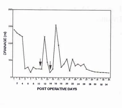
the liver. The sub-sequent daily drainage is shown in (Fig.1).It
reducedto about 400 ml per 24 hr. by the 5th day.
Sclerotherapy with intracavitory tetracycline solution
was attempted
on
llth
andl4th
days but rebound increaseof
daily outputto
>
12OO ml per 24hr. andoccurence of fever and pain prompted abandoning
of
this therapy. Subsequent drainage fluid cultured E.coli,
but no treatment was given as patient had no
constitu-tional symptoms. A radiocontrast cavitogram through
the Malecot catheter after 3 wks showed a small cavity.
Except for discomfort of the tube, the patient tolerated
the therapy well. She was discharged from the hospital
after 5 weeks with a daily drainage
of <
l0O ml. andMed J Indones
2 a 6 a to 12 t4t6 la â 2 2a æ ao eil
POST OPERATIVE DAYTS
Fig 1. Daily Drainage Chart
adequate instructions for tube care, and to record the daily tube output.
Two
months later, the patient cameto
the surgeryoutpatient department and stated that the tube had slipped out and that the daily output was < 20 ml at that point
of
time.A
repeat ultrasound showed irregularhyperechoic zone over the right lobe
of
liver withoutany cystic area-possibly scar tissue.
CONCLUSION
For simple uncomplicated symptomatic cysts
of
the liver the methodology of external drainage, preferablydependent, is a good proposition. Reviewing the above
case, the externally draining catheter could have been
easily placed through the services of an interventional
radiologist, thus avoiding the aMominal scar.
It
is, therefore, possible to advocate this form of treatmentas first line of therapy.ll
All
advantages of this mini-mal intervention are apparent except that the patienthas to bear the tube coming out of the aMomen for an
approximate period
of
3 months; to which the patientreadily agrees as laparotomy under anaesthesia is for-gone.
REFERENCES
1. Haded AR, Westbrook KC, Graham GG, Morris WD,
Campbell GS. Symptomatic paracytic liver cysts. Am J Surg
1977; 134:73944.
2. Petroski D, Stafford C, Coodley EC. Cystic liverdisease with
.associated hypernephroma. Am J Gastroentrol L973;59: 255.
3. Willeamson RCN, Ramns NI, Shorey BA. Congenital solitary cyst ofthe liver and spleen. Br I Surg 1978; 65: 871.
[image:2.595.346.561.81.270.2]Vol 5, No 3, July - September 1996
4. Fredman M. Polycystic disease of liver. Am J Gastroenterol 1958; 29: 83-6.
5. Von Mayenburg H. Uiber die cysten leber beitr: path Anat
l9l8;64:477.
6. Moschowitz E. Non paracytic cyst (congenital) of liver with a study of aberrant bile ducts. Am J Med Sci 1906;
l3l:
674-99.
7. Sanfelippo Plvt, Beahrs OH, Weiland LH. Cystic disease of liver. Ann Surg 1974; L79:922-5.
8, Burch JS, Jones HE. Large nonparacytic,cyst of liver
simulating ovarian cyst. Am
I
Obstet Gynecol 1952 63:44t-4.
9. Ackman FD, Rhea LJ. Non paracytic cyst of liver; their clinical and pathological aspects. BrJ Surg l9l3; lg: &g-54.
10. Goldstein HM, Carlygle DR, Nelson RS. Treatrnent of symptomatic hepatic cyst by percutaneous instillation of Pantopaque. Am I Radiol L976; 127:85G3.
ll.
Romer CE, Fencci IT, Mueller pR, Simeone IF, van Son-nenbrg E, WittennbergI.
Hepatic cysts: Diagnôsis and therapy by sonographic needle aspiration. Am I Radiol 19gl;
136: 1065-70.12. Flagg RS, Robinson DW. Solitary non paracytic hepatic 'cysts. Arch Surg 1967;95:964.
13. Goldsmith HS. Omental transplantation. Rev Surg L967;24: 379.
14. Americks f, Appleman H, Frey C, Àfalignant non paracytic Cyst of the liver. Ann Surg 1972; 176: 7 L3.