Vol7, No 7, Jan:.uary - March 1998 Aspirated pearutt 49
Chest
X
Ray
Examination
of
Aspirated
Peanut
in
Children:
Is
it
Imprortant
?
Efiaty
Arsyacl
SoepardiAbstrak
Metode diagnostik yang alarat untuk menegakkan diagnosis aspirasi kacang pada anak sangat iliperlukan bagi ahli TIIT. Telah
dilala*an suatu penelitian ràrospektif untuk menilni peranan pemeriksaan foto toraks sebelum tindalan bronkoskopi dilahtkan Tujuan penelitian
ki
arialah untuk menintukan sensitivitas ilan spesifisitas pemeriksaan foto toralcs sebelum dan sesudah 24 jam teraspirasi -korongyang dikonfirmasikan dengan adanya kacang yang teilihat walûu bronlaskopi. Delapan
puluh
sanbilan penderita aspirasi*or.oij aolong
ke BagianTIIT FKUI, Jakarta, dari bulanJanuari 1986 sampai denganDaçember 7996. Usia mereka berkisar antara s sanpai SA bulan. pemeriksaan foto toraks tid,ak dilakukan pada 5 penderita karena adanya gawat napas pada 3 kasus dan 2 penderita menoiakuntuk dilalaùan tindakanbronkoskopi, olehkarena itu tak diitatkan dalam penelitian ini.Analisis statbtik menunjukkanbahwasensitivitas pemeiksaan foto toraks 5
j,lVo,
spesifisbas j j,3Vo, nilai dugaan positif 95,6Vo dan nihi dugaan negatif 2,6Vo.Abstract
An accurate method of detecting peanut aspiration in children's tracheobronchial tree, is very important Ûo otolaryngologist'
A
retrospective study to analyze the role of chest X-ray examination as a diagnostic test betore bronchoscopy was conducted. The objectiveof this study is to determine the sensitivity and specificity of plain chest X-ray examination in diagnosing Peanut aspiration which was confirmed by bronchoscopy, before and after ?A hours of aspiration. Eighty nine cases of aspirated peanut in children were found during 1l- years period, from January 1986 to December 1996, in the Department of Otorhinolaryngology, Faculty of Medicine, University of
Indlnesia, Jakarta. Their ages varied from 3 to 58 months. Chest X-ray was not performed in 5 patients because of violate respiratory distress in 3 patients and 2 patients refused the bronchoscopy procedure, therefore they were excluded from the investigation. Statistical analysis revealed that the sensitivity of chest X-ray examinati onwas 53.lVo, the specificity was 33,3Vo, the positive predictive value was 95.6Vo an.d the negative predictive valuewas 2.6Vo.
Keywords: Aspiration, bronchoscopy
The diagnosis
of
peanutaspiration
in
children is
still
a big problem for otorhinolaryngologist. Thechildren
themselves
could not give
anexact
historical
review
and the
symptoms
of
respiratory obstruction might
bedisappeared
when
they
arrived
atthe hospital.
Peanutitself is a râdioluscent foreign body,
and can
not be
detected
by
radiological
examination
while
the
bronchoscopy
is
an invasive diagnostic
Procedure.Since the
peanut
can occlude the airway
suddenly,early diagnosis
andprompt treatment is
mandatory.A
long-period obstruction
due toit
might
cause edemaof
the mucosa and constriction
of
the lumen
of
thetracheobronchial
tree, and thenfollowed by lung
com-plication.
Department of Olorhinolaryngology, Faculty of Medicine, (Jniversily of IndonesialDr. C ipto Mangunkusumo Hospital, Jakarta, Indonesia
The objective of
this study is to
determine
the
sen-sitiviy
andspecificity
of
chest
X-ray
asa
diagnostic
tool in
aspirated
peanutbefore
and
after
Z
hours
of.
aspiration and
to asses
the diagnostic
accuracy
of
a peanutin
the
airway by
comparing
plain
chestX-ray
and bronchoscopy findings.
Both
procedures are
evaluated
in
the
fint,
second andthird
day
of
aspira-tion.
METHODS
A
retrospective
study
analyzedthe role
of plain
chestX-ray
as adiagnostic
testin
suspected aspirated peanutin
thetracheobronchial
tree. Themedical
recordscon-taining the patient's identity, the history
of
peanutaspiration,
the physical examination including
signsand
symptoms,
otorhinolaryngological
examination,
and radiological and bronchoscopy findings
were
reviewed.
Time interval
between
aspiration and
the50 Soepardi
recorded.
Since peanutis
aradioluscent foreign body,
it
can
not
be
detected
in
radiological
examination;
hence
the
search
of
the
subsequent
result
i.e.
lung
complication
such as atelectasis, emphysema, infiltrateor
pneumonia was
conducted
by a
radiologist.
Radiological
examination
wasclassified
aspositive
if
there
was lung
complication caused
by
thelodging
of
peanut
in
thetracheobronchial
tree, and negativeif the
lung was normal. Positive
bronchoscopy meant that
there
was a
peanut
in
the
tracheobronchial
tree and
negative if
it
was
not
found on bronchoscopy.
Eighty
nine
cases
were found during 11
years,
between
January
tg8e
to
December 1996
in
our institution.
Chest
X-ray and
bronchoscopy
procedureswere
per-formed
in
only
84 cases.
Three patients undenvent
bronchoscopy
without
prior
chest
X-ray
becauseof
violate
respiratory distress and
2
patients
refused
bronchoscopy
procedure,
hence
they were
excluded
from
this
investigation.In
the 84 cases the chestX-ray
was doneon
admission
atthe Department
of
Radiol-ogy,
andthe bronchoscopy
u/asperformed
by
using
arigid
bronchoscope
under general aenesthesiaby
otor-hinolaringologist.
RESULTS
Eighty four patients which
meet
the criteria
of
this
study
were analyzed.
They were
45boys
(53.67o) and 39 girls (46.47o),thus
the sexratio was
1.1.:
1. Thesepatients'
age ranged
from
3 to
58
months,
with
the meian age of ?Â3months.
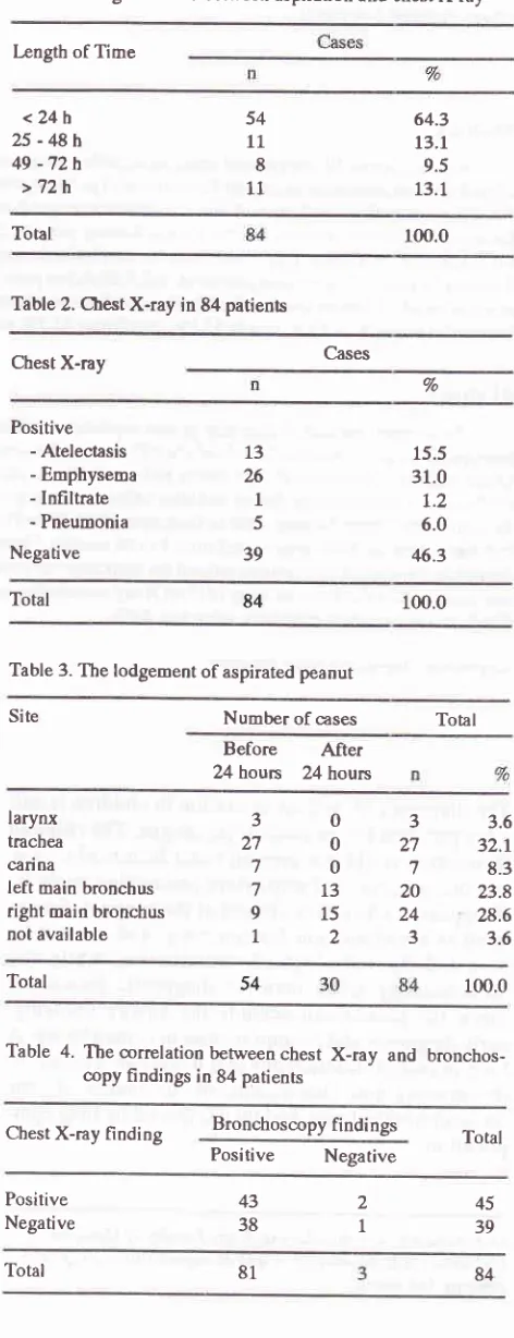
Fifty
four
patients (6a3Vo)
camein
the
first
Z
hours(0-24 hours), 11 patients
(l3.lVo)
in
the
second
Z
houn (25 up to 48
hours),
8 patienûs(9.5%)in
thethird
24 hours (49
up to
72
houn) and
the rest
11 patients(l3.l7o)
came after 72 hoursof
aspiration (Table
1).The
radiological
examination
revealedpositive
result
in
45
patients
(53.7Vo)
and negative result
in
39patients
(46.3Vo)(Table
2).Bronchoscopy
which was carried out
in
all
patientsindicate
the position
of
the peanut, however
in
3patients
the
peanut was
not
found
in
the
tracheobronchial tree (Table
3).Table 4 shows the
correlationbetween
chestX-ray
andbronchoscopy
findings
in
84
patients.
True
positive
was
found in
43 patients, false
positive in 2 patients,
true negative
in I
patient and false
negative
in
38 patients.Diagnostic
test analysis revealed that thesen-sitivity
of
chest
X-ray
examination
compared
with
bronchoscopy investigation as a gold
standard
was 53.I7o, the specificitywas
33.3 7o,positive predictive
Med J Indones
value was 95,6Vo
and negative
predictive value
was 2.67o.Table 1. tængth of time between aspiration and ,r:hest X-ray
Length of Time
<24h
25 - 48h 49 -72h >72h
64.3 13.1 9.5 13.1
54
11
8
11
Total 100.0
Table 2. Chest X-ray in 84 patients
Cases Chest X-ray
Vo
Positive
- Atelectasis
- Emphysema
- Infiltrate
- Pneumonia
Negative
13 26
1
5
39
15.5
31.0
L.2
6.0
46.3
Total 84 100.0
Table 3. Tbe lodgement of aspirated peanut
Site Number of cases Total
Before
After 24 hours 24 hourslarynx
trachea
carina
left main bronchus
right main bronchus
not available
303
27027
707
71320
9L524
L23
3.6
32.r 8.3
23.8 28.6
3.6
Total 54 100.0
Table 4. Tbe correlation between chest
copy findings in 84 patients
X-ray
andbronchos-Cbest X-ray finding Bronchoscopy findings
Positive
Negative TotalPositive
Negative 4338 2
I
4539 [image:2.595.303.539.131.745.2]VoI 7, No 1, January - March 1998
Table
5 showsthe
54patients
that camein
the first 24 hours.Of
those cases chestX-ray
revealed 17positive
and37 negative
findings; on the
other
handbronchos-copy
resulted
in
53 positive findings
and L
negativefinding. Analysis of
diagnostic
test in the firstZ
hours revealed that thesensitivity
of
chestX-ray
was 30.zEo,specificity
was O7o, positive predictive value was
94.17o
and negative
predictive
value was
0%'
In
the
thirty
patients
who
came aftet 24
hoursof
aspiration,
the chest
X-ray
examination
results werepositive in
28patients
and negative
in
2
patients.
Bronchoscopy
results were
positive
in28
patients
andnegative
in 2
patients. Diagnostic analysis
revealed
that the
sen-sitivity of chest X-ray
after
24 hoursof aspiration
was 92.97o,specificity
was
O7o,positive predictive value
was 92.97o and
negative predictive value
was 0%.Table 6 shows the
correlation between
chestX-ray
ands, and
the lodgment duration
of
obronchiql
tree.
CbestX-raY
of
ame
in
25 up to 48
hours after
aspiration
was 9positive
and 2 pegative, andbronchos-copy
findings
showed
L1 posidive and none wasnega-tive.
Sensitivity of
chestX-ray
in
25-48houn after
thelodgement of
a peanut was 81.87o. ChestX-ray results
of
the
8
patients that
came
in
49
to
72 hours
after
aspiration was
8 positive and nonewas negative,
andbronchoscopy showed
7positive
and
1negative.
Sen-Aspirated
peanut
51sitivity
of
a chestX-ray
in 49-72
hoursafter
the lodge-ment of a peanut was lOO7o. Eleven patientswho
came after 72 hours afteraspiration,
the chest X-ray showed11
positive findings and none
was negative,
while
onbronchoscopy
10 was positive and 1 was
negative'
Chest
X-ray sensitivity after
72 hoursof
thelodgement
of
a peanutwas
L00%.The
sensitivity of chest
X-ray
in
the first day was 30.27o, the second day was 81.87o,the third day
was
IOO% and after 72 hourswas lo07o'
DISCUSSION
Peanut
apiration
cases are themost
fr-equent caseof
all
foreign body aspiration in children.l
'2'l'4
Apeanut
hasa high
concentration
of
fat
and
is
hygroscopic,
there-fore
its
lodgement
in
the tracheobronchial tree
will
cause mucosal edema and the
airway can be
occluded
in
a
short
time.
A
peanut
in
the larynx
and
tracheausually
can occlude theairway
directly
andrespiratory
distress
can occur suddenly. However,
a
peanut
in
carinawill
causelung complication
in the affected sideafter several days. Therefore, the radiographic
ex-amination
may
be misleading
if
it
is
normal.
The peanutmay not produce
sufficient
obstruction,
or the
obstructive
phase may not have developed.Frequently
only
obstruction of
a segmenof lung
is
demonstrated,because the peanut ciluses enough
reaction
to occlude
a branch
of
thetracheobronchial
tree.Table 5. Chest X-ray and bronchoscopy findings bpfore and after 24 hours of aspiration
Bronchoscopy findings
Chest X-ray findings Before 24 hours After 24 hours
n (+) n (-) n (+) n (-)
Positive Negative
16
37
1
0
2 0 26
2
[image:3.595.48.476.625.741.2]Total
Table 6. The correlation between chest X-ray, and bronchoscopy findings, and the lodgement duration of peanuts
Bronchoscopy findings
Chest X-ray [indings o
-24h
25 - 48h 49-72h
>72h
28 53
(,
(+) (-)(+) (-)
(+) (-)
(+)
1
0
9077
2000
10
t6
37
10 0 Positive
Negative
52 Soepardi
Sakurai et
all
from
OsakaMedical
School reported that 55.3 Voof
the casesof
foreign body aspiration
showedradioluscent foreign body and the majority was
apeanut.
Mu
etal' in
their
studiesduring
7 years,found
that 677o
of
the 378 casesof
foreign body aspiration
atthe
Shengyang
Hospital
in China aspirated a
peanutand
6O%
showed abnormalities after more than
24hours
of
aspiration.
Esclamando
et
al3 reported
that 677oof
peanut
aspiration_caseswill
suffer
lung
com-plication
after 24 irours.a'5' 6This study evaluated
therole
of
chestX-ray
examina-tion in
detecting
a peanutin
the
airway. In this
studylung abnormalities were found
in
63.67oof
cases, andthen
we analyzed
the sensitivity and specificity of
chestX-ray
as adiagnostic
test.Diagnosis
of aspirated peanutin
children by
chestX-ray.examination did not
show
high sensitivity
and
specificity. Its sersitiviy
was
53.17o andspecificity
was
33.37o. Thediagnostic
sensitivity
was
increasedaccording to
theduration
of
aspiration,
on thefirst
day
itwas
30.2%o, on the secondday
it
was
81.87o and on thethird
day
it
was l0Ù7o.Although plain
chest
X-ray provide a good
tool
for
studying pulmonary
complication,
fluoroscopy
would
be better in
diagnosing
a peanut in thetracheobronchial
tree.' Inspiratory
andexpiratory anteroposterior
chestX-ray
are suggested,while
on thecontrary
it
isimpos-sible
to
askchildren in
respiratory
distressto
do deepinspiration and expiration. Therefore
bronchoscopyshould substitute those mentioned
procedures
as
adiagnostic
tool
in
diagnosing
a
peanut
in
the
tracheobronchial tree,
within
the
first
24
hours of
aspiration.
Med J Indones
In
conclusion, abnormal chest
X-rays were found in
29.62%
of
patients
who
aspirated
apeanut
within
thefirst Vl.houn,
but abnormal
chestX-ray
was found
in
86.67o
of
patients after
24hours.
plain
chestX-ray
is better done in aspirated peanut inchildren which occur
more than
24
hours. Bronchoscopy should be
per-formed
immediately in
caseswith airway
obstruction
that are warranted
by history
and
physical
examina-tion.
REFERENCES
1.
SakuraiK
Iwata S, Takasu A, Takeda N, Kaso R. Foreignbodies in the esophagus, trachea and bronchi handled in our clinic. In: I-ee, Inouye T, Fukuda H, Sato T, editors. Recent advanæs
in
bronchology. Amsterdam: Elsevier,l99O:449-50.
2.
Iskandar N. Ingested and inhaled foreign body in Dr. CiptoMarigunkusumo Hospital Jakarta. Indones Med
J
Otor-hi nola ryngol I99 4:31,1-8.3.
Mu
L,
SunD,
He P. Radiological diagnosisof
aspiratedforeign bodies in children. Review of343 cases. J laryogol Otol 1990;104:778-82.
4.
Esclamando RM, Richardson MA. Laryngotracheal foreign bodiesin
children:A
comparisonwith
bronchial foreignbodies. Am J Dis Child t987;I4:259-62.
5.
Kirks DR. The pediatric ER chest: What every radiologist should know. In: Nash DCH, pefterson H, editdrs. pediatric radiology. l-ondon : Meri t Communi cati ons, 1992: 15 4 -j L6.
Healy GB. Management of tracheobronchial foreign bodiesin
children:An
update. Ann Otol Rhinol Laryngol 1990;