Proceedings
The Joint Meeting of Conference and Congress
of Asian Society of Veterinary Pathology
(ASVP) 2011
&
) セ@The 10
th
Scientific Symposium of Indonesian
E
Society of Veterinary Pathology (lSVP) 2011
The Role of Veterinary Pathology Animal Health for
Improving Eco-Health
IPB International Convention Centre
Bogar, Indonesia, l'Jovember 2011
Faculty of Veterinary Medicine
Bogor Agricultural University 20 I I
All right reserved. No part of this book may be reproduced in any form without
pennission in writing from the publisher, except by reviewer who wishes to quote brief
passages in review written for inclusion in magazine or newspaper.
Perpustakaan Nasional Indonesia Cataloguing inPublication Data
The Joint Meeting of the 5th Conference and Congress of Asian Society of Vetrinary
Pathology (ASVP) and the 10th Scientific n Symposium of Indonesian Society of
Veterinary Pathology (ISVP), 2011,
The Proceedings of the Joint Meeting of the 5th Conference and Congress of ASVP and
the 10th Scientific n Symposium of ISVP, November 2224,2011,
IPB International Convention Centre, Bogor Indonesia.
ISBN; 9789794933718
Typest and Printed by
Penerbit IPB Press
Contact Address
ASVP 2011 Organizing Committee
Division of Veterinary Pathology
Department of Veterinary Clinic, Reproduction
&
Pathology
Faculty of Veterinary Medicine
Bogor Agricultural University
Wing 6th
&
7th, 1st Fl.
.II.
Agatis, Campus IPB of Darmaga
Bogor 16680, INDONESIA
Phone/Fax. +622518421807
Email ;[email protected]
Website: http://asvpbogor201I.event.ipb.ac.id
Invited Speaker
Page
OPS 1
OPS:2
OPS 3
OP54
OPS 5
OPS 6
OPS 7
POI P 02I'
03
P 04
I' 05
I'
06
TGFbeta Niche Regulation for Better Therapy
jinKyu Park! , EUI1lv[i Leel !, Ah Young Kin/!, Sang Young You!!. s・ッョMyッャャャQセ@ HOI/! . ElinJoo Le/ c, and Kl'llShik
Jeong l :' '
Pathological findings and Molecular charcteristics ofPRRSV
isolated from infected in North of Vietnam
Ng/lyen Thi Lanl. Nglll'en HUll
Na1/1
L. Ngl;yenBa
Hierl Le ,Huynh Thanh P/lIIong
L NgllyenThi
hッ。セN@Ryoji YOll1agllchi-Rat Tumor Models and their Applications, Pariicularly
for Renal and Hepatic Fibrosis
Jyoji
YalllateVascular Pathology Associated with Experimental
Pastellrel/a IIlItltocida
serotype: B:2 Infection in Calves
S. J(lSni.
E.lY1.
AlIIl1(l.At
Z(JI/Iri-Sa(/d. Z Zakario, S.S. Arslwd.A. R. Ol/wr
andT
I.
A:::llliChallcnges in safety evaluation ofbiomaterials. biomedical
devices and engineered tissue constructs
TV Ani/kllmar. R Deepa (ll1d
Al
JaseerDiagnostic Classification and Detection of Minimal Residual
Disease (MRD) in Canine Lymphoma
セbャャャQァウゥーゥーッエL@ N :\4wwc//(/i. J Clwyapollg, S. Wangl1uifhol/l
ond S.
TechollgulIIs/l\\'(//i
The Development of Porcine Reproductive and Respiratory
Syndrome Virus Vaccine
Min Yuan Chid', Hui Ting Chan", Yi Yin Do",
PllngLing Huang". Victor Fei Pang", ChianRen
Participant
Pathology and Veterinary Pathology ChallengeFuture
Pcrspective
1.1. A/
SlIlfatlUltra structural Characteristic of Blood Cells in Giants
Freshwatcr Stingray
(Hil//(/lIfllnt clwop/iruya)A.Sailasl/to. Y. ;\Iat/lIJra,
N.
CI/(/I1SII(,Analysis of
1'53and PCNA Expression in the Genital and
Extra genital CTVT Microarray
S. Jon/alltsa.
K.
T(lIIgfrogoon.P.
Kli%fha,S.
Bolflwisollf!,.S. iVallgnait!II//II. P. TO/lgkOll'Olfana, A. Rl/ngsipl/)(It. S.
To ngkOlI'atto110
Comparing Expression ofHERR2 in Malignant Feline
Mammary Tumors
Tissue Microarray
tV
RlIngrass(/l/Iee. P. Tml'Ornpanich. W. Jilltowes, S.Bulfhaisong, S. Wal1gnaifhlll1l. P. Tangkmraffmw. A.
Rllngsipipai,
S. TClllgkall'affonaInvestigation of the Death of Java Rhinoceros (Rhinoceros
/
sondaicus) in Ujung Kulon National Park
Adhi Racl1llI(Jf Slidraiat h。イセャGo、ゥN@ Halldayani, Agl/s
pイセャGoャiiィャャ、ゥL@ Ridlmn Seti(JlI'{I/I
Pathogenicity of a Pathogenic Filed Isolate NDV/Bali
1107in
Commercial Chicken Strain ISA brown
Anak Agllng
Ani /vliwh
Adi,iHade
Kardel1a. AセイッOャャ。ャャ@ ,HanfikAsfmi'(J Yoshihiro Hayashi.
YaslIl70hll
AlotSl/lllotoP07 Immunohistochemical Characterization of Macrophages and 37 Myofibroblasts in Biliary Cirrhosis due to Liver Fluke
(Fasciola spp) Infestation in Cattle
HM Golbar. V Junianfifo.
C.
Ichikawa. M. Tanaka.T.
[::.m!'a.Af KlIlI'amlira. OJ. 'lamale
P 08 Expression of Metallothionein Protein in Malignant Feline 40
Mammary Tumors by Using Tissue Microarray P. Siffhicharoenchai.
P.
Alarlow. P. Prufthifhmrorn. S. Sirivaidyapong.S
Wangnaithllll1 . .S
Tangkall'utfana, A. RUl1gsipiputP 09 Genetic Characterization and Phy logenetic Analysis of Thai 43
Canine Distemper Virus (CDV) Isolates
S
TechangamslI\\'{ln, N Charoensiml. A. Radranakatikanol7. A. RlIl1gsl/JI/)(ItPIO In Vitro Antimicrobial Activity of Saccharomyces cerevisae 46
Against Streprococclis agalactiae Isolated from Nile Tilapia.
K Pinpill1ai. P. Kaywlsalllrlwj.
C.
Rodkhwl1. A. POl1pornpisit. LV. pirarat.PI! Necrotizing Granulomatous Lesions of Uterus in a Dog by 48
Nematode Larvae in the family ofOnchocercidae
JK Park A
Y
Killl, El'" Lee.l.:.J.
Lee, SY. YOII. SY. Han. DAf KH·ak. TH Kim. KS. JeongPI2 Phylogenetic and RFLP analysis of Canine Distemper Virus 51
Fusion (F) Gene from Domestic Dogs in Thailand
A. Rac/tanakatikanon. N. Clwroensivu/. J. Kemrchoroen. K. Oraveeraklil. K Sliwo/lnokaril.
r
Poovorowo!1.S
Techongwl/s 11\\'(//1
P 13 Immunohistochemical Detection of Macrophages and 54
Myolibroblasts in rat Model of BleomycinInduced Scleroderma
Vetlli::.ah Jl/lliO/lto. Cliisa [chikm\'(/. Takeshi 1::'<7\m. lv[itslirII Kill \'(/11 /(/I'll. Jroji 'l(II11ate.
PI4 High Pathogenic Avian Influenza Infection in Wild Birds of 56
Korea.
OlinKl'Ong 1"'0011. WooHee Park kyャャャQァMヲセ|Giiョ@ Lee. InSoon Rooh. Moo/1 Yo ling Rhyoo. Hl'eRyoung Kim. YOII Chan Bae. OSoo Lee
P 15 Ganglioneuritis of Indonesian Abalone 57
Dewi Ratill AglIllgprimno. Mm\'aI'Sllballgkit. Agus SlIl1al'to
P
16 Canine Multiple Epithelioid Hemangioma in Labial Mucosa 59Elv!. Lee. A
Y.
Kil1l. EJ. Lee. BS. Joo. SY. YOII. SY.Han.
YHYOOI1. KS. Jeong
PI7 Histopathological Changes and Apoptosis Detection in Canine 62
Myxomatous Mitral Valve Disease Using Tissue Microarray Technique
T. Jiranantasak, A. Rungsipipat, S. Disatian
PI8 Case Report: Infectious Canine Hepatitis and Colibacilosis in 65
Malayan Sun Bear (He/arc/os Afalayanlls)
Agustin [ndrm\'afi
P 19 Generation of Hepatocellular Carcinoma animal Model in 67
Sparoglle Dmrfey Rats
Aglls Setil'Ol1o. [ndriayzmi Prahastllti. Elpita Br Tarigall. fJkterono
Dlt'i
BlI(Zvati. Andi [llamaP
20 Case Report: Ovarian Lipoma in a Dog 70JH Park A
Y.
Kim. EJI Lee.sr.
YoII. SY. Han. EJ. Lee. BS.100. AI Mlln. I'll.. Yoon, KS Jeong
P 21 Decreased Myostatin Moves Up Fetal Myogenesis in Somatic 72
Cell Nuclear Transferred Dog
P 22
Ceruminous Adenocarcinoma with Psammoma Bodies in
75
External Auditory Canal of a Dog
A Y Kim, E/v1 Lee, SY YOIl, SY Han, YR. Yoon. KS Jeong
P 23
Production and Characterization of Plyclonal Anti DomainI
78
AlphaFetoprotein Antibody
AglIs Setiro/lo, Elpita BI' Tarigan, IlldriaYlini Pralwsfufi. , Akferono Dwl Blldmfi, Andi Ufallla
P 24
Pesticide Poisoning in Korean Wild Birds from 2008 to 20
II 81OIlI1-Kl'o/lllg Moon, Moon- rOllng Rhyoo, Kyung-Hl'lIl1 Lee, In-Soon Roh. Woo-Hee Park, Dong-GYII Kim, Jin- YOllllg Shin, HYIlI1-Jeong Kl1'li/l, Clwe-III; Lim, O-Soo Lee
P 25
The Effect of Blackseed
(Nigel!([ satim)Oil Extracts on Mice
82(Mus musculus) Testicle Histopathology,
A, Rahllli, D,R, AglI!1gpr(I'0I10, S Estllnlllgsiit
P 26
Splenic Angiomyxoma in a Dog: The First Reported Case
85
BS, Jo, A Y Kim, Elf Lee, SY YOll, SY. Hcm, EJ. Lee, JR.
Park. AL MIIII, YR. YOOII, KS Jeong
P27
A Case of Hemangiosarcoma in a Jindo Dog
H7AL M'III, A
Y.
Kim, EM Lee, SY. Yo II, St'. Han. fJ Lee, BS.JO(), JR. Pork. KS Jeong
P 28
Foot and Mouth Disease Virus Infection in Young Korean
Black Goat
(Capra itirclIs)in Korea
KI'II/Ig-Hmn Lee, In-Sool1 Roil, Woo-Hee Park. MoolI- YOllllg R!tl'Oo, YOlIlIg-JOOII Ko, JOllg-Hl'eoll Pork, OIlIl-KnJllng Moo/l, O-Soo Lee
P 29
Acute Severe Melena Caused by Salmonella Paratyphii C
90
Infection in Javan Slow Loris Nycticebus Javanicus (A Case
Report)
[//fall Citronillgplltri, S//(/rlllilli JP, Kat/"e/e {S, rVendi P
P 30
A Case of Bacterial Septicaemia in a Malayan Tapir (Tapirlls
92indiclls)
Sr/\'(/lIlo, S, S//(//,il"lldill, :'vI. ZWllri-Saad
P 31
Effect of Garlic
(Allilllll sol/mill L)to the Immunity of Carp
94
Fish
(Cl'prillllS (,lII1)io)Infected by
EdH'ordsiel/a tonfa Kllmi({sih, Sya/'itiu/din TotoThe Application of Immunohistochemistry to Detect Prion
96
Protein (Prp) of
B(m'lIe Spongitiml/ El7cepjaiopafhy(BS E) in
Feedlot Cattle to Anticipate BSE Emergence in Indonesia
Rilli /)unwyanti
P 33
Mammary Adenocarcinoma with PanCytokeratin and Alpha
98
Estrogen Recepto Immunohistochemical Detection in Captive
African Hedgehog
Haエ・ャ・イjセLH@ a/hit'ellf!'is)kHOエイセ|G。@ ClwnkOlI', KOlllkiell' Pinpimai, Poison Tiellt/wi, Nopadul1 Pirarof.
P 34
The Suspect Atlatoxicosis in Toxicity Case Caused by
100
Aspergillus Fungi
WohYllni, Hadi PUrl/WIIO セヲOL@
P 35
Gross Pathology Report of a WildBorn Captive Sumatran
102Rhino
(Dicerorhinlls Sli17 W frellS is ),Torgamba
DR. aァオャWァーイセャGoャiッL@
AI
Suhangkil, S. EStlll1illgsih,D,
Condra, 4ndricll1syahP 36
Pulmonary Lesions of an Aged Sumatran Rhinoceros
105
Wilrin Wil7o!'sih, Vetni::ah Junianto, DeH'i Rati"
Agungpn)'Ono, Dac/wl D. SlIbrata.
P 37
Widespread Fibrosis Due to Uremia in an Aged Sumatran
107Rhinoceros
(Dicerorhinlls slIl1Iatrensis)Vetni:;ah JlIl1ianfo, Aglis Selil'Ono, BalJlbong Pontjo
PANCREATIC BETA CELLS EVALUATION AFTER TREATED
BY
Plwleria macrocarpa
(Mahkota Dewa) FRUIT EXTRACT IN DIABETIC MONKEY
E. Sulistiawati\ I. H. Supartol•2, M. Bintang1, 1. Indraswaril, S.A. Prabandari l
Iprimate Research Center, Bogor Agricultural University
'Department of Chemistry and 3Department of Biochemistry Faculty of Mathematics and Natural Science, Bogor Agricultural University
Keyword: Phaleria macrocarpa, Diebetes Mdlitus. ,\/acaca/ascicularis
Introduction
Phaleria macrocarpa, is a medicinal plants originated from Papua. Empirically, it is capable to control \arious health problems including diabetes mellitus. There is growing evidence that excess generation of highly reactive free radicals largely due to oxidative stress (hyperglycemia) causing increase blood level. This further exacerbates the development and progression of diabetes and its complications. Based on previous studies, Phaleria macrocarpa contained antioxidant of phenolic glycoside (Oshimi, et al., 2008) and lignans pinoresinol, lariciresinol, and matairesinol (Saufi et aI, 2008). The aim of this study was to evaluate the numhcr of heta cell pancreas and to detect antigen-antihody reaction of セ@ cells after treated Phaleria macrocarpa fruit extract in diabetic monkey (DM) induccd with streptozotocin (STZ).
Materials and Methods
Pancreatic tissues from fifteen diabetic adults male Macaca fascicularis were collected at necropsy and preserved in 4% paraformaldehyde as fixative solution. Each pancreatic tissues were trimmcd at three difTercnt areas; caput, corpus and cauda, processed for immunohistochemical staining, then evaluated and calculated under microscope. Monkeys werc induced by single intravenous injection of STZ (55 mg/kg BW) to be DM. All DM were divided randomly into threc groups (n = 5 anilllals). First group, DM treated
only with distilled water as control, second and third groups were treated with Phaleria macrocarpa fruit extract of 1000 and 500 mg/kg BW, respcctively. All experimental procedure on theses animals were conducted in compliance with the guideline estahlished hy the Institutional Animal Care and Use Committee.
Results
Pancreatic
l3
cells of the Langerhans Islets distributed randomly in three different areas, caput, corpus, and cauda pancreas. The mean of numberl3
cells on the caput pancreas of the control DM was higher than both of treated DM group. While the mean of the numberl3
cells on the corpus and caudapancreas of DM which had received extract of 500 mg/kgBW was higher than the control and animals with 1000 mg/kgI3W (Figure. 1).
Immunohistochemical staining method showed various color intensity which depends on the concentration of antigenantibody binding, tissue preparation and other factors. In this study, the hrown color intensity indicated the amount of insulin that sccreted by pancreatic
l3
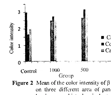
cells. Factors afTccting the color intensity of the antibodyantigen binding specificity closely related to the concentration of primary antibody used in the process. Optimization of antiinsulin concentration was performed and revealed that the concentration of I: 1500 \\'as the optimal concentration with unstained background. supported by Gobel (20 II).I3ascd on the data of the color intensity of pancreatic セ@ cell evaluation, the caput pancreas of the control DM had the lowest of color intensity compared to both treated groups. Corpus and cauda areas of animals treated with 500 mg/kg BW showed highcr intensity compared to the other groups (Figure 2).
Disscussion
Detection of pancreatic
l3
cells in Langerhans Islet hy immunohistochemistry indicate the presence of セ@ cell damage in diabetes mellitus. Pancreatic tissue of untreated animals had lowest pancreaticl3
cエセQQ@ compared to treated animals. This may he caused hy ad\'anced セ@ cell damaged due to cytotoxic cffect of STZ. Glucose uptake through glucose transporters (GLUT2) caused the STZ intoセ@ cdls, resulting in DNA alkylation. DNA was fi'agmented so that activating poly (ADPribose) synthetase, the enzyme that polymerizes to form the ADPrihose poly (ADPribose), and activation uf the ATP and NAD+ reduction. Decreased production of ATP and NAD+ led to the opening of K+ channels and the plasma membrane hyperpolarization. Furthermore, closure of the gate voltage reduced Ca2+ concentration and insulin secretion resulting in
l3
cell death (Elsner et al.1000 500 Group • Caput • Corpus '" Cauda Control
Figure I Mean of the number of
p
cells on three different areas of pancreas by immuno histochemical staining evaluationFruit extract improved total number of
p
cell, modestly higher in the lowest dosage (500 mglkg BW). This was supported by Suparlo CI 01. (20 I0), that Phaleria macrocarpa tj'uit e;;tract increased insulin secretion on diabetic cynomolgus monkeys. This fruit extract was already available in the market with dosage of 500 mg/capsule. It revealed that dose of 500 mg/kgBW was more appropriate to improve hyperglycemic condition or to reduce blood glucose leve!'Pancreatic
p
cells distribution in three different areas of untreated animals showed that the caput and the corpus pancreas had higherp
cells than the cauda pancreas. In the highest dosage, the corpus area showed higher numbers ofP
cells than in the caput and caudal pancreas. Total number ofp
cells for the lowest dosage showed higher number in the corpus and the cauda compared to caput pancreas. This result 'was supported by Oahmiarti (2000), that the distribution of diabetic pancreasp
cells located at the periphery of thc islets of Langerhans. However, according to Sundler and Harkanson (1988),P
cell distribution diflcred from each species and the composition of the islets of Langerhans dif1ered from each area of the pancreas. The Langerhans islets located in the cauda pancreas seemed II) has tendency to be more numerous.The color intensity of pancreatic
p
cells were highest in treated animal with 500 mg/kg BW. This result was also indicated by increasing in the number of pancreaticP
cells. J\ dark brown color on the Langcrhans Islets was resulted from the aflinity of antiinsulin with high insulin and vice versa. Affinity of antibody was the strength of the bond of a side of antibody binding (paratop) with antigenic detenninants (epitopes) (Vara 2005). So, it can be stated that the number of pancreatic セ@ cells ,vas directly proportional to the color intensity obtained from the reaction of antigen and antibody binding by immunohistochemical staining methods.Conclusion
Phaleria marcocarpa fruit extract with dosage of 500 mglkgBW orallY was the best dose to improve pancreatic beta cells damage in DM.
3
.-5'
a
2'"
• Caput5
.5
• corpus
1
"0 IliiCauda
u
.-0
GイセMMセイセBBGMセ@Control 1000 500 Group
Figure 2 Mean ofthe color intensity of
P
cells on three different area of pancreas by immuno histochemical staining evaluationAcknowledgement
This study was partly supported by
the
Ministry
of Research and Technology fund (RT20091388) and partly by internal research funds of Primate Research Center of Bogor Agricultural University.References
Elsner M. Guldbakke B. Tledge M, Munday R, Len:aen S. 2000. Relative importance of transport and alkylation for pancreatic betacell toxicity of streptozotocin. Diabctologia.43: 15281533.
Gobel S. 20 II. Optimasi antiinsulin pada sel セ@
pancreas Macaw fascicularis secara imunohistokimia [Laporan]. Bogor: Program Diploma, Institut Pertanian Bogor.
Harmanto N. 2003. Afahkola Dcwa Dbat Pusaka Para Dewa. Jakarta: Agromedia Pusaka.
Oshimi, S., Zaima, K., Matsuno. Y. Hirasawa, Y., Iizuka,
:r,
Studiawan, H., Indrayanto, G., Zaini,N.C., Morita, H 2008. Studies on the constituents from the fruits ofPhaleria macrocOlpa,
Nat Med(Tokyo), 62(2), pp. 207210
Saufi, A, von Heimendah!. c.B., Alfermann, AW., Fuss, E. 2008. Stereochemistry of lignans in
Phaleria macrocarpa (Schell) Boer!. Z
Nalurforsch, 63(12), Pl'. 1316
Suparto IH et at 2009. Pha/aia macrocarpa fruit extract as insulinotropic agent in streptozotocin-induced diabetic cynomolgus monkeys (Macaca
fascicu!aris). Oi dalam: Proceeding of
International Conference on Medicinal Plants,
Surabaya, 2122 Juli 20 I O.
Sundler J, Hakanson R. 1988. Handbook of Chemical Neuroanatomy Vol: 6. The peripheral Nervous System Elsevier Science Publisher BV.