HAYATI Journal of Biosciences March 2011 Vol. 18 No. 1, p 33-36
EISSN: 2086-4094
Available online at: http://journal.ipb.ac.id/index.php/hayati DOI: 10.4308/hjb.18.1.33
Methoxyacetic Acid Induced Apoptosis on the
Forelimb Bud of Swiss Webster Mice
AGUS HARYONO1∗∗∗∗∗, TIEN WIATI SURJONO2, LIEN ALINA SUTASURYA2, SRI SUDARWATI2
1Study Program of Biology Education, Palangkaraya University, Jalan Yos Sudarso C-11,
Kampus UNPAR Tanjung Nyahu, Palangka Raya 73112, Indonesia 2School of Life Sciences and Technology, Bandung Instute of Technology,
Jalan Ganesha 10, Bandung 40132, Indonesia
Received May 19, 2010/Accepted March 4, 2011
Methoxyacetic acid (MAA) causes digit malformations of mice when it was given orally on gestation day 11. Previous observation showed that malformation was caused by cell death. The aims of the research were to determine the types of cell death, first time of cell death and their distribution pattern on forelimb bud of Swiss Webster (SW) mice. Ten mM/kg of body weight (bw) of MAA were administered by gavage to SW mice on gestation day 11. Forelimb bud of mouse embryos of gestation day 11 + 0, 1, 2, 3, 4, 5 hours were processed with paraffin method and were made plantar section. Cell death at plantar section were colored with 4,6-diamino-2-phenylindole hydrochloride (DAPI) and hematoxylin. The result showed, that digit malformations initially by apoptosis mesenchymal cell at proximal of axial mesoderm in around of primary axial artery has done one hour after treatment. Apoptosis at the axial area, the site formation of digital ray III distributed to preaxial area where digits I and II are formed, and to the site formation of digits IV and V. The number of mesenchyme cell of digital rays II, III, and V was decrease by the increasing of gestation day, while digital ray was not formed and finally digits I, II, III, and V were missing. The reduction number of cell of digital ray IV were delayed time to be formed and its small size. Thereby it can be concluded, that MAA induced digit malformations of SW mice started by apoptosis which is occurrence has been increase in area of digital ray formation, so that digital ray can not be formed, but when formed it will not developed.
Key words: apoptosis; limb teratogenicity; methoxyacetic acid
___________________________________________________________________________
INTRODUCTION
Dimethoxyethyl Phthlate (DMEP) and 2-methoxyethanol (2-ME) are used progressively in various industries. Both DMEP and 2-ME can be pollutant in environment and enter to body through respiration or by oral, later in the body as well as can come into embryo. In the body, DMEP and 2-ME will be metabolism become to Methoxyacetic acid or MAA (Kitagawa et al. 2000).
MAA were embryotoxic and teratogenic agent to animal laboratories, 5 mmol/kg bw of MAA could inhibit development of Swiss Webster mice preimplantation embryo (Sumarsono 2001), 10 mM/kg bw of MAA given by gavage to Jcl/ICR mice on gestation day 11 induced 94.46% of fetuses are digit malformation (Rasjad et al. 1991). Previous observation the effect of MAA causes digit malformations in CD-1 mice (Greene et al. 1987), Jcl/ ICR (Rasjad et al. 1991) and at A/J mice (Sudarwati et al. 1995) were caused by intensive cell death at the limb bud. But that way, type of cell death, first time of cell death and how the distribution pattern of cell death at early limb development which was caused by MAA still unclear. To know the mentioned was conducted investigation the effect of MAA on early forelimb malformation of SW mice.
MATERIALS AND METHODS
Experimental Animal. Swiss webster (SW) mice purchased from Biological Department, ITB and locked in animal cage of Biology of ITB at room temperature 27.2 + 0.6 oC and humidity 84.9 + 10.3% with periode light 12 hours and dark 12 hours. Animal feed (Charoen Pokphand 551) and drinking water given ad libitum. Nulliparous female (25-30 g) were paired 1:1 in the home cage of the males. Vaginal plugs were designated as gestation day 0 (Taylor 1986).
Materials, Dosage, and Sampel Collection. MAA (CH3OCH2COOH) were obtained from Wako Pure Chemical Industries Ltd. No. Code Production is 135-07762 with molecular weight of 90.08 and purity of 97%. As a solvent used aquabidest sterile, dose of MAA is 10 mM/kg bw administered by gavage on gestation day 11. Forelimb bud of SW mice was collected at 0, 1, 2, 3, 4, 5 hours after treatment.
Detection of Apoptosis at Forelimb Bud of SW Mice. Fifteen dams of control and 15 dams of treated group were scarified by cervical dislocation. Forelimb bud were collected from pregnant mice of gestation day 11 was given MAA dose 10 mM/kg of bw by gavage and were observed at 0, 1, 2, 3, 4, 5 hours after treatment and processed with paraffin method to be made by plantar _________________
∗ ∗ ∗ ∗
∗Corresponding author. Phone: +62-536-3229117,
section. Plantar section of 5-7 µm was stained with DAPI and Hematoxylin. Cell death was determined from plantar section that was colored with DAPI and used fluorescence microscope (Aharony et al. 1995). Then its result was verified using microscope at the same section that was colored with hematoxylin (Humason 1967; Jolly et al. 1997). Determination of cell death based on of apoptosis and of necrosis criteria according to Bowen and Bowen (1990). The first time of cell death determined from result of observation time start 1 until 5 hours after treatment, while its distribution were determined at the area of limb bud including preaxial, axial, and postaxial areas.
RESULTS
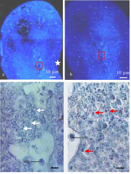
The Type of Cell Death. All section was colored with DAPI, both treated and control group were examined and showed the existence of cells that was strong fluorescence. To determine type of cell death were used forelimb plantar section of gestation day 11 + 3 hours, because at the axial mesoderm of forelimb bud was strong fluorescence. Verification result of same plantar section colored with Hematoxylin of cells that was strongly fluorescence with DAPI (Figure 1b) represent fragment of piknotic cell with marking of apoptosis cell death (Figure 1d), and also embryonic blood cell. At this area and all region of limb bud was not found necrosis cell death. Based on this result of this perception can be concluded that the type of cell death which is induction by MAA is apoptosis.
The First Time of Cell Death. Apoptosis at the control and treated groups, perceived at limb bud on gestation day 11 + 0, 1, 2, 3, 4, 5 hours showed that apoptosis only occur at treatment group and not at the control group (Table 1). Apoptosis at treatment group started to be detected at gestation day 11 day + 1 hour in proximal mesoderm of axial mesoderm especially at surrounding of primary axial artery. Thereby, the first time of apoptosis a 10 µm b 10 µm
c d sde
sde
Figure 1. Type of death cell at fore limb bud of SW mice gestation day 11 day + 3 hours. a. Control group (DAPI stained), b. Treated group (DAPI stained), c. Box of A enlarged, colored with Hematoxilyn show normal cell (white arrow) and embryonic blood cell (sde), d. Box at figure. b. Apoptotic bodies with piknotic nuclei as apoptosis (red arrow). : Preaxial side; White arrow of figure c. as normal cell: Red arrow at figure d. as Apoptotic cell; Sde: Embryonic blood cell.
Table 1. Distribution of apoptosis at fore limb bud of SW mice from control and treated dams of gestation day 11 and observation at various time after treatment (gestation day 11 + X Hour)
Zone of cell death Mount of apoptosis +: 1-5 cells, +: more than 5 cell (still countable), +++: many (uncountable), ++++: abudance.
sde
Figure 2. Distribution of apoptosis at fore limb bud of SW mice gestation day 11 + 4 hours stained by DAPI. a. Control group, Fluorescence’s cell as embryonic blood cell; b. Treated group, Apoptotic bodies (ap) with strong fluorescence were detected at axial and preaxial areas; Aap: Primary axial artery; INZ: Interior necrotic zone; SM: Sinus marginalis; sde: embryonic blood cell.
induction by MAA at forelimb bud of SW mice was at 1 hour after treatment.
Distribution of Cell Death. Distribution of apoptosis at the limb bud was perceived at gestation day 11 + 0, 1, 2, 3, 4, 5 hours (Table 1). All perception regions at control did not show the existence of apoptosis. At the treatment group in ectoderm area, apoptosis started to be detected in preaxial area at gestation day 11 + 4 hours (Figure 2b) and its remain were detected at 5 hours after treatment, at the postaxial just detected at 5 hours after treatment. At mesoderm, apoptosis started to be detected at 1 hour after treatment in axial area of the limb bud, at the proximal mesoderm around primary axial artery. Apoptosis in this area were increased, especially at mesoderm in surrounding primary axial artery. At preaxial areas, apoptotic cell death was detected at 2 hours after treatment and progressively mount at gestation day 11 day + 5 hours disseminating up at edge of limb bud. At postaxial area, a few apoptosis just detected at gestation day 11 + 5 hours. Thereby, apoptosis induced by MAA at the limb bud of Swiss Webster mice, initially from mesoderm proximal area, around of primary axial artery, then distribute to mesoderm preaxial area and finally to postaxial mesoderm.
DISCUSSION
Based on to criteria of cell death by Bowen and Bowen (1990) and Jolly et al. (1997), type of cell death of limb bud mesoderm induced with MAA at this research is apoptosis, cell showing characteristic cell death of necrosis was not found at all. Cell death induced with 2-ME or MAA also occurred at CD and A/J mice, cell death is expressed as necrotic (Greene et al. 1987; Rasjad et al.1991), when its cell death clarification by morphology, type of cell death is apoptosis. Ambroso et al. (1998) characterized of cell death which was induced by 2-ME at neuroepithelial cell of neural groove mice, that cell death represent as physiological cell death which is known taking place to through mechanism of apoptosis.
Apoptotic body formation during apoptosis take place between 30-60 minutes (Thornberry & Lazebnik 1998). Concentration of MAA in plasma of embryo one hour after treatment reached 1.2 times in the plasma of dams (Sleet et al. 1988). At this research, one hour after treatment with MAA was found apoptotic bodies in proximal area of mesoderm especially at the around of primary axial artery. It was suggested that existence of MAA and apoptosis of mesoderm at this research started at less than one hour after treatment. TUNEL method needed to detection of DNA fragmentation to know precisely checking process of apoptosis furthermore.
Apoptosis physiological cell death which recognized as Programmed Cell Death (PCD) also can be induced with various toxicant. 2-ME or MAA are cause apoptosis at Rat spermatocyte (Ku et al. 1995). Ku and Chapin (1994) suggested, that apoptosis at spermotocyte caused by increasing of membran permeability, so that concentration of Ca2+ in cell increase and endonuclease cleavage of DNA (Li et al. 1995). Increase of concentration of Ca2+ can be
pursued by calcium channel blocker and of apoptosis prevention (Li et al. 1997). According to Green and Reed (1998), increasing of Ca2+ concentration in mitochondria by agent can be trigger cytochrome-c released to sitosol and activated procaspase become caspase.
The effect of MAA to activate procapase become caspase is still unclear. MAA come into cell so that
increasing of Ca2+ concentration or MAA directly
induce mitochondria release cytochrome-c. MAA in mitochondria will be combined with acetyl~CoA form methoxyacetyl~CoA and into Krebs cycle so that it can disturb ATP synthesize (Mebus et al. 1992). Bowen and Bowen (1990), increase of Ca2+ concentration in cell can be triggered by reduction of ATP synthesize. Ruyani et al. (2003) suggested that down regulation of LBP-p40 after MAA treatment intensively induced apoptotic in mesoderm and finally caused limb malformation.
2-ME or MAA can cause apoptosis at cell which is active to proliferation and expand, for example at neural groove (Ambroso et al. 1998), mesenchymal cell of mouse limb bud (Greene et al. 1987; Rasjad et al. 1991 and at this research). Rao and Shaha (2002), MAA-induced apoptosis at germ cell of mouse was resulted of increasing caspase-9 and caspase-3 concentration, and also its degradation of content of glutathione. Apoptosis was induced by hyperthermia (Umpierre et al. 2002), with analogue of cyclophosphamide (Huang & Hales 2002), with MAA and MALD (Takagi et al. 2002) cause the increasing of concentration caspase-3. It was suggested that apoptosis cell death by MAA at this research also induce activation of caspase-3 and required more detail check.
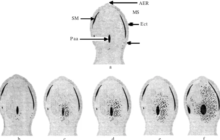
Limb malformation at early development of induction by MAA at A/J mice was caused by mesenchymal cell death (Sudarwati et al. 1995). At this research, limb malformation caused by apoptosis cell death of mesenchymal cells was in proximal mesoderm area around primary axial artery (Figure 3b). The occurrence of apoptosis disseminated to mesoderm (the site of digital ray III formation), to preaxial areas (the site of formation of digital rays I, II) and to postaxial area (the place of digital rays IV and of V formation) (Figure 3cdef). Distribution pattern of apoptosis of this research was the same as reported by Rasjad et al. (1991). Therefore malformation of the digit was ectrodactily of digit I, II, III, and V. The amount of mesenchymal cells migrated to place of digital ray IV was decreased, so that digit IV was formed small in a size. Besides amount of cell influenced condensation of mesenchymal cell on formation of digital ray, there were other factors influencing extracellular matrix component. To understand that, it needs a perception of pattern attendance of ECM component which play a role on digit chondrogenesis.
MAA induced limb malformation was initiated by apoptosis followed by increasing of its occurrence in the site of digital ray and it caused no formation of digital ray. The formed digits did not expand because of mesenchymal cell of the limb bud was decreased caused by very little amount of cell which shlould form condensation of digital ray.
AER
MS
Ect SM
Paa
a
b c d e f
Figure 3. Diagram of apoptosis distribution pattern induced by MAA on forelimb bud of Swiss Webster mice. a: Control group; b: Treated group gestation day 11 + 1 hour; c: 11 gestation day + 2 hours; d: gestation day 11 + 3 hours; e: gestation day 11 + 4 hours; f: gestation day 11 + 5 hours. Paa: Primary axial artery; AER: Apical Ectodermal Ridge; ANZ: Anterior Necrotic Zone; Ect: Ectoderm; MS: Mesoderm; SM: Sinus marginalis; Preaxial side; : Apoptosis cell death.
REFERENCES
Aharony D, Danttes A, Oren M, Amsterdam A. 1995. cAMP-mediated signal as determinants for apoptosis in primary granulosa cells. Exp Cell Res 218:271-282. doi:10.1006/ excr.1995.1156
Ambroso JL, Stedman BD, Elswick BA, Welsch F. 1998. Characterization of cell death induced by 2-methoxyethanol in CD-1 mouse embryos on gestation day 8. Teratology 58:231-240. doi:10.1002/(SICI)1096-9926(199812)58:6<231::AID-TERA4>3.0.CO;2-X
Bowen IB, Bowen SM. 1990. Programmed cell death in tumors and tissues. London: Chapman and Hall.
Green DR, Reed JC. 1998. Mitochondria and apoptosis. Science
281:1309-1312. doi:10.1126/science.281.5381.1309
Greene JA, Sleet RB, Morgan KT, Welsch F. 1987. Cytotoxic effect of ethylene glycol monoethyl ether in the forelimb bud of the mouse embryo. Teratology 3:23-34. doi:10.1002/ tera.1420360105
Huang C, Hales BF. 2002. Role of caspases in murine limb bud cell death induced by 4-hydroperoxycyclophosphamide, an activated analog of cyclophosphamide. Teratology 66:288-299. doi:10.1002/tera.10100
Humason GL. 1967. Animal tissue techniques. 2nd Ed. W. H. Freeman, Company, USA.
Jolly PD, Smith PR, Heath DA, Hudson NL, Lun S, Still LA, Watts CH, McNatty KP. 1997. Morphological evidence of apoptosis and the prevalence of apoptotic versus mitotic cells in the membrana granulosa of ovarian follicles during spontaneous and induced atresia in ewes. Biol Reprod 56:837-846. doi:10.1095/biolreprod56.4.837
Kitagawa K, Kawamoto T, Kunugita N, Tsukiyama T, Okamoto K, Yoshida A, Nakayama K. 2002. Aldehyde dehydrogenase (ALDH) 2 Associates with oxidation of methoxyacetaldehyde; in vitro analysis with liver subcellular fraction derived from human and Aldh2 gene targeting mouse. Fed Euro Biochem Soc 476:306-311.
Ku WW, Chapin RE. 1994. Spermatocyte toxicity of 2-methoxyethanol in vivo and in vitro: Requirement for an intact seminiferuous tubule structure for germ cell degeneration.
Toxicol In Vitro 8:1191-1202.
doi:10.1016/0887-2333(94)90109-0
Ku WW, Ghanayem, BI, Wine RN, Chapin. 1995. Spermatocyte toxicity of 2-methoxyethanol (ME) in rats and guinea pigs: Evidence for the induction of apoptosis. Toxicol Appl Pharmacol 134:100-110. doi:10.1006/taap.1995.1173
Li LH, Wine RN, Chapin RE. 1995. 2-methoxyacetic acid (MAA)-induced spermatocyte apoptosis in human and rat testes: an in vitro comparison. J Androl 17:538-549.
Li LH, Wine RN, Miller SD, Reece JM, Smith M, Chapin RE. 1997. Protection against methoxyacetic acid-induced spermatocyte apoptosis with calcium channel blocker in culture rat seminiferous tubules: Possible mechanism. Toxicol Appl Pharmacol 144:105-119. doi:10.1006/taap.1997.8129
Mebus CA, Clarke OD, Stedman DB, Welsch F. 1992. Methoxyethanol metabolism in pregnant CD-1 mice and embryos. Toxicol Appl Pharmacol 112:87-94. doi:10.1016/ 0041-008X(92)90283-X
Rao AV, Shaha C. 2002. N-acetylcysteine prevents MAA induced male germ cell apoptosis: role of gluthione and cytochrom c.
FEBS Lett 527:133-137. doi:10.1016/S0014-5793(02)03196-4
Rasjad C, Yamashita K, Datu AR, Yasuda M. 1991. Pathogenesis of limb malformations induced by methoxyacetic acid. J Med Sci 40:101-107.
Ruyani A, Sudarwati S, Sutasurya LA, Sumarsono H, Gloe F. 2003. The laminin binding protein p40 is involved inducing limb abnormality of mouse fetuses as the effects of methoxyacetic acid treatment. Toxicol Sci 75:148-153. doi:10.1093/toxsci/ kfg159
Sleet RB, Greene JA, Welsch F. 1988. The relation of embryotoxicity of disposition of 2-methoxyethanol in mice.
Toxicol Appl Pharmacol 95:195-207. doi:10.1016/0041-008X(88)90120-2
Sudarwati S, Surjono TW, Yusuf AT. 1995. Kelainan perkembangan awal anggota depan mencit A/J yang diinduksi asam metoksiasetat (MAA). JMS (Supl H) 8:60-70.
Sumarsono H, Adelina M, Kusumaningtyas H. 2001. Asam metoksiasetat menurunkan kualitas embrio mencit Swiss Webster tahap praimplantasi. Hayati 8:62-65.
Takagi A, Yamada T, Hayashi K, Nakade Y, Kojima T, Takamatsu J, Shibata E, Takeuchi Y, Murate T. 2002. Involvement of caspase 3 mediated apoptosis in hematopoietic cytotoxicity of metabolites of ethylene glycol monomethyl ether. Industrial Healt 40: 371- 374. doi:10.2486/indhealth.40.371
Taylor P. 1986. Practical teratology. London: Acad Pr. Thornberry NA, Lazebnik Y. 1998. Caspases: Enemies Within.
Science 126:1312-1317. doi:10.1126/science.281.5381.1312
Umpierre CC, Little SA, Mirkes PE. 2002. Co-localization of active caspase-3 and DNA fragmentation (TUNEL) in normal and hyperthermia-induced abnormal mouse development.
Teratology 63:134-143. doi:10.1002/tera.1024