Anatomy and Morphometry of Reproductive Organ of Male
Mouse Deer (
Tragulus javanicus
)
Najamudin1,*, Amrozi2, Srihadi Agungpriyono3, & Tuty Laswardi Yusuf2
1 Department of Animal Science, Faculty of Agriculture, Tadulako University, Palu, Indonesia,
*e-mail: [email protected]
2 Department of Clinic, Reproduction and Pathology,
3Department of Anatomy, Physiology and Pharmacology, Faculty of Veterinary Medicine, Bogor Agricultural University (IPB), Bogor, Indonesia
Abstract
number of rotations was two and a half and showed branch form, which the function was not yet known.
Key words: Male mouse deer, reproductive organ, anatomy
Introducton
Mouse deer (Tragulus javanicus) s the smallest rumnant anmals n the world.
Ths anmal s only found n tropcal forests n southern Asa, ncludng the slands of Java, Sumatra, and Kalmantan. The populaton of mouse deer s thought to declne due to habtat destructon and converson nto agrcultural uses and huntng actvty as well as the threat of predators that can prey on t.
Natural breedng of mouse deer n captvty has not succeeded yet. Factors causng the falure of mouse deer breedng n captvty are not known, but most lkely t s caused by a lack of knowledge or nformaton about the reproductve bology of ths anmal.
Informaton about the anatomy and morphometry of male mouse deer repro-ductve organs has not been wdely reported. Male reprorepro-ductve system s an m-portant factor n the success of anmal breedng or anmal reproducton technol-ogy. Knowledge of such nformaton s one of the mportant and decsve factors n achevng the success of anmal breedng n captvty. The purpose of ths study was to examne the anatomy and morphometry of the male mouse deer reproduc-tve organs to support the anmal breedng and reproducton for conservaton and cultvaton.
Materals and Methods
Time and Location
Ths study was conducted n May and October 2009 at the Feld Laboratory of Reproductve Rehabltaton Unt (URR), Department of Clnc, Reproducton, and Pathology, Bogor Agrcultural Unversty.
Materials
Three healthy and mature (had canne teeth) male mouse deer wth body weght ranged 1.8-2.0 kg were used n the experment.
Measurement and Weighing of the Reproductive Organs
These organs were cleaned from fat and lad n ts orgnal poston n the body. They were observed, documented, measured, and weghed and the data were tabulated and analyzed descrptvely accordng to Toelhere (1993).
Testes. The length of the testes was measured by placng a measurng tape at the end of the testes from one sde to another wth or wthout the caput and cauda epddyms. Dameter of the testes was measured by usng a mcrocalper at the largest part of the testes, and then weghed usng an analytcal balance.
Vas deferens. The length of the vas deferens was measured by placng the measurng tape at the end of the cauda epddyms, and the tape was then pulled untl t reached the end before the enlargement of the vas deferens to the ampulla. Vas deferens dameter was measured by usng mcrocalper before t was weghed.
Ampulla of vas deferens. Ampulla of vas deferens length was measured from the ntal enlargement of the vas deferens to the border wth vesculars gland. The dameter of ampulla was measured at the largest part before t was weghed.
Vesicularis gland. The longest part of the vesculars gland was consdered as the length, whle the shortest was consdered as the wdth. Dameter of the vesculars gland was measured wth a mcrocalper. The two glands had been separated from the man organ before they were weghed.
Prostate. Measurement of prostate length, dameter, and weght were smlar to those of vesculars gland.
Bulbourethralis (Couper). Measurements of bulbourethrals were smlar to those of vesculars glands.
Penis. Measurements of total pens length was started from the base of the pens (the radx) up to the pens free end, and length measurement was also conducted for pens parts such as the pens glans and prepuce. Dameter of the pens was measured at the largest part of the organ.
Data obtaned from each measurement were tabulated and the average was calculated and analyzed descrptvely.
Results and Dscusson
testes), complementary sex glands (the ampulla, the vesculars gland, the prostate, and the bulbourethrals), and channels consstng of the epddyms, the vas deferens and the urethra, and the external gental organs or copulators organ called the pens.
Testes. Male mouse deer had a par of testcles whch were wrapped by the tunca albugena protected by the scrotum on the outsde. The testes functons to produce spermatozoa n a process called spermatogeness and produces testosterone hormone n the ntersttal cells (Leydg) (Hafez & Hafez 2000; Toelhere 1987). Mouse deer testes length was 12.33 ± 2.89 mm, dameter was 8.20± 1.92 mm, and weght was 0.81± 0.17 g (Table 1).
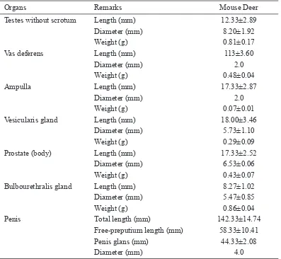
Table 1 Morphometry of male mouse deer reproductve organs
Organs Remarks Mouse Deer
Testes wthout scrotum Length (mm) 12.33±2.89
Dameter (mm) 8.20±1.92
Weght (g) 0.81±0.17
Vas deferens Length (mm) 113±3.60
Dameter (mm) 2.0
Weght (g) 0.48±0.04
Ampulla Length (mm) 17.33±2.87
Dameter (mm) 2.0
Weght (g) 0.07±0.01
Vesculars gland Length (mm) 18.00±3.46
Dameter (mm) 5.73±1.10
Weght (g) 0.29±0.09
Prostate (body) Length (mm) 17.33±2.52
Dameter (mm) 6.53±0.06
Weght (g) 0.43±0.07
Bulbourethrals gland Length (mm) 8.27±1.02
Dameter (mm) 5.47±0.85
Weght (g) 0.86±0.04
Pens Total length (mm) 142.33±14.74
Free-preputum length (mm) 58.33±10.41
Pens glans (mm) 44.33±2.08
Epididymis. The epddyms s a structure that s elongated and tghtly attached to the tests. The mouse deer epddyms conssted of caput n the anteror, corpus n the dorsal, and cauda epddyms n the posteror.
Vas deferens. Vas deferens connected the epddyms wth the accessoryes glands, serves as a channel of transport of spermatozoa from the epddyms to the ampulla. The length of the mouse deer vas deferens n ths study was 113 ± 3.60 mm whch was shorter than that of tmor deer (452.0 ± 0:44 mm) (Nalley 2006) or sheep (24 cm) (Hafez 1987).
Ampulla of vas deferens. Ampulla of vas deferens s the magnfcaton of the end of the vas deferens before vesculars gland. The length of the mouse deer ampulla (17.33 ± 2.87 mm) was smaller than that of Tmor deer (72.53 ± 2.39 mm) (Nalley 2006) or sheep (70 mm) (Hafez 1987).
Vesicularis gland. There was a par of vesculars glands attached to the dorsolateral edge of vesca urnary neck. Mouse deer vesculars gland length and dameter were 18.00 ± 3.46 mm and 5.73 ± 1.10 mm, respectvely. Ths was smaller than the vesculars gland of tmor deer (45.36 ± 1.42 mm) or sheep (40 mm).
Bulbourethralis gland (Cowper). In the mouse deer, there was a par of bulbourethrals glands wth a length of 8.26 ± 1.02 mm, dameter of 5.47±0.85 mm and weght of 0.86 ± 0:04 g. Bulbourethrals gland s also very clearly seen n horses and pgs. Nalley (2006) reported that these glands were not found n Tmor deer.
Penis. The pens s a male copulaton organ and s establshed by the erectle tssue. The pens of mouse deer was fbroelastc whch s smlar to that of cattle, so that the pens corpus and glans were only slghtly enlarged durng erecton.
Concluson
Mouse deer pens s characterzed by a flexura sgmodea, fbroblastc type wth a very dstnctve pens tp, whch forms a spral wth a spn number of two and a half n clockwse drecton.
Acknowledgements
The authors are ndebted to the Feld Laboratory of Reproductve Rehabltaton Unt, and the Anatomcal Research Laboratory, Faculty of Veternary Scence, Bogor Agrcultural Unversty..
References
Hafez ESE. 1987. Anatomy of male reproducton n: Reproduction in Farm
Ani-mals. Ed. 7th Ed. Lea and Febger. Phladelpha.
Hafez ESE and Hafez B. 2000. Anatomy of female reproducton n: Reproduction
in Farm Animals. Ed. 7th Ed. Lppncott Wllams & Wllams.
Nalley WMM. 2006. Kajan Bolog Reproduks dan Penerapan Teknolog
Insem-nas Buatan pada Rusa Tmor (Cervus timorensis). [dsertas] Bogor. Sekolah
Pascasarjana Insttut Pertanan Bogor.