Various factors affecting the bacterial corneal ulcer healing: a
4-years study in referral tertiary eye hospital in Indonesia
Keywords: bacterial corneal ulcer, fluoroquinolone, Pseudomonas sp.
pISSN: 0853-1773 • eISSN: 2252-8083 • http://dx.doi.org/10.13181/mji.v24i3.1044 • Med J Indones. 2015;24:150-5
• Received 03 Sep 2014 • Accepted 08 Jul 2015
Correspondence author: Made Susiyanti, [email protected]
Copyright @ 2015 Authors. This is an open access article distributed under the terms of the Creative Commons Attribution-NonCommercial-ShareAlike 4.0 International License (http://creativecommons.org/licenses/by-nc-sa/4.0/), which permits unrestricted non-commercial use, distribution, and reproduction in any medium, provided the original author and source are properly cited.
Muhammad Asroruddin,1
Rina L.D. Nora,2 Lukman Edwar,2 Soedarman Sjamsoe,2 Made Susiyanti2
1 Department of Ophthalmology, Faculty of Medicine, University of Tanjungpura, Tanjungpura University Hospital, Pontianak,
Indonesia
2 Department of Ophthalmology, Faculty of Medicine, Universitas Indonesia, Cipto Mangunkusumo Hospital, Jakarta, Indonesia C l i n i c a l Re s e a rc h
ABSTRAK
Latar belakang: Ulkus kornea merupakan salah satu penyebab utama gangguan penglihatan dan kebutaan di seluruh dunia. Penelitian ini bertujuan mengevaluasi faktor-faktor yang mempengaruhi penyembuhan ulkus kornea bakteri, termasuk faktor predisposisi, organisme penyebab, sensitivitas antibiotik, dan hasil terapi.
Metode: Semua data diambil secara retrospektif berdasarkan rekam medis pasien ulkus selama 4 tahun (2008-2011) di Rumah Sakit Cipto Mangunkusumo, Jakarta. Dilakukan apusan kornea untuk pemeriksaan gram dan/atau kultur. Hasil terapi ulkus dianalisis menggunakan uji kai kuadrat dan
one-way ANOVA, serta post-hoc analysis.
Hasil: Sebanyak 220 kasus ulkus kornea bakteri ditemukan berasal dari 216 pasien. Faktor risiko ulkus yang paling sering ditemukan adalah trauma okuler (45,8%). Kokus gram-positive ditemukan pada 65,7% kasus. Pseudomonas sp. (25,0%) dan Staphylococcus epidermidis (18,4%) merupakan spesies yang paling banyak ditemukan, dan sensitif terhadap hampir semua jenis antibiotik. Sekitar 83,0% (106 kasus) membaik dengan pemberian antibiotik saja, sisanya tidak membaik dan memburuk. Rerata masa penyembuhan ulkus yang sempurna adalah 17,5 ± 8,9 hari dan ulkus ringan mengalami masa penyembuhan tercepat. Ulkus yang diterapi dengan tetes mata fluorokuinolon menyembuh lebih cepat dari regimen lain yaitu dalam waktu 14 hari.
Kesimpulan: Trauma okuler merupakan faktor risiko ulkus kornea yang paling sering. Penyebab mikroorganisme tersering adalah Pseudomonas sp. Sebagian besar kasus membaik dengan pemberian antibiotik saja. Kasus yang diberikan fluorokuinolon menyembuh lebih cepat dibanding jenis lain. Rerata masa penyembuhan ulkus adalah sekitar 17,5 hari.
ABSTRACT
Background: Corneal ulcer is one of the most common causes of visual acuity impairment and blindness all over the world. The aim of the study was to evaluate various factors affecting the bacterial corneal ulcers healing, including the predisposing factors, causative organisms, antibiotic sensitivity, as well as the treatment outcomes.
Methods: All data were taken retrospectively from medical records of patients who underwent corneal scraping for Gram examination and/or culture over a-4-year period (2008-2011) at the Cipto Mangunkusumo Hospital Jakarta. Treatment outcome were analyzed using Chi-square test, one-way ANOVA, and post-hoc analysis. Mean time required for complete epithelial healing was also investigated.
Results: 220 cases of bacterial corneal ulcers in 216 patients were included. The most common risk factors were ocular trauma (45.8%). Gram-positive coccus were found in 65.7% cases other than other microbes. Pseudomonas sp. (25,0%) and Staphylococcus epidermidis (18.4%) were the most common isolates, sensitive to almost all kinds of antibiotics. About 83.0% (106 cases) were improved with antibiotics only, the rest were not improved and worsened. Mean time for complete epithelial healing was 17.5 ± 8.9 days with mild ulcer had the most rapid recovery. Eyes treated with fluoroquinolone eyedrops were healed in 14 days, faster than other regiments.
Bacterial keratitis is now a world-wide leading cause of visual loss and blindness. Ulcerative keratitis (corneal ulcers) can be caused by bacteria, fungi, viruses, or parasites.1-4 The
incidence or number of cases varies between western and developing countries due to different predisposing factors. For example, Cipto Mangunkusumo Hospital (CMH) Jakarta has recorded 88 cases of 202 new corneal ulcers (43.6%)5 and 132 cases of 262 new cases
(50.38%)6 in different terms. These numbers
slightly differ from Thailand7 and India.8,9
The most leading causes of corneal ulcers are
Streptococcus, Pseudomonas, and Staphylococcus.
Study at CMH in last 20 years found Pseudomonas
aeruginosa (49%) and Staphyloccus epidermidis
(24%) as the causes.10 However, in last ten
years, ulcer was dominated by Staphylococcus
epidermidis.5,6,11
Predisposing factors of bacterial corneal ulcers are commonly ocular trauma, contact lens users, ocular surface diseases, and as well systemic factors like diabetes mellitus and immunocompromise. Corneal ulcers are emergency cases, sight-threatening, and sometimes progressive, and also potentially resulting complications such as corneal perforation and endoftalmitis, that would need immediate surgery. Thus, this needs early diagnosis-including Gram examination and culture of corneal scraping-and agressive prompt treatment.1-4,12,13
This study was aimed to periodically evaluate the predisposing factors, patient demographics, clinical characteristics, causative organisms, antibiotic sensitivity and resistance, as well as the treatment outcomes of bacterial corneal ulcers at CMH. This study would obtain the empirical evidences that would be the guidelines in treating bacterial corneal ulcer.
METHODS
A retrospective analysis of all patients with positive results of gram examination and/ or culture proven bacterial corneal ulcer of corneal scraping was performed. A total of 216 patients (220 eyes) registered during period of November 2012 to January 2013 at CMH were analyzed. Ulcers caused by mixed infection or immunological type were excluded.
We collected all data related to socio-demographic features, duration of symptoms, predisposing factors (history of trauma, diabetes mellitus, steroid use), prior therapy received including traditional medicine, contact lens use, and ocular and systemic disease. Initial visual acuity at the time of presentation, laterality, and all clinical findings including ulcers size and depth, inflammation reaction in anterior chamber, hypopion, and corneal scraping results were also collected. Disease severity was graded to mild, moderate, and severe based on modified Jones criteria.14
Antibiotics resistance and treatment outcomes
including mean time required for of complete
epithelial healing were also assessed. The data were analyzed using SPSS 16.0 and Microsoft
Excel 2007. Chi-square test and one-way
ANOVA were used to analyze the treatment outcome, which p-value less than 0.05 regards as statistically significant. All data taken from medical records of patients. Confidentiality of subjects identity were guaranteed.
RESULTS
Two hundred and sixteen cases (220 eyes) or 32.9% of 656 new corneal ulcers cases were
identified as bacterial ulcers during 1 January
2008 – 31 December 2011 at CMH. Others were caused by fungi, virus, Achantamoeba, and noninfectious ulcers.
Demographics, predisposing factors, and clinical features
Out of 216 patients, 145 (67.1%) were males and 71 (32.9%) were females. Age of patients of over 40 years old (48.6%) dominated, with range between three months old to 83 years. Demographic and clinical features are described in table 1.
Most patients did not have systemic predisposing factors (91.6%), but there were patients with diabetes mellitus, leukemia, and human
immunodeficiency virus/acquired immune deficiency syndrome (HIV/AIDS).
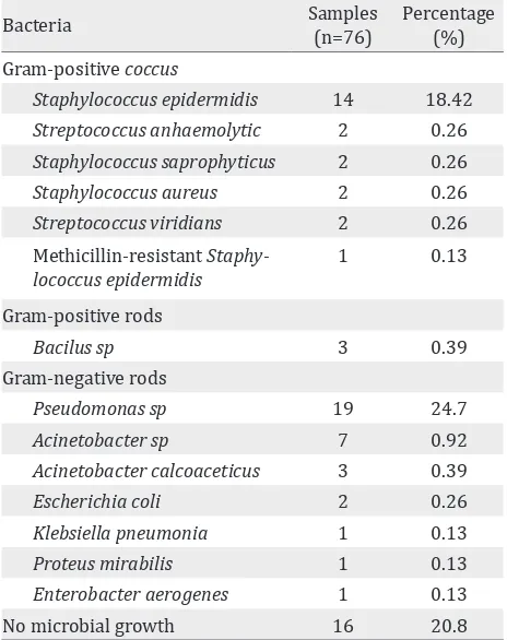
Causative bacteria
Table 2 shows that microbial culture of corneal scraping were dominated by Gram-negative rods,
Gram-Variables Number of patients/eyes
Percentage (%) Sex (n=216 patients)
Male 145 67.1
Female 71 32.9
Age
< 18 years 27 12.5
19 – 40 years 84 38.9
> 40 years 105 48.6
Local predisposing factors (n=216 patients)
External
Surgical and non-surgical trauma
99 45.8
Contact lens use 22 10.2 Eyelid and tears dysfunction 29 12.4
Abnormal cornea 3 1.3
Topical drugs 2 0.1
Unspecified (red eye, pain, etc)
10 4.6
No records 51 23.6
Systemic predisposing factors
Diabetes mellitus 10 4.6
HIV/AIDS 3 1.4
Immunological disorders 5 2.3 No influencing factors 198 91.67 Laterality (n=220 eyes)
RE (right eye) 111 51.4
OU (both eyes) 4 1.9
LE (left eye) 101 46.8
Ulcer location
Central 121 54.6
Paracentral 67 30.6
Peripheral 26 12.0
No records 6 2.8
Ulcer size
< 2 mm 45 20.4
2 – 6 mm 118 54.2
> 6 mm 43 19.4
No records 14 6.0
Mean (n=203 eyes) 4.1 mm ± 0.2
Ulcer depth (stromal)
< 1/3 41 19.0
1/3 - 2/3 108 49.1
> 2/3 10 4.6
Perforation 43 19.9
No records 18 8.3
Table 1. Demographics, predisposing factors, and clinical features of bacterial corneal ulcers at CMH
positive coccus, Staphylococcus epidermidis, 14 isolates (18.42%). Culture was not performed in eight patients because of small lesion and evidence of clinical improvement after given prior therapy.
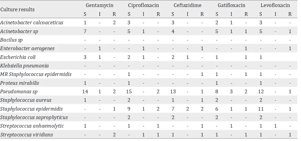
Pattern of bacterial resistance and sensitivity to some antibiotics are shown in table 3 below.
Treatment outcome
Topical antibiotic was given to all subjects in this study as the initial therapy. Treatment outcome of
bacterial corneal ulcer is classified into improved,
not improved (steady), and worsen. Clinical improvement was noted in 106 patients, in which 62 of them followed the treatment until corneal ulcers healed. The remaining 44 patients were lost to follow up. No improvement and clinical worsening were experienced by four and 18 patients, respectively, and they were further underwent surgery such as amniotic membrane transplantation (AMT) one patient, penetrating keratoplasty eight patients, periosteal graft three patients, and evisceration two patients (Table 4).
Out of 62 patients who followed the treatment until formation of corneal scarring, mean days of epithelial healing or corneal scarring form could
Bacteria Samples
(n=76)
Percentage (%) Gram-positive coccus
Staphylococcus epidermidis 14 18.42
Streptococcus anhaemolytic 2 0.26
Staphylococcus saprophyticus 2 0.26
Staphylococcus aureus 2 0.26
Streptococcus viridians 2 0.26
Methicillin-resistant Staphy-lococcus epidermidis
1 0.13
Gram-positive rods
Bacilus sp 3 0.39
Gram-negative rods
Pseudomonas sp 19 24.7
Acinetobacter sp 7 0.92
Acinetobacter calcoaceticus 3 0.39
Escherichia coli 2 0.26
Klebsiella pneumonia 1 0.13
Proteus mirabilis 1 0.13
Enterobacter aerogenes 1 0.13
No microbial growth 16 20.8
Culture results Gentamycin Ciprofloxacin Ceftazidime Gatifloxacin Levofloxacin
S I R S I R S I R S I R S I R
Acinetobacter calcoaceticus 1 - 2 3 - - 3 - - 2 1 - 3 -
-Acinetobacter sp 7 - - 5 1 - 4 - - 5 1 1 5 - 1
Bacilus sp - - -
-Enterobacter aerogenes - 1 - - 1 - - - 1 - - 1 - - 1
Escherichia coli 3 1 - 2 1 - 2 1 - 1 1 1
Klebsiella pneumonia - - -
-MR Staphylococcus epidermidis - - - 1 - - - - 1 1 - 1 1 -
-Proteus mirabilis 1 - - 1 - - - 1 - - 1 -
-Pseudomonas sp 14 1 2 15 - 2 13 - 1 8 3 2 12 - 1
Staphylococcus aureus 1 - - 2 - - 1 - 1 2 - - 2 -
-Staphylococcus epidermidis - - 1 9 1 2 7 2 2 6 1 1 11 - 1
Staphylococcus saprophyticus - - - 2 - - 2 - - 2 - - 2 -
-Streptococcus anhaemolytic 1 - - 1 - 1 - - 1 - 1 - 1 1
-Streptococcus viridians - - 2 - 1 1 1 - 1 1 - 1 1 - 1
Table 3. Culture pattern of bacteria with some antibiotics
S : Sensitive; I : Intermediate; R : Resistent
Ulcers Grading
Treatment Outcome
Total Improved
(%)
Not improved
(%)
Worsen (%)
Mild 11 (100) 0 (0) 0 (0) 11 Moderate 59 (89) 2 (3) 5 (8) 66 Severe 36 (71) 2 (4) 13 (26) 51 Total 106 (83) 4 (3) 18 (14) 128 Antibiotics
Fluoroquinolone 73 (88) 2 (2) 8 (10) 83 Aminoglycoside 20 (69) 2 (7) 7 (24) 29 Cephalosporine 10 (100) 0 (0) 0 (0) 10 Combination 3 (50) 0 (0) 3 (50) 6
Table 4. Treatment outcomes of corneal ulcer according to disease severity and types of antibiotics
Variable Patients
(n=48)
Mean time (days) Ulcer grading
Mild 5 7 (3-30)
Moderate 28 15.8 ± 8.4
Severe 15 21.9 ± 7.9
Total 48 17.5 ± 8.9
Antibiotics group
Fluoroquinolone 29 14 (3-30) Aminoglycoside 11 19.2 ± 8.6 Cephalosporine 7 19.6 ± 1.08
Table 5. Mean time for epithelial healing based on ulcers grading and antibiotics group
be analyzed in 48 patients (mild 25 pts, moderate 28 pts, severe 15 pts) whose. Fourteen patients were excluded because they had predisposing factors that could inhibit healing process such as lagophtalmus and corneal exposure, diabetes mellitus, HIV/AIDS, persistent trichiasis, chemical injury, and immunocompromise in leukemic patients.
Regardless of disease severity, it is shown that the fastest time for corneal healing was found
in fluoroquinolone group (14 days), followed
by aminoglycoside group (19.2 + 8.6 days), and cephalosporine group (19.6 ± 1.08 days, table 5).
DISCUSSION
Microbial corneal ulcers are the major cause of visual loss and blindness in Indonesia. The male versus female ratio in this study was 2:1. This was similar to other study conducted by Sirikul, et al7 in Thailand. This is probably men have more
common predisposing factor in our study was ocular trauma. However, our ratio was different from other study that conducted by Fong, et al15
in Taiwan dan Marangon, et al16 in New York
(1:1), Bharathi, et al8 in southern India (1.5:1),
and Shalchi, et al17 (1:1.5). Corneal ulcer in our
study affected mostly aged over 40 years of old
and young adults. This study was quite similar
to study performed by Srinivasan, et al9 (61.3%),
and Narsani, et al18 in Pakistan.
Ocular trauma was the leading predisposing factor for corneal ulcer in our study. This result was similar to study by Damayanti and Sitompul5,
Bharathi, et al8, and Srinivasan, et al.9 However,
our result was different from study by Fong, et al15
and Keay, et al19 in Australia that found contact
lens use (44.3%) as the most common factor, and trauma (23.8%), as well as by Mah-Sadorra, et al13
and Bourcier, et al.20
The causative bacterium found in our study was
Pseudomonas sp. This finding was similar to study
performed by Damayanti and Sitompul5 and
Fong, et al.15 However, Narsani, et al18 in Pakistan
found Staphylococcus aureus as the most common isolate, followed by Pseudomonas aeruginosa dan
Streptococcus pneumoniae. On the other way,
Chawla, et al21 found Staphylococcus epidermidis
(77,0%) as the predominant isolate. Pseudomonas sp. as the most commonly found bacterium,
mostly sensitive to gentamycin, ciprofloxacin, ceftazidime, gatifloxacin, dan levofloxacin. This
result was different from study by Sitompul, et al10 in last decade that 27% Pseudomonas had
already resistant to gentamycin. Staphylococcus
epidermidis was also dominantly sensitive to
vancomycin, levofloxacin, dan gatifloxacin except gentamycin. Resistance to fluoroquinolone
group had been partly encountered, primarily to
ciprofloxacin, levofloxacin, dan gatifloxacin. This
supports similar result in study by Damayanti and Sitompul5, Shalchi, et al17, and Maske, et al.22
Our treatment outcome which gained up to 80% patients with clinical improvement was better than study conducted by Hussain, et al23 that
reached 68.4% patients, and Damayanti and Sitompul5 during 2005-2007 that reached 74,0%.
The most common used antibiotics in our study were topical fluoroquinolone and aminoglycoside eyedrops with proper concentration. Single used antibiotics (monotherapy) was given more than
combined formula. The standard dose was also
given more than fortified dose. Fluoroquinolone is used frequently because of its good sensitivity for
all groups of bacteria, positive and Gram-negative, including good ocular penetration and longer half-life. In our study, most patients treated
with fluoroquionolone revealed better clinical
improvement than treated by other groups. Type
of antibiotics were commonly ofloxacin and levofloxacin, other than moxifloxacin as fourth
generation quinolones. However, Shah, et al24 dan
Kowalski, et al25 in their clinical trial found that moxifloxacin or gatifloxacin gave better potency
in combating bacteria, even for resistent bacteria
by second and third generation quinolones.
Besides fluoroquinolones, some patients in
our study were also given combined therapy of cefazolin 10% + tobramycin 1.5%. This therapy suits WHO guidelines for bacterial corneal ulcer in Southeast Asia region. Study by Khokhar, et al26 found that this combination compared to ofloxacin had no different clinical
outcomes. Mean day for epithelial healing was approximately 15 days, as well as study by Panda, et al27 dan Constantinou, et al28.
However, these two groups were not applicable to our study since only one patient included.
Nevertheless, regarding the efficacy and safety,
good availability and cost-effective, and issue of kitchen pharmacy, clinicians prefer to give
fluoroquinolones as initial empirical therapy
for bacterial corneal ulcers in last decades. The mean days of epithelial healing in our study, 17.5 days, was better than results in study of Damayanti and Sitompul5 which was 19 days.
Healing in moderate corneal ulcer was 15.8 days, which is also better than study of Damayanti and Sitompul5, 17 days. Our result was quite similar
to study of Khokhar, et al26 and Panda, et al27 with
the similar method.
This study has some limitations including the design as retrospective study, in which not all medical records can be included, high rate of lost to follow-up (35.2%), and low rate of bacterial culture (< 50%).
In conclusion, the most common risk factor for bacterial corneal ulcers in this study was trauma, and the most commonly isolated organism was
Pseudomonas sp. Most cases were improved with
faster healing. Complete epithelial healing occurred in 17.5 days. These factors affected features of the corneal ulcers healing in this study.
Conflict of Interest
The authors affirm there is no conflict of interest
in this study.
REFERENCES
1. Weisenthal RW. 2013-2014 Basic and clinical science course, section 8: external disease and cornea. American Academy of Ophthalmology; San Fransisco, 2010. p. 22-30.
2. Garg Prashant, rao GN. Corneal ulcers: diagnosis and management. Community Eye Health. 1999;12(30):21-3.
3. aao.org [internet]. San Fransisco: Cornea/external disease panel members. Preferred practice pattern guidelines. Bacterial keratitis. [Update Sept 2012; Cited 2012 Dec]. Available from: www.aao.org
4. World Health Organization. Guidelines for the management of corneal ulcer at primary, secondary, and tertiary care health facilities in the South-East Asia Region. WHO Regional Office for South East Asia; 2004.
5. Damayanti Y, Sitompul R. Karakteristik klinis dan hasil terapi ulkus kornea bakteri di Poliklinik Mata Rumah Sakit Dr. Cipto Mangunkusumo Periode 1 Juli 2005 - 30 Juni 2007. Thesis. Jakarta: Fakultas Kedokteran Universitas Indonesia; 2008. Indonesian.
6. Wirastana. Karakteristik ulkus kornea jamur di Divisi Infeksi dan Imunologi Polimata RSCM tahun 2007-2009. Thesis. Jakarta: Fakultas Kedokteran Universitas Indonesia; 2010.
7. Sirikul T, Prabriputaloong T, Smathivat A, Chuck RS, Vongthongsri A. Predisposing factors and etiologic diagnosis of ulcerative keratitis. Cornea. 2008;27(3):283-7.
8. Bharathi MJ, Ramakrishnan R, Meenakshi R, Padmavathy S, Shivkumar C, Srinivasan M. Microbial keratitis in South India: influence of risk factors, climate, and geographical variation. Ophthalmic Epidemiol. 2007;14(2):61-9. 9. Srinivasan M, Gonzales CA, George C, Cevallos V,
Mascarenhas JM, Asokan B, et al. Epidemiology and aetiological diagnosis of corneal ulceration in Madurai, south India. Br J Ophthalmol. 1997;81(11):965-71. 10. Sitompul R, Asyari F, Sjamsoe S, Marsetio M.
Perbandingan efektifitas dibekasin dan gentamisin sebagai terapi pada ulkus kornea bakteri. Ophthalmologica Indonesiana. 1997;17:47-52. Indonesian.
11. Rahmadhani SFS. Peranan kultur dan uji kepekaan dalam penalaksanaan ulkus kornea bakteri di Sub Bagian Infeksi dan Imunologi Ilmu Penyakit Mata RS Cipto Mangunkusumo 1995-1998. Thesis. Jakarta: Fakultas Kedokteran Universitas Indonesia; 1999. Indonesian.
12. Green M, Apel A, Stapleton F. Risk factors and causative organisms in microbial keratitis. Cornea. 2008;27(1):22-7. 13. Mah-Sadorra JH, Yavuz SG, Najjar DM, Laibson PR,
Rapuano CJ, Cohen EJ. Trends in contact lens-related corneal ulcers. Cornea. 2005;24(1):51-8.
14. Jones DB. Decision-making in the management of microbial keratitis. Ophthalmology. 1981;88(8):814-20. 15. Fong CF, Tseng CH, Hu FR, Wang IJ, Chen WL, Hou
YC. Clinical characteristic of microbial keratitis in a university hospital in Taiwan. Am J Ophthalmol. 2004;137(2):329-36.
16. Marangon FB, Miller D, Alfonso EC. Impact of prior therapy on the recovery and frequency of corneal pathogens. Cornea. 2004;23(2):158-64.
17. Shalchi Z, Gurbaxani A, Baker M, Nash J. Antibiotic resistance in microbial keratitis: ten-year experience of corneal scrapes in the United Kingdom. Ophthalmology. 2011;118(11):2161-5.
18. Narsani AK, Jatoi SM, Lohana MK, Dabir SA, Gul S, Khanzada MA. Hospital -base epidemiology, risk factors and microbiological diagnosis of bacterial corneal ulcer. Int J Ophthalmol. 2009;2(4):362-6.
19. Keay L, Edwards K, Naduvilath T, Taylor HR, Snibson GR, Forde K, et al. Microbial keratitis, predisposing factors and morbidity. Ophthalmology. 2006;113(1):109-16. 20. Bourcier T, Thomas F, Borderie V, Chaumeil C, Laroche
L. Bacterial keratitis: predisposing factors, clinical and microbiological review of 300 cases. Br J Ophthalmol 2003;87(7):834-8.
21. Chawla B, Agarwal P, Tandon R, Titiyal JS, Sharma N, Agarwal T, et al. In vitro susceptibility of bacterial keratitis isolates to fourth-generation fluoroquinolones. Eur J Ophthalmol. 2010;20(2):300-5.
22. Maske R, Hill JC, Oliver SP. Management of bacterial corneal ulcers. Br J Ophthalmol. 1986;70(3):199-201. 23. Hussain I, Khan BS, Soni M, Iqbal M, Habibullah. Non-viral
microbial keratitis: etiology, clinical features and visual outcome. J Coll Physicians Surg Pak. 2012;22(3):151-4. 24. Shah VM, Tandon R, Satpathy G, Nayak N, Chawla
B, Agarwal T, et al. Randomized clinical study for comparative evaluation of fourth-generation fluoroquinolones with the combination of fortified antibiotics in the treatment of bacterial corneal ulcers. Cornea. 2010;29(7):751-7.
25. Kowalski RP, Dhaliwal DK, Karenchak LM, Romanowski EG, Mah FS, Ritterband DC, et al. Gatifloxacin and moxifloxacin: an in vitro susceptibility comparison to levofloxacin, ciprofloxacin, and ofloxacin using bacterial keratitis isolates. Am J Ophthalmol. 2003;136(3):500-5. 26. Khokhar S, Sindhu N, Mirdha BR. Comparison of
topical 0.3% ofloxacin to fortified tobramycin-cefazolin in the therapy of bacterial keratitis. Infection. 2000;28(3):149-52.
27. Panda A, Ahuja R, Sastry SS. Comparison of topical 0.3% ofloxacin with fortified tobramycin plus cefazolin in the treatment of bacterial keratitis. Eye (Lond). 1999;13(Pt 6):744-7.