The Profile of Thrombocytosis in Pediatric Intensive Care Unit
Selvi Nafianti
Department of Child Health, Medical School University of Sumatera Utara/ Haji Adam Malik Hospital Medan
archipelago country.
Objective: To describe the distribution of thrombocytosis in pediatric intensive care unit
.
Methods: A rectrospective study reviewed medical chart of all children in Pediatric Intensive Care Unit (PICU) Haji Adam Malik Hospital with all diagnosis during January 2007 – December 2007. All the patients who diagnosed as thrombocytosis were enrolled to this study. The data was collected include age, sex, diagnosis in PICU will be reported descriptively.
Results: From the total 319 cases admitted to PICU, there were 65 had thrombocytosis. Most thrombocytosis 29(65) were in group age 1-5 years, and 21(65) in group 5-10 years, and 15(65) in group age more than 10 years. Boys more than girls (35 vs 30). Thrombocytosis occurred mostly in complicated surgical patients, head injury and infectious diseases. Conclusion: Thrombocytosis is common problem in critically ill patient. It is required a longitudinal study to determine the cause of thrombocytosis in critically ill patients.
Keywords: thrombocytosis, critically ill patients, PICU
Abstrak: Latar belakang: Pada pasien yang dirawat di PICU trombositosis biasa terjadi secara sekunder atau merupakan reaksi terhadap beberapa factor penyebab. Trombositosis reaktif biasanya disebabkan peningkatan pelepasan sejumlah sitokin sebagai respon terhadap infeksi, inflamasi, vaskulitis, trauma jaringan, dan factor-faktor penyebab lain. Tidak ada bukti yang menyatakan bahwa kejadian berbeda secara signifikan antara satu negara, ataupun antar etnik yang berbeda maupun akibat perbedaan letak seperti geografis Indonesia sebagai negara kepulauan.
Tujuan: Menggambarkan kejadian trombositosis pada pasien sakit berat yang dirawat di PICU. Metode: Penelitian ini merupakan penelitian retrospektif dengan membuka rekam medik seluruh pasien anak yang dirawat di PICU dengan berbagai penyebab selama periode Januari 2007 – Desember 2007. Seluruh pasien yang menderita trombositosis diikutkan dalam penelitian ini. Seluruh data akan dicatat termasuk data usia, jenis kelamin, diagnosis selama perawatan di PICU, dan data akan disajikan secara deskriptif.
Hasil: Dari sebanyak 319 pasien yang dirawat di PICU, terdapat sebanyak 65 kasus mengalami trombositosis. Jumlah terbanyak trombositosis terjdi pada usia 1-5 tahun sebanyak 29 kasus, dan 21 kasus pada usis 5 – 10 tahun serta 15 kasusu trombositosis berusia di atas 10 tahun. Anak laki- laki lebih banyak dari perempuan dengan jumlah masing – masing 35 dan 30 kasus. Thrombosis terjadi terbanyak pada pasien dengan komplikasi operasi, cedera kepala dan penyakit infeksi. Kesimpulan: Trombositosis merupakan masalah yang sering terjadi pada pasien yang sakit berat seperti pasien – pasien yang memerlukan perawatan di PICU. Perlu dilakukan penelitian lebih lanjut untuk menjelaskan penyebab trombositosis pada pasien sakit berat.
INTRODUCTION
Thrombocytosis is classified as either primary or secondary. Thrombocytosis in infancy and childhood is not an uncommon condition. There are very few local paediatric literature on its incidence and clinical significance. This is probably because platelet count involves tedious manual counting.1-4
Furthermore, primary thrombocytosis is extremely rare in childhood. It is usually secondary to other underlying conditions. The physiologic reference range of platelet counts is 150-400 X 109
/L. A platelet count exceeding the upper limit is called thrombocytosis. Primary thrombocytosis is caused by autonomous production of platelets unregulated by the physiologic feedback mechanism to keep the count within the reference range. Primary thrombocytosis is a component of a myeloproliferative disorder (eg, essential thrombocythemia, myelofibrosis with myeloid metaplasia, polycythemia vera, chronic myelocytic leukemia [rare]) or, in rare cases, of acute myelocytic leukemia.5-7
In contrast to primary thrombocytosis, secondary thrombocytosis is an exaggerated physiologic response to a primary problem, such as an infection. In pediatrics, primary thrombocytosis is exceedingly rare, whereas secondary, or reactive, thrombocytosis is common, particularly in infants. Secondary thrombocytosis (the term reactive thrombocytosis is used in all subsequent discussions) usually is transient and subsides when the primary stimulus ceases. In spite of the strikingly high platelet count thrombotic and/or hemorrhagic complications are highly exceptional.8-12
This is in contrast to thrombosis and bleeding that develop more commonly as complications of primary thrombocythemia. Dame and Sutor stated that the annual incidence of newly diagnosed primary thrombocytosis in childhood is 1 case per 10 million population. According to these authors, about 75 children with primary thrombocytosis were reported from 1966-2000.3-5
Dror et al published the results of an analysis of 36 children with essential thromboctyosis. The frequency of reactive
thrombocytosis is far more common than essential thrombocytosis and depends on age. Rates are highest during the first 3 months of life. Preterm infants have higher frequencies than those of term infants.1-3
According to Sutor's summarization of the findings from several studies, 3-13% of hospitalized pediatric patients had a thrombocyte count of more than 500 X 109
/L. In one study, 0.5% of hospitalized children had a platelet count more than 800 X 109
/L. No evidence suggests that the incidences of either primary or reactive thrombocytosis vary significantly from one country to another or from one ethnic group to another. In patients with primary thrombocytosis, which is a myeloproliferative disorder, the frequency of thrombosis and/or hemorrhage widely varies among various reports (20-84% for thrombotic complications and 4-41% for bleeding complications).2-5
However, these statistics are for adult patients, and incidences of hemorrhagic and thrombotic complications in primary thrombocytosis of children are not known.1-6
There is only limited study reported thrombocytosis in the Pediatric Intensive Care Unit (PICU) patients. This study objective is to describe the profile of trombocytosis in PICU in Haji Adam Malik Hospital Medan.
METHODS
A rectrospective study reviewed medical chart of all children in Pediatric Intensive Care Unit (PICU) Haji Adam Malik Hospital with all diagnosis during January 2007 – December 2007. All the patients who diagnosed as thrombocytosis were enrolled to this study. The data was collected include age, sex, diagnosis in PICU will be reported descriptively.
RESULTS
Table 1.
Distribution of platelet status in PICU patients
Platelet status N=329
Thrombocytopenia 60
Normal 204
Thrombocytosis 65
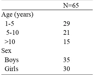
Table 2 showed that thrombocytosis occurred most in group age 1 – 5 years, followed by group age 5 – 10 years and > 10 years in 29 cases, 21 cases and 15 cases consecutively. There were 35 boys had thrombocytosis compared to 30 cases in girls.
Table 2.
Distribution of thrombocytosis based on age and sex
N=65 Age (years)
1-5 29
5-10 21
>10 15
Sex
Boys 35
Girls 30
Table 3.
Underlying diseases in thrombocytosis
N=65
Complicated surgery 31
Infectious diseases 21
Head injury 10
Others 3
The underlying disease of thrombocytosis in this study were complicated surgery in 31 cases, infectious diseases in 21 cases, head injury in 10 cases and others in 3 cases.
DISCUSSION Denton4
et al, reported that extreme thrombocytosis (ExtThr; platelet count
1000x109
/l) isuncommon but may have an increased occurrence in critically ill children.
The incidence of ExtThr for children on the PaediatricIntensive Care Unit at Bristol Royal Hospital for Children betweenJanuary 2001 and December 2004 was calculated, and the notesof children identified with ExtThr were reviewed for possible common aetiological factors, potential treatment regimes and outcome. Vora6
et al estimated the incidence and causes of secondary thrombocytosis in children, a 12 month study of all patients attending a children's hospitaland discovered to have a platelet count over two times the upper normal limit (> 800 x 10(9)/l) was undertaken. Data so obtained were analysed both separately and together with those from two previous studies to gain as broad a perspective as possible. Of 7916 children who had platelet counts during the study period, 36 (0.5%) produced a value > 800 x10(9)/l; there were 19 boys and 17 girls. There was a preponderance ofyoung infants (median age 13 months). Twenty seven of the 36 had some sortof associated infection, bacterial in 18 and viral in nine. The other nine were either recovering from anti-neoplastic chemotherapy (n = 6), werepost-operative (n = 2), or simply iron deficient (n = 1). Combining these patients with those described in previous studies allowed a review of 139 unselected children with very high platelet counts. Fifty three (38%) had infections, 29 (20%) had traumatic or surgical tissue damage, 16 (11%)
had malignant disease undergoing
chemotherapy or surgery, and 13 (9%) had connective tissue or autoimmune disorders. Secondary thrombocytosis is not rare and is most frequently seen in very young infants after infection. Itcan arise in a wide variety of other circumstances including rebound from myelosuppression, iron lack, or as part of an acute phase response. It is clinically unimportant in terms of morbidity and requires no treatmentother than that for the primary condition. Yohannan6
et al, study in six hundred sixty-three children aged 1 to 16 years with thrombocytosis (defined as a platelet count of more than 500 X 109
disorders (4.1 %), and malignancy (2%). Thrombocytosis associated with multiple, simultaneous causative factors was seen in 3.3% of cases. Among all patients with infections, osteomyelitis and septic arthritis were associated with higher platelet counts
than other infections (P<. 0001).
Thrombocytosis secondary to infections was significantlymore common in children under 5 years of age, whereas chronicinflammation, malignancy, and renal disorders were more commoncauses of thrombocytosis in children over 5 years of age. Thrombocytosis of 1 million or more platelets was seen in 13 (2%)
children. No thrombocytosis-related
complications were seen in any children,and none required any specific treatment. Thrombocytosis is a frequent finding in children. It is due to a variety of etiologic factors and is of little clinical discriminatory value. It is often due to an acute-phase phenomenon in response to infection, tissue damage, blood loss, or anemia, and is rarely due to malignancy.Vannucchi7
et al, studied about the role of elevated platelet counts in thrombosis, which represent the predominant complicationof CMPD, significantly affecting prognosis and quality of life as well as, paradoxically, in the pathogenesis of the hemorrhagicmanifestations, will be discussed. Established and novel potentialrisk factors for thrombosis, including the clinical relevanceof the JAK2V617F mutation, and current management strategiesfor thrombocytosis are also briefly discussed. Shafer6
, noted in his study that the challenge of correctly identifying the cause of thrombocytosisin an individual patient becomes particularly critical
when the clinician is confronted with
treatment decisions. Patients with secondary (reactive) thrombocytosis do not require platelet-lowering or antiplatelet treatment because their abnormal platelet count itself does not place them at risk for hemostaticor vascular events. It is crucial, however, to
identify the cause of their secondary
thrombocytosis, even when it is clinically inapparent, so that treatment can be directed
to the underlying disease. A normal
erythrocyte sedimentation rate and a normal level of C-reactive protein may help to rule out an underlyinginflammatory disorder. The search for occult cancer should involve a
thorough physical examination, including examination of stool specimens for occult blood, chest radiography, and further testing as indicated by systemic and localizing symptoms and signs.
Chan8
et al, reported the introduction of the newer generation of electronic cell counters allows the routine reporting of platelet numbers when the peripheral blood count is requested. In a 12-month period,100 episodes of marked thrombocytosis (platelet count more than 900 x 109
/L) were found among 94 children. These patients wereyoung (median age 9 months). All but one episode of marked thrombocytosis occurred as a phenomenon secondary to a variety of disease states. Infections, especially those involving
the central nervous systems were the
commonest cause of an elevated platelet count in this series. Malignant diseases alone were rarely associated withthrombocytosis of this magnitude. The elevated platelet count began to decline at a mean of 3 days after diagnosis, and nothrombotic or hemorrhagic complications were encountered. Marked thrombocytosis is a benign, common phenomenon in young children, but specific treatment is not required. Natalie9
et al, on their study objecyive to estimate the frequency of primary and secondary thrombocytosis in children. To describe the diseases associated with secondary thrombocytosis. To relate the magnitude of the thrombocitosis and the different diagnoses. Resulted that 584 cases of thrombocytosis were found and their study, representing 32.4% of the blood counts. 334 clinical case notes were reviewed, 62% male. 3 patients 0.9% had a platelet count over 1,000,000, 2 of them had primary thrombocytosis (essential thrombocythaemia and chronic myeloid leukaemia) and the third had a bacterial meningitis. The diseases associated with secondary thombocytosis were infection 48.8% (respiratory 70%), iron deficiency 18.6% tissue damage (burns and surgery) 12.6%. Concluded that the frequency of primary thrombocytosis is low, when it is less than 1,000,000 a secondary aetiology is most likely. Heng10
process. The mechanism by which the acute inflammation causes thrombocytosis is not fully understood. Two humoral factors, thrombopoietin and megakaryocyte colony stimulating factor appear to regulate the production and the numbers of circulating platelets. There is a wide variety of causes associated with thrombocytosis. In our study, bacterial infection in particular pneumonia was the predominant cause of thrombocytosis. Other common conditions included urinary tract infection, gastroenteritis and Kawasaki’s disease. Secondary thrombocytosis is a benign and self-limiting condition. None of the cases developed any complications associated with the high platelet level. This could have accounted for the low proportion of followed-up platelet count especially when the child had recovered from the underlying illness. The onset of thrombocytosis occurred within the first week of illness in majority of the illness. The normalisation time depends on the severity of inflammation. Generally, the normalisation time is longer in bacterial infections compared to the viral infections.
Kousaku11
et al, determine the incidence and etiology of childhood thrombocytosis, over 15,000 platelet counts in 7,539 patients performed at a single regional hospital were reviewed. When thrombocytosis was defined as ≥500 x 109/l ofplatelet counts, the
condition could be diagnosed in 6.0% (456 cases) of the patients. All patients were classified as having secondary thrombocytosis. The incidence of thrombocytosis dramatically changed throughout child development; it was 12.5% in neonates, peaked to 35.8% in 1-month-old infants and then returned to 12.9% in 6- to 11-month-old infants. Thereafter, it gradually decreased with age to only 0.6% in 11- to 15-year-old children. Frequent causes of thrombocytosis included infection (67.5%), Kawasaki disease (9.4%), prematurity (7.7%) and iron deficiency anemia (6.4%). Thrombocytosis was an incidental finding in a substantial population of early infants. Thrombocytosis as a reaction to several types of infection and Kawasaki disease was more common in children under 7 years old, while autoimmune disease and tissue damage were major causes in children aged 11-15 years. No child had thromboembolic complications. These findings indicate that childhood
thrombocytosis is a benign condition and its incidence and etiology seem to depend on age. Conclusion: Thrombocytosis is common problem in critically ill patient. It is required a longitudinal study to determine the cause of thrombocytosis in critically ill patients.
REFERENCES
1. Kaushansky K. Regulation of
megakaryopoiesis. In: Loscalzo J, Schafer AI, eds. Thrombosis and hemorrhage. 3rd ed. Philadelphia: Lippincott Williams & Wilkins, 2003:120-39.
2. Cheung MC, Hicks LK, Pendergrast J, Shafer AI. Trombocytosis. N Engl Med J 2004;350:2524-5.
3. Denton A, Davis P. Extreme
thrombocytosis in admission to pediatric intensive care: no requirement for treatment. Arch Dis Child 2007;92:515-516.
4. Vora AJ, Lilleyman JS. Secondary
thrombocytosis. Arch Dis Child 1993;68:88-90.
5. Yohannan D, Higgy KE, Al-Mashhadani SA, Santosh-Kumar CR. Thrombocytosis. Clin Pediatr 1994;33(6):340-3
6. Shafer AI. Trombocytosis. N Engl J Med 2004;350:1211-9.
7. A. M. Vannucchi and T. Barbui.
Thrombocytosis and thrombosis. Hematol 2007; 2007(1): 363 - 370.
8. Chan KW, Kaikov Y, Wadsworth LD.
Thrombocytosis in childhood: A survey of 94 patients. Pediatr 1989;84(6):1064-7.
9. Natalie RZ, Juan TC, Veronica SA.
Trombocytosis in children. Rev Child Pediatr 2000;71(4):307-10.
10. Heng JT, Tan AM. Thrombocytosis in childhood. Singapore Med J 1998;39(11):485-7.