Role of PvdQ in
Pseudomonas aeruginosa
virulence under iron-limiting conditions
Pol Nadal Jimenez,
13
Gudrun Koch,
13
Evelina Papaioannou,
1Mariana Wahjudi,
1,2Joanna Krzeslak,
1Tom Coenye,
3Robbert H. Cool
1and Wim J. Quax
1Correspondence Wim J. Quax [email protected]
1
Department of Pharmaceutical Biology, University of Groningen, 9713 AV Groningen, The Netherlands
2
Faculty of Pharmacy and Faculty of Technobiology, University of Surabaya, Indonesia 3
Laboratory for Pharmaceutical Microbiology, Ghent University, 9000 Ghent, Belgium
Received 17 May 2009 Revised 10 September 2009 Accepted 18 September 2009
PvdQ, an acylase fromPseudomonas aeruginosaPAO1, has been shown to have at least two functions. It can act as a quorum quencher due to its ability to degrade long-chainN
-acylhomoserine lactones (AHLs), e.g. 3-oxo-C12-HSL, leading to a decrease in virulence factors. In addition, PvdQ is involved in iron homeostasis by playing a role in the biosynthesis of pyoverdine, the major siderophore ofP. aeruginosa. In accordance with earlier studies on RNA level, we could show at the protein level that PvdQ is only expressed when iron is present at very low concentrations. We therefore set out to investigate the two functions of PvdQ under iron-limiting conditions. Gene deletion ofpvdQdoes not affect growth ofP. aeruginosabut abrogates pyoverdine production, and results in an accumulation of 3-oxo-C12-HSL. Phenotypic analyses of ourDpvdQmutant at low iron concentrations revealed that this mutant is impaired in swarming motility and biofilm formation. Additionally, a plant and aCaenorhabditis elegansinfection model demonstrated that the deletion ofpvdQresulted in reduced virulence. None of the phenotypes in the present study could be linked to the presence or absence of AHLs. These results clearly indicate that under iron-limiting conditions PvdQ plays a major role in swarming motility, in biofilm development and in infection that is more likely to be linked to the pyoverdine pathway rather than the LasI/LasR/3-oxo-C12-HSL quorum-sensing circuit.
INTRODUCTION
Iron, which is essential for bacterial life, is not freely available and in many environments is present below the concentration required for bacterial growth (Braun & Hantke, 1997). Bacteria have established a system to sequester iron. They secrete iron-scavenging molecules, siderophores, that chelate iron from the environment, and transport it into the cells by binding to specific receptors on the cell surface (Neilands, 1993, 1995). Also multicellular organisms, e.g. mammals, have developed systems to strictly regulate iron homeostasis; as well as the need to scavenge iron ions present only at low concentrations, cells have to be protected from the damaging radicals that can be formed in the presence of excess iron (Carpenter et al., 2009; Miethke & Marahiel, 2007). Molecules that regulate the exchangeable pool of iron (Freestone et al.,
2000, 2002, 2003), e.g. lactoferrin and transferrin, cause the concentration of free iron in serum to be as low as 10224 M. Upon infection with pathogenic bacteria, the infected host and the pathogen will therefore start a fierce battle for iron. Studies of a pathogen such asPseudomonas aeruginosa under iron-limiting conditions therefore seem appropriate to better understand its behaviour.
P. aeruginosais an opportunistic pathogen infecting mainly immunocompromised individuals, such as HIV patients, as well as those suffering from burn wounds and cystic fibrosis (Holder, 1993). This bacterium produces two well-characterized siderophores. Pyoverdine has a high affinity for iron, whereas pyochelin, the second siderophore, has only a low iron affinity (Cox et al., 1981; Cox & Adams, 1985; Poole et al., 1996).
Pyoverdine is a complex molecule composed of a fluorescent chromophore linked to a peptide moiety (Meyer, 2000; Wendenbaumet al., 1983). This siderophore is considered a virulence factor, capable of enhancing P. aeruginosa infection and virulence. Pyoverdine has been shown not only to increase its own expression, but also to Abbreviations: AHL, N-acylhomoserine lactone; EDDHA,
ethylenedia-mine di(o-hydroxy)phenylacetic acid; HSL, homoserine lactone. 3These authors contributed equally to this work.
influence the expression of at least two virulence genes: those encoding exotoxin A and PrpL protease (Beareet al., 2003; Lamontet al., 2002). Therefore, pyoverdine synthesis needs to be tightly regulated. Ferric uptake regulator (Fur) can be seen as the major suppressor of the expression of iron-regulated genes (Prince et al., 1991, 1993; Vasil & Ochsner, 1999). Under low-iron conditions Fur is released from promoter regions, allowing transcription (Escolar et al., 1999; Neilands, 1990). One of the Fur-regulated genes, the sigma factor genepvdS, is the regulator of some genes involved in pyoverdine biosynthesis (Cunliffeet al., 1995; Miyazakiet al., 1995; Ochsneret al., 1995; Viscaet al., 2002).
Pyoverdine and subsequently iron acquisition are important forP. aeruginosato develop different lifestyles. It has been shown that iron can serve as a signal for biofilm development (Banin et al., 2005; Singhet al., 2002; Yang et al., 2007). Particularly iron limitation compromises biofilm formation (Banin et al., 2005; Patriquin et al., 2008). Mere iron diffusion is not enough to allow biofilm formation; to form biofilms, a functional iron-uptake system is required (Baninet al., 2005). Low iron concentra-tions induce P. aeruginosa twitching motility, suggesting iron to be one of the links between biofilm formation and this type of motility (Singhet al., 2002; Singh, 2004).
ThepvdQgene (PA2385 in strain PAO1 and PA14_33820 in strain PA14) is located within the pyoverdine (Pvd) locus, but its potential role in pyoverdine biosynthesis inP. aeruginosais still unclear. PvdQ, which has homology to b-lactam acylases (Sio & Quax, 2004), belongs to the N-terminal nucleophile hydrolase (Ntn) superfamily and has been shown to degrade some N-acylhomoserine lactones (AHLs) (Sio et al., 2006), the major communication molecules in Gram-negative bacteria. As for most Pvd genes, expression of this acylase occurs under iron
starvation (Lamont & Martin, 2003; Ochsner et al., 2002), but little is known about the effects of the enzyme under those conditions. Therefore, we set out to investigate the role ofpvdQ in P. aeruginosa PA14. A pvdQ deletion strain did not show any growth impairment compared to the wild-type. The deletion strain was analysed for phenotypes associated with iron, such as biofilm formation and motility. AHL levels were measured to see whether they correlated with the phenotypes observed. Our data suggest that PvdQ is a key enzyme regulating virulence in P. aeruginosa. At low iron concentration, PvdQ decreases the levels of 3-oxo-C12-HSL; it also controls pyoverdine production and swarming motility, increases virulence via the pyoverdine/iron pathway, and regulates biofilm formation via an as yet unidentified mechanism.
METHODS
Bacterial strains and growth conditions.The bacterial strains and plasmids used in this study are listed in Table 1.Escherichia coliS17-1 lpir was used as the donor strain in bacterial conjugation (Simon
et al., 1983).P. aeruginosacompetent cells were prepared as described by Choiet al.(2006). Bacteria were grown at 37uC in Luria–Bertani (LB) medium or on LB agar plates (Sambrook et al., 2001). For plasmid selection and maintenance, antibiotics were added to growth media at the following concentrations:E. coli– gentamicin, 10 mg l21; tetracycline, 10 mg l21;P. aeruginosa– gentamicin, 25 mg l21; tetracycline, 200 mg l21.
General DNA manipulations.DNA manipulation was performed using standard techniques (Sambrooket al., 2001). PCR fragments were purified using the QIAquick PCR Purification kit (Qiagen). DNA fragments were purified from agarose gels with the QIAquick Gel Extraction kit according to the manufacturer’s instructions. Genomic DNA from P. aeruginosa strains was isolated using a genomic DNA isolation kit (GenElute bacterial genomic DNA kit, Sigma-Aldrich). Plasmid isolation was performed using Nucleospin Plasmid Isolation kit (Macherey-Nagel). DNA sequencing was carried out by Macrogen.
Table 1. Strains and plasmids used in this study
Strain or plasmid Description Reference
E. coli
DH10B pMCT-pvdQ Sioet al.(2006)
S17-1lpir galU galK rpsL(StrR)endA1 nupG thi pro hsdR hsdM+recA(RP4-2Tc : : Mu Km : : Tn7)lpir
Simonet al.(1983)
OP50 A uracil auxotroph derived fromE. coliB Brenner (1974)
P. aeruginosa
UCBPP-PA14 Clinical isolate; referred to as PA14 Leeet al.(2006)
PA14DpvdQ DpvdQchromosomal deletion mutant of PA14 This study
PA14DpvdQ: : pME6032-pvdQ This study
PA14pvdQmutant ID27758 PA14 transposon insertion mutant Liberatiet al.(2006)
Plasmids
pEX18Gm Suicide plasmid carryingsacBR, GmR Hoanget al.(1998)
pME6032 lacIQ-Ptac expression vector; pVS1-p15A shuttle vector TetR Heebet al.(2002)
pME6032-pvdQ pvdQin pME6032 Sioet al.(2006)
pSB1075 lasR lasl9(P. aeruginosaPAO1) : :luxCDABE(Photorhabdus luminescensATCC 29999) fusion in pUC18 ApR, acyl-HSL biosensor producing bioluminescence
Construction of apvdQdeletion mutant.An in-frame deletion of
pvdQwas obtained via splicing by overlapping extension PCR (SOE-PCR: Horton et al., 1989). Briefly, approximately 1 kb fragments located upstream and downstream ofpvdQwith an additional short sequence of overlap (given in bold letters in their sequences below) were amplified from genomic DNA using primer pair ForA/RevA [ForA, 59-GACAAGCTTGGTGTCGCAGAGCGAGTT-39, containing aHindIII restriction site (underlined); RevA, 59- CATGAGACACGC-GTCCCCATCGATGTCGTTTC-39] and primer pair ForB/RevB [ForB, 59-GGGACGCGTGTCTCATGATAAGCAATGCCTATC-39; RevB, 59-CAGGAATTCGGCCATCGGTAGCA-39, containing an
EcoRI restriction site (underlined)]. Next, the two DNA fragments were joined together, completed and the final product boosted by a third PCR using primers ForA and RevB. The resulting fragment was cloned into pEX18Gm carrying a sacB sucrose-sensitivity gene (Hoang et al., 1998) using the EcoRI and HindIII restriction sites. This plasmid was transformed intoE. coliS17-1lpir and conjugated intoP. aeruginosato generate an in-frame deletion of thepvdQgene in the PA14 strain by allelic exchange. Gentamicin-resistant, sucrose-sensitiveP. aeruginosastrains were selected, followed by selection of double recombinants on Vogel–Bonner minimal medium (Schweizer, 1991) containing 5 % (w/v) sucrose. The deletion was confirmed by PCR of thepvdQgene and Southern blot analysis of digested genomic DNA by using a DIG High Prime DNA labelling and detection starter kit I (Roche) according to the manufacturer’s instructions.
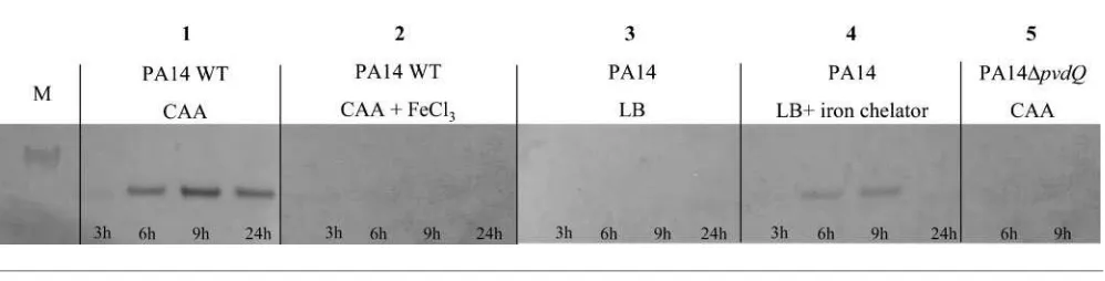
Western blot assay.Purified protein was used to produce polyclonal PvdQ antibodies in rabbits (Eurogentec).P. aeruginosaPA14 and its DpvdQmutant were grown in CAA medium (containing, per litre: 5 g low-iron Bacto Casamino Acids (Difco), 1.54 g K2HPO4.3H2O, 0.25 g
MgSO4.7H2O), LB medium and LB medium supplemented with the
iron chelator 2,29-dipyridyl (300mM). Samples were taken 3, 6, 9 and 24 h after inoculation. Cultures were spun down for 10 min at 13 000g and the pellet resuspended in Bug Buster lysis buffer (Novagen). The lysate was boiled for 10 min, and 20ml was subsequently separated on a 4–12 % polyacrylamide gel (Invitrogen). The proteins were transferred to a nitrocellulose membrane, blocked with 5 % milk, and probed with rabbit polyclonal PvdQ antibody (1 : 1000 dilution in TBS-T). Proteins were detected with goat anti-rabbit antibody (1 : 5000 dilution in TBS-T) conjugated to alkaline phosphatase.
Twitching motility assay. Twitching motility was assayed by the subsurface agar method (Alm & Mattick, 1995). A 2ml aliquot of aP. aeruginosa overnight culture was stab-inoculated through a 1 % LB agar plate. The zone of twitching was visualized 24 and 48 h after incubation at 30uC and 37uC by staining with Coomassie brilliant blue R250 (Pierce). Twitching motility was also assayed after addition of 100mM 2,29-dipyridyl.
Swimming assay. Swimming assays were performed using BM2 glucose minimal medium [62 mM potassium phosphate buffer (pH 7), 0.5 mM MgSO4, 10mM Fe(II) sulfate, 0.5 % Casamino acids
and 0.4 % glucose](Overhage et al., 2007) containing 0.3 % (w/v) agar. Plates were spot-inoculated with 2ml of an overnight culture and the swimming zone was measured after incubation for 24 and 48 h at 30uC or 37uC.
Swarming assay. Swarming assays were done using BM2 glucose minimal medium containing 0.5 % (w/v) agar. Approximately 8 h after they were poured, the plates were inoculated with 2.5ml of an overnight culture in triplicate. Comparisons were made only among plates poured from the same batch of agar. Swarming media were also supplemented with one of the following iron sources: Fe(II) sulfate (10–300mM), Fe(III) sulfate (10–300mM), Fe(III) chloride (18– 300mM) and Fe(II) citrate (10–300mM). Swarming motility was also tested on media containing the iron chelator 2,29-dipyridyl (50mM– 1 mM), 3-oxo-C12-HSL, C4-HSL and partially purified pyoverdine
(as described by Koedamet al., 1994). Compounds were added on a sterile disc at various concentrations.
Biofilm formation. Cultures ofP. aeruginosaPA14 and theDpvdQ
mutant were grown overnight in CAA medium and diluted in the same medium to OD6000.1. To test the effects of iron on theDpvdQmutant,
the same iron sources that were tested in the swarming motility assay were added to the medium at 100mM, a concentration proven to restore swarming motility in theDpvdQmutant. In order to minimize the intrinsic variability of the crystal violet assay (Peeterset al., 2008) each test was performed in.20 wells of a round-bottomed polystyrene 96-well plate (Greinier Bio-One). A 100ml aliquot of the diluted cultures was added to each well and the plates were incubated at 30uC (static biofilm). After 24 h of adhesion, the supernatant was removed and the wells were extensively rinsed with sterile physiological saline. Then 100ml of fresh medium was added to the wells and the plates were incubated for 24, 48 or 72 h. Biofilm biomass was quantified using the crystal violet method described by Christensenet al.(1985) with minor modifications (Peeters et al., 2008). Briefly, after removal of the supernatant and extensive washing with 0.9 % NaCl, 100ml of 99 % methanol was added to each well. After 15 min of fixation, the methanol was removed and the plates were air-dried. Then 100ml crystal violet (10 mg ml21; Merck) was added to the wells and the plates were incubated at room temperature for 20 min. The excess of crystal violet was removed under tap water and the plates were subsequently air-dried. To release the crystal violet, 150ml of 33 % acetic acid was added to the plates and absorbance was measured at 600 nm. For the iron complementation studies, the different iron salts tested in the motility assays were added to CAA medium to a final concentration of 100mM. After washing the biofilm, fresh CAA medium containing the same amount of iron salts was added to the wells. The following steps were identical to those described above.
Determination of autoinducer production.The amount of 3-oxo-C12-HSL produced was determined using the biosensor E. coli(pSB1075), which produces light in response to long-chain AHLs (Winson et al., 1998). 3-Oxo-C12-HSL concentrations were determined at different stages of biofilm grown in round-bottomed polystyrene 96-well plates. Biofilm supernatants of wild-type PA14 and thepvdQdeletion mutant were collected after 24, 48 and 72 h. After 10 min centrifugation of the cultures, the supernatants were filtered using a 0.2mm pore filter (Whatman) and stored at220uC for later analysis. After collecting all the samples, a bioassay was started at 37uC by adding 180ml of a 1/100 dilution of an overnight
E. coli(pSB1075) culture and 20ml of each supernatant sample. The amount of light produced by the biosensor was read every hour during a 20 h time course in a multifunctional microplate reader (FLUOstar Omega, BMG Labtech). Data points obtained immediately prior to maximum light production were used for comparisons (about 10 h after initiation of the bioassay).
TestingP. aeruginosavirulence in a plant model.In order to develop a simple screening mechanism for infection byP. aeruginosa,
we exploited the opportunistic plant infectious behaviour of this bacterium. Various plants were tested and potato (Solanum tuberosum) was selected due to easy handling and rapid visualization of infection. The potato tuber surface was sterilized with 70 % ethanol to reduce microbial contamination. Slices about 3–5 mm thick were placed in sterile Petri dishes on paper dampened with sterile water. The slices were inoculated with 10ml of an overnight culture previously adjusted to OD600 0.3–0.4. Infection development was
Caenorhabditis eleganskilling assay.AC. eleganskilling assay was performed to test the toxicity of PA14 and thepvdQdeletion mutant.P. aeruginosastrains were grown overnight at 37uC in CAA medium (supplemented with Fe(II) sulfate, Fe(III) chloride or Fe(II) citrate where necessary) and then diluted 100-fold into fresh broth. Nematode growth medium (NGM; Brenner, 1974) plates (59 mm diameter) were then spread with 80ml of the respective cultures. The plates were incubated at 37uC for 24 h and allowed to equilibrate to room temperature for 30 min, then 40 L4 nematodes from stock plates were transferred onto theP. aeruginosalawn. The plates were incubated at 24uC and scored for living and dead worms every 3–4 h for 6 days. For statistical purposes a minimum of three replicates per trial were performed. The usualC. elegansfood bacteriumE. coliOP50 was used as a negative control to evaluate background levels of worm death. A worm was considered to be dead when it failed to respond to plate tapping or gentle touching with a platinum wire. Worms that died as a result of getting stuck to the wall of the plate were excluded from the analysis. Results are presented as the percentage of living nematodes on the killing plates compared to their survival on theE. coliOP50 control strain.
RESULTS
Expression ofPvdQunder iron-limiting conditions
pvdQ expression under iron-limiting conditions was verified by growing strain PA14 in CAA medium with or without additional Fe(III) chloride (100mM) as well as in LB medium and LB medium supplemented with the iron chelator 2,29-dipyridyl (300mM) (Fig. 1). As a control, we grew theDpvdQmutant in the same media (only shown for CAA medium). Our results demonstrate that PvdQ can only be detected on Western blots when the bacteria are grown in media with low amounts of iron, indicating iron-dependent regulation of PvdQ production.
Influence ofpvdQdeletion on growth in
iron-depleted conditions
Pyoverdine-negative strains normally show growth impair-ment under low-iron conditions. Hence, we compared PA14 to the DpvdQ mutant. Both were grown in CAA medium and samples were taken over 24 h. OD600 was
measured to compare growth, whereas absorbance of the supernatant at 405 nm (A405) was measured to examine
pyoverdine production. Fig. 2 shows no difference in growth during low-iron conditions, but clearly demon-strates the absence of pyoverdine in the deletion strain. It should be noted here that the addition of 0.5 g l21of the strong chelator ethylenediamine di(o-hydroxy)phenylacetic acid (EDDHA) impaired growth severely in the pvdQ deletion strain but only slightly in the wild-type strain (see Supplementary Fig. S1, available with the online version of this paper).
Influence of PvdQ on motility
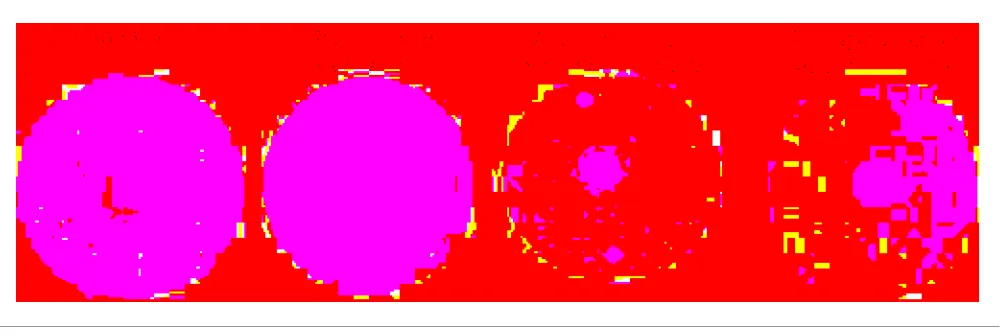
Motility has been associated with nutrient availability (Deziel et al., 2003; Kohler et al., 2000; Rashid & Kornberg, 2000; Singhet al., 2002). We examined the effects ofpvdQdeletion inP. aeruginosaon three different types of motility: flagellar-mediated swimming motility, swarming, and type IV pili-mediated twitching motility. Swimming and swarming motility assays were performed in the standard BM2 medium or BM2 medium without Fe(II) sulfate. No difference was observed in twitching and swimming motility between wild-type and deletion mutant (data not shown). In contrast to the wild-type strain, no swarming motility could be observed for the pvdQ deletion strain (Fig. 3a). Plasmid-borne expression of pvdQ or addition of partially purified pyoverdine allowed complementation of the mutant strain, restoring the swarming level to that of the parent strain (Fig. 3b, c). Addition of 3-oxo-C12-HSL and C4-HSL did not restore swarming motility in the mutant (not shown).
In a recent study, apvdQtransposon mutant from the P. aeruginosa PA14 mutant library (Liberati et al., 2006) showed a decrease in swarming motility (~75 % dimin-ished compared to wild-type) (Overhageet al., 2008). This P. aeruginosa PA14 pvdQ transposon mutant (ID27758) was compared to ourpvdQdeletion strain on the same type of swarming plates. Interestingly, the transposon mutant also showed total absence of swarming motility (Fig. 3d). The differences seen in the earlier study (Overhage et al.,
2008) can thus be attributed to laboratory conditions and not to any polar effects from the transposon.
To investigate whether the reduced swarming motility was influenced by differences in production of rhamnolipids (Caiazzaet al., 2005; Dezielet al., 2003; Kohleret al., 2000; Overhage et al., 2007), an orcinol test and a TLC analysis (as described by Wilhelmet al., 2007) were conducted with our parent and mutant strain. However, no differences were observed (data not shown).
Role of iron in swarming motility
Based on the lack of pyoverdine production in our deletion strain and the evidence that low levels of iron stimulate
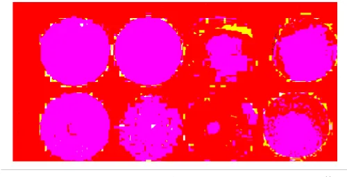
surface motility (Singhet al., 2002; Singh, 2004), we tested the influence of different iron sources on the swarming behaviour of the DpvdQ mutant. Fe(II) sulfate, Fe(III) sulfate, Fe(III) chloride and Fe(II) citrate were used as individual iron sources. Additionally, swarming behaviour was studied on iron-depleted swarming agar (containing 2,29-dipyridyl). Individual addition of each iron com-pound (shown for Fe(III) chloride in Fig. Fig. 4b) resulted in increasing swarming motility in the deletion mutant (up to wild-type level; Fig. 4a). Addition of 2,29-dipyridyl resulted in inhibition of swarming of the wild-type PA14 (Fig. 4c).
We also tested whether addition of a partially purified batch of pyoverdine had any effect on swarming motility (Fig. 4d). Indeed swarming motility was restored, indic-ating a direct relation between PvdQ and pyoverdine. These experiments show that sufficient iron is needed for swarming motility.
PvdQ plays a role in iron uptake and biofilm formation
Staining with crystal violet revealed a significant 32-fold reduction in biofilm production in CAA medium in the DpvdQ mutant in comparison to the parental strain (Fig. 5). To test the hypothesis that iron was directly responsible for these differences, we added the four abovementioned iron sources. At the concentrations tested (10–300 mM) these iron salts did not restore biofilm formation (Fig. 5: data shown only for Fe(III) chloride). These results indicate that iron alone does not account for the differences in the amount of biofilm produced by the DpvdQ mutant, suggesting that PvdQ plays a crucial role in biofilm formation, which seems to be independent of its role in the iron/pyoverdine pathway.
Fig. 3. Swarming motility of PA14DpvdQ. Swarming motility was assayed on BM2 medium solidified with 0.5 % agar. (a) Swarming impairment in PA14DpvdQ.(b, c) Swarming motility can be fully restored by plasmid-borne gene expression (b) or by addition of partially purified pyoverdine (c). ThepvdQtransposon mutant (ID27758) shows the same impairment as the clean deletion used in our studies (d).
1.2
1.0
0.8
OD
600
or
A405
0.6
0.4
0.2
2 4 6 8
Time (h)
10 12 o/n
Fig. 2. Growth of wild-type PA14 (black diamonds) and theDpvdQ
mutant (grey squares). Growth was monitored in iron-limited CAA medium. Wild-type and deletion strain show no differences in growth (full lines). However, when measuring theA405of the
Detection of 3-oxo-C12-HSL under iron-limiting conditions
Analysis of the cell-free supernatants of biofilms formed in CAA minimal medium by the wild-type strain and the DpvdQ mutant in a bioassay indicated a significant decrease in 3-oxo-C12-HSL concentration in the wild-type strain (Fig. 6), which can be attributed to the degradation of this autoinducer following PvdQ production. Reduced levels of 3-oxo-C12-HSL were observed by other authors
while studying the amounts of AHLs in the cystic fibrosis lung (Singhet al., 2000). A link between PvdQ and low 3-oxo-C12-HSL levels has been suggested as a possible explanation for the observed autoinducer decrease (Hentzeret al., 2005).
In vivoeffect of PvdQ in a plant infection model
The effects of PvdQ on virulence were studied in a plant model system. Potato slices inoculated with the DpvdQ strain exhibited a pronounced decrease in infection compared to the parental strain, for which clear infection was observed 48 h after incubation at 30uC (Fig. 7). Complementation of the pvdQ mutant with plasmid pME6032-pvdQ or addition of partially purified pyover-dine restored infection to the wild-type level (Fig. 7). Restoration with iron sources could not be performed, as iron alone already caused fouling of the potato surface.
In vivoeffect ofpvdQexpression in a C. elegans
infection model under iron-limiting conditions
C. elegansnematodes were exposed to PA14 and theDpvdQ mutant in order to study the effect of PvdQ under iron-limiting conditions in vivo. The nematodes were trans-ferred to CAA plates with lawns of the respective bacterial strains and monitored over a 6 day period. It should be noted that CAA medium is different from the normally used C. elegans infection medium (Papaioannou et al., 2009).
Fig. 4. Influence of iron on swarming motility. PA14 DpvdQ is impaired in swarming motility compared to wild-type (a). However, this swarming motility can be restored by the addition of iron (Fe(III) chloride) (b). Enough iron needs to be present in order for the cells to perform swarming motility; addition of iron chelators also inhibits swarming in the wild-type (c). Partially purified pyoverdine can restore swarming motility in the deletion strain (d).
7
6
5
A600
4
3
2
1
PA14 PA14DpvdQ PA14DpvdQ
+FeCl3
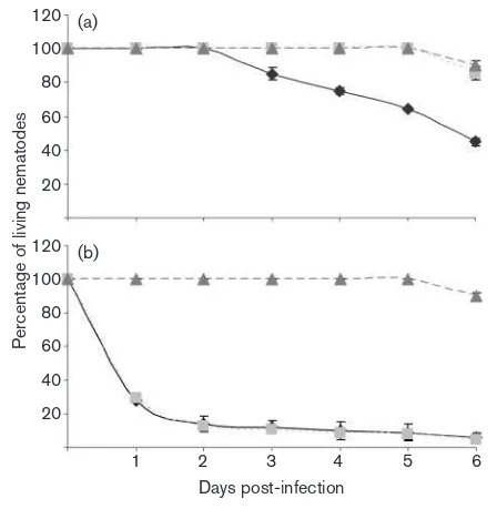
During the first 2 days the nematodes did not show any alterations in their behaviour in any of the plates. However, on the third day post-infection the animals exposed to the wild-type strain showed locomotion problems and their pharyngeal pumping rate started to decrease (Fig. 8a). The number of eggs present on the plates after 4 days was very limited. The few offspring that could be seen on the plates showed a reduced growth rate and by the fifth day, the LT50 was reached. In
contrast to this, the DpvdQ mutant was avirulent to the nematodes, which showed no disease-like symptoms through-out the period of the assay. Hundreds of eggs were present on the plates by the third day and the offspring went through their life cycle without any complications. No decrease in movement or pharyngeal pumping rates was observed. After the completion of the assay, bacterial lawns on the DpvdQ mutant plates were almost completely consumed.
To test if the different iron sources restore the toxicity of the DpvdQmutant, we added one of the following iron sources to the CAA plates: Fe(II) sulfate, Fe(III) chloride or Fe(II) citrate. Each of these iron compounds restored the toxicity of the deletion strain to at least wild-type level (shown for Fe(III) chloride in Fig. 8b). In many cases the animals showed egg-laying defects and the eggs were hatching inside
the adult. Medium supplemented with Fe(III) chloride appeared to result in the highest overall toxicity levels, compared to all other media used: after 1 day of exposure to this medium only 30 % of the animals were alive (Fig. 8b). It should be noted here that PA14 was more virulent under all the iron-supplemented conditions tested compared to the iron-limiting conditions.
All of the above assays were performed usingE. coliOP50 as a negative control to evaluate the background death levels of the worms. This strain was avirulent in all the assays performed under these conditions, and the nema-todes fed on this strain went through a normal life cycle without any complications.
DISCUSSION
Studies on quorum-quenching acylases have focused on the potential of these enzymes to target infections by a broad range of Gram-negative pathogens. As a recent exampleP. aeruginosa has been shown to produce PvdQ, an acylase capable of degrading its own quorum-sensing molecule (3-oxo-C12-HSL) (Huang et al., 2003; Sio et al., 2006). 12000
10000
8000
6000
4000
2000
PA14
24 h
PA14DpvdQ PA14 PA14DpvdQ PA14 PA14DpvdQ
Luminescence
48 h 72 h
Fig. 6. Quantification of 3-oxo-C12-HSL. Strains PA14 and PA14DpvdQ were grown in a biofilm, and cell-free extracts prepared on three consecutive days were analysed using the biosensor strain E. coli(pSB1075). Light produced in response to 3-oxo-C12-HSL was quantified. For comparison, light values prior to the exponential phase were selected. All values are the means±SEfrom at least three
independent experiments.
Fig. 7. PvdQ stimulatesP. aeruginosavirulence in a plant model system. Infection ofP. aeruginosaPA14 and theDpvdQ
However, little is known about the physiological role of this enzyme inP. aeruginosa. Interestingly,pvdQis part of the Pvd locus and as such is involved in biosynthesis of pyoverdine, the major siderophore of this bacterium. As with most Pvd genes, pvdQis only expressed under iron-limiting conditions as shown by microarray studies (Ochsner et al., 2002). The consequence of this is that, apart from the involvement in pyoverdine biosynthesis, the quorum-quenching capabilities of PvdQ are likely to become apparent only when low concentrations of available iron are present in the environment.
To study the control of quorum quenching and side-rophore production by one enzyme we set out to investigate the phenotypes affected by PvdQ. We con-firmed that pvdQ is only expressed under low-iron conditions (Fig. 1). This result strongly suggests that PvdQ is only produced, and thus fulfils a physiological role, when the iron concentration is low. The deletion of pvdQleads to the lack of pyoverdine synthesis as judged by the absence of the typical green colour that is present in the parent strain grown in CAA medium (Fig. 2) and as analysed by HPLC (unpublished results).
In CAA medium the deletion strain grew at a rate similar to the parent strain (Fig. 2). The absence of a growth defect in our pyoverdine mutant suggests that in the absence of a strong iron chelator, the other siderophore pyochelin is sufficient for unaltered growth of thepvdQmutant under the conditions tested. This hypothesis was confirmed by addition of the strong iron chelator EDDHA to the growth medium, which resulted in a growth defect for theDpvdQ mutant (Supplementary Fig. S1), supporting the obser-vation made by others that pyoverdine is necessary for growth in the presence of strong iron chelators (Lamont & Martin, 2003; Ochsneret al., 2002).
Interestingly, we could not observe any change in twitching or swimming motility, although low iron concentrations have been shown to enhance twitching motility (Patriquin et al., 2008; Singhet al., 2002; Singh, 2004). Similar results where obtained by others (Banin et al., 2005), where biofilm formation and twitching motility showed no correlation.
PvdQ plays a role in swarming motility ofP. aeruginosa, as shown by the observation that thepvdQdeletion strain was impaired in swarming motility (Fig. 3a), and that expression of plasmid-borne pvdQ was able to restore swarming to the wild-type level (Fig. 3b). Addition of 3-oxo-C12-HSL and C4-HSL had no effect on restoration of swarming motility, neither could any difference in rhamnolipid production be observed, strongly suggesting that this phenotype is quorum quenching independent. Addition of different iron sources (Fig. 4b) or partially purified pyoverdine (Fig. 4d) restored swarming motility, indicating that this phenotype is under control of iron. These results are consistent with the observations made in Pseudomonas putida, where swarming could be restored in a pyoverdine mutant ofP. putidaKT2440 by addition of iron or pyoverdine (Matillaet al., 2007).
Biofilm formation has been shown in previous studies to be disrupted in pyoverdine-negative strains; these biofilm defects could be restored by addition of Fe(II) citrate or Fe(III) chloride (Baninet al., 2005; Patriquinet al., 2008). Interestingly, most biofilm-deficient mutants were demon-strated to have enhanced swarming motility, suggesting that these two phenotypes are inversely regulated (Caiazza et al., 2007). In our case, theDpvdQdeletion strain is not able to form biofilms in low-iron medium (CAA). Addition of different iron sources to the medium could not rescue this phenotype in our mutant strain (Fig. 5). These observations give us an indication that in P. aeruginosa, PvdQ plays a role in biofilm formation that goes beyond the acquisition of iron.
To rule out the possibility of the quorum-quenching ability of PvdQ having an influence on the results observed, 3-oxo-C12-HSL levels were measured. These levels were higher in the DpvdQ mutant than in the wild-type, corroborating the enzyme’s capability to degrade in vivo long-chain AHLs (Fig. 6). Two other studies indicate a low level of 3-oxo-C12-HSL in biofilm-forming P. aeruginosa 120 (a)
Fig. 8. Effects of deletion ofpvdQonP. aeruginosatoxicity under iron-limiting conditions in the C. elegans infection model and complementation by Fe(III) chloride.P. aeruginosaPA14 and its
DpvdQ mutant were screened for virulence in the C. elegans
model. (a) Under iron-limiting conditions (CAA medium) the
cells: low levels of 3-oxo-C12-HSL were found in the sputum of cystic fibrosis patients (Singhet al., 2000), and in a more recent study microarray data linked the reduction in 3-oxo-C12-HSL to an increase in pvdQ expression inP. aeruginosabiofilms (Hentzeret al., 2005). However, it seems more likely that the 3-oxo-C12-HSL levels are simply a consequence of the presence of PvdQ, but not instrumental in biofilm formation. Overall, a relationship between PvdQ and biofilm formation seems clear from all the reported evidence.
Studies in a burned mouse infection model revealed that pyoverdine/iron acquisition is important for virulence (Meyer et al., 1996). But what role does PvdQ play? If deletion ofpvdQhas an opposite effect on biofilm formation under low-iron conditions compared to rich medium, then what about its effect on virulence? Our findings show that the DpvdQ mutant is strictly avirulent in two different models, a plant model and a C. elegans model. When applying wild-type P. aeruginosa strain on potato slices, fouling lesions were evident, while infection was suppressed in theDpvdQmutant (Fig. 7). These results are in line with the biofilm results, especially because infection could be restored bypvdQcomplementation with plasmid pME6032-pvdQ (Fig. 7). Under iron-limiting conditions, PvdQ positively influences a number of virulence phenotypes. In aC. elegansslow-killing assay, lack of iron in the medium resulted in the PA14DpvdQmutant being avirulent to the nematodes whereas the PA14 wild-type, under the same conditions, had toxic effects. This observation confirms the important role that PvdQ has in virulence. However, the virulence enhancement can only be observed under iron-limiting conditions. Once the media are supplemented with iron sources, the results obtained from the slow-killing assays dramatically change. Addition of any one of the three iron compounds, Fe(II) sulfate, Fe(III) chloride and Fe(II) citrate, to CAA medium resulted in the PA14DpvdQmutant reaching toxicity levels comparable to the PA14 wild-type (shown for Fe(III) chloride in Fig. 8b). Taking the results together, we can conclude that the use of the PvdQ protein as a quorum-quenching agent for therapeutic purposes (Papaioannouet al., 2009) should only be considered under conditions where enough iron is present.
The recent postulation that quorum sensing and iron uptake are related in a complicated and nutritionally conditioned manner (Shroutet al., 2006) is in line with the results from our experiments demonstrating that the effect of PvdQ under iron-limiting conditions is different from that in rich medium. Deletion of the gene does not lead to improved biofilm formation or virulence as would be expected by the resulting higher levels of AHLs. Complementation of pvdQ-related phenotypes in swarm-ing and virulence by addition of iron compounds leads to the conclusion that the role of PvdQ in the iron-uptake pathway overrules its deacylase activity under iron-limiting conditions. However, the inability of iron compounds to restore biofilm formation in the DpvdQ mutant and the parallel swarming complementation (by addition of iron
sources) suggest that PvdQ is a key enzyme where these – and possibly more – pathways are involved in a complicated interplay that needs further elucidation.
ACKNOWLEDGEMENTS
We gratefully acknowledge Miguel Ca´mara and Paul Williams for providing the biosensor strain pSB1075 and 3-oxo-C12-HSL, and Fred Ausubel and Elina Drenkard for providing the PA14 transposon strain ID27758. We also thank Sandra Matthijs and Pierre Cornelis for helpful discussion, Nele Matthijs for excellent technical assistance, and Diane Black for critically reading the manuscript. This research was partly funded by EU grant Antibiotarget MEST-CT-2005-020278 to P. N. J., G. K. and E. P.
REFERENCES
Alm, R. A. & Mattick, J. S. (1995). Identification of a gene, pilV, required for type 4 fimbrial biogenesis inPseudomonas aeruginosa, whose product possesses a pre-pilin-like leader sequence. Mol Microbiol16, 485–496.
Banin, E., Vasil, M. L. & Greenberg, E. P. (2005). Iron and
Pseudomonas aeruginosa biofilm formation. Proc Natl Acad Sci U S A102, 11076–11081.
Beare, P. A., For, R. J., Martin, L. W. & Lamont, I. L. (2003). Siderophore-mediated cell signalling in Pseudomonas aeruginosa: divergent pathways regulate virulence factor production and sidero-phore receptor synthesis.Mol Microbiol47, 195–207.
Braun, V. & Hantke, K. (1997). Receptor-mediated bacterial iron transport. InTransition Metals in Microbial Metabolism, pp. 81–101. Edited by G. Winkelmann & C. J. Carrano. Amsterdam: Harwood Academic Publishers.
Brenner, S. (1974).The genetics ofCaenorhabditis elegans. Genetics 77, 71–94.
Caiazza, N. C., Shanks, R. M. & O’Toole, G. A. (2005).Rhamnolipids modulate swarming motility patterns of Pseudomonas aeruginosa. J Bacteriol187, 7351–7361.
Caiazza, N. C., Merritt, J. H., Brothers, K. M. & O’Toole, G. A. (2007). Inverse regulation of biofilm formation and swarming motility by
Pseudomonas aeruginosaPA14.J Bacteriol189, 3603–3612.
Carpenter, B. M., Whitmire, J. M. & Merrell, D. S. (2009).This is not your mother’s repressor: the complex role of fur in pathogenesis.
Infect Immun77, 2590–2601.
Choi, K. H., Kumar, A. & Schweizer, H. P. (2006).A 10-min method for preparation of highly electrocompetent Pseudomonas aeruginosa
cells: application for DNA fragment transfer between chromosomes and plasmid transformation.J Microbiol Methods64, 391–397. Christensen, G. D., Simpson, W. A., Younger, J. J., Baddour, L. M., Barrett, F. F., Melton, D. M. & Beachey, E. H. (1985).Adherence of coagulase-negative staphylococci to plastic tissue culture plates: a quantitative model for the adherence of staphylococci to medical devices.J Clin Microbiol22, 996–1006.
Cox, C. D. & Adams, P. (1985).Siderophore activity of pyoverdin for
Pseudomonas aeruginosa.Infect Immun48, 130–138.
Cox, C. D., Rinehart, K. L., Jr, Moore, M. L. & Cook, J. C., Jr (1981). Pyochelin: novel structure of an iron-chelating growth promoter for Pseudomonas aeruginosa. Proc Natl Acad Sci U S A 78, 4256– 4260.
Pseudomonas aeruginosa: PvdS is probably an alternative sigma factor.
J Bacteriol177, 2744–2750.
Deziel, E., Lepine, F., Milot, S. & Villemur, R. (2003).rhlAis required for the production of a novel biosurfactant promoting swarming motility in Pseudomonas aeruginosa: 3-(3-hydroxyalkanoyloxy)alk-anoic acids (HAAs), the precursors of rhamnolipids. Microbiology 149, 2005–2013.
Escolar, L., Perez-Martin, J. & de Lorenzo, V. (1999).Opening the iron box: transcriptional metalloregulation by the Fur protein.
J Bacteriol181, 6223–6229.
Freestone, P. P., Lyte, M., Neal, C. P., Maggs, A. F., Haigh, R. D. & Williams, P. H. (2000).The mammalian neuroendocrine hormone norepinephrine supplies iron for bacterial growth in the presence of transferrin or lactoferrin.J Bacteriol182, 6091–6098.
Freestone, P. P., Williams, P. H., Haigh, R. D., Maggs, A. F., Neal, C. P. & Lyte, M. (2002). Growth stimulation of intestinal commensal
Escherichia coliby catecholamines: a possible contributory factor in trauma-induced sepsis.Shock18, 465–470.
Freestone, P. P., Haigh, R. D., Williams, P. H. & Lyte, M. (2003). Involvement of enterobactin in norepinephrine-mediated iron supply from transferrin to enterohaemorrhagic Escherichia coli. FEMS Microbiol Lett222, 39–43.
Heeb, S., Blumer, C. & Haas, D. (2002).Regulatory RNA as mediator in GacA/RsmA-dependent global control of exoproduct formation in
Pseudomonas fluorescensCHA0.J Bacteriol184, 1046–1056. Hentzer, M., Eberl, L. & Givskov, M. (2005).Transcriptome analysis of
Pseudomonas aeruginosabiofilm development: anaerobic respiration and iron limitation.Biofilms2, 37–61.
Hoang, T. T., Karkhoff-Schweizer, R. R., Kutchma, A. J. & Schweizer, H. P. (1998).A broad-host-range Flp-FRT recombination system for site-specific excision of chromosomally-located DNA sequences: application for isolation of unmarked Pseudomonas aeruginosa
mutants.Gene212, 77–86.
Holder, I. A. (1993). Pseudomonas aeruginosa burn infections: pathogenesis and treatment. In Pseudomonas aeruginosa as an Opportunistic Pathogen, pp. 275–295. Edited by M. Campa, M. Bendinelli & H. Friedman. New York: Plenum Press.
Horton, R. M., Hunt, H. D., Ho, S. N., Pullen, J. K. & Pease, L. R. (1989). Engineering hybrid genes without the use of restriction enzymes: gene splicing by overlap extension.Gene77, 61–68. Huang, J. J., Han, J. I., Zhang, L. H. & Leadbetter, J. R. (2003). Utilization of acyl-homoserine lactone quorum signals for growth by a soil pseudomonad andPseudomonas aeruginosaPAO1.Appl Environ Microbiol69, 5941–5949.
Koedam, N., Wittouck, E., Gaballa, A., Gillis, A., Hofte, M. & Cornelis, P. (1994).Detection and differentiation of microbial siderophores by isoelectric focusing and chrome azurol S overlay.Biometals7, 287–291. Kohler, T., Curty, L. K., Barja, F., van Delden, C. & Pechere, J. C. (2000).Swarming ofPseudomonas aeruginosais dependent on cell-to-cell signaling and requires flagella and pili.J Bacteriol182, 5990–5996. Lamont, I. L. & Martin, L. W. (2003).Identification and characteriza-tion of novel pyoverdine synthesis genes inPseudomonas aeruginosa. Microbiology149, 833–842.
Lamont, I. L., Beare, P. A., Ochsner, U., Vasil, A. I. & Vasil, M. L. (2002). Siderophore-mediated signaling regulates virulence factor production inPseudomonas aeruginosa. Proc Natl Acad Sci U S A99, 7072–7077.
Lee, D. G., Urbach, J. M., Wu, G., Liberati, N. T., Feinbaum, R. L., Miyata, S., Diggins, L. T., He, J., Saucier, M. & other authors (2006). Genomic analysis reveals that Pseudomonas aeruginosa virulence is combinatorial.Genome Biol7, R90.
Liberati, N. T., Urbach, J. M., Miyata, S., Lee, D. G., Drenkard, E., Wu, G., Villanueva, J., Wei, T. & Ausubel, F. M. (2006).An ordered, nonredundant library of Pseudomonas aeruginosa strain PA14 transposon insertion mutants.Proc Natl Acad Sci U S A103, 2833– 2838.
Matilla, M. A., Ramos, J. L., Duque, E., de Dios, A. J., Espinosa-Urgel, M. & Ramos-Gonzalez, M. I. (2007). Temperature and pyoverdine-mediated iron acquisition control surface motility of Pseudomonas putida. Environ Microbiol9, 1842–1850.
Meyer, J. M. (2000). Pyoverdines: pigments, siderophores and potential taxonomic markers of fluorescent Pseudomonas species.
Arch Microbiol174, 135–142.
Meyer, J. M., Neely, A., Stintzi, A., Georges, C. & Holder, I. A. (1996). Pyoverdin is essential for virulence ofPseudomonas aeruginosa. Infect Immun64, 518–523.
Miethke, M. & Marahiel, M. A. (2007). Siderophore-based iron acquisition and pathogen control.Microbiol Mol Biol Rev71, 413–451. Miyazaki, H., Kato, H., Nakazawa, T. & Tsuda, M. (1995).A positive regulatory gene,pvdS, for expression of pyoverdin biosynthetic genes inPseudomonas aeruginosaPAO.Mol Gen Genet248, 17–24. Neilands, J. B. (1990).Molecular aspects of regulation of high affinity iron absorption in microorganisms.Adv Inorg Biochem8, 63–90. Neilands, J. B. (1993).Siderophores.Arch Biochem Biophys302, 1–3. Neilands, J. B. (1995). Siderophores: structure and function of microbial iron transport compounds.J Biol Chem270, 26723–26726. Ochsner, U. A., Vasil, A. I. & Vasil, M. L. (1995).Role of the ferric uptake regulator of Pseudomonas aeruginosa in the regulation of siderophores and exotoxin A expression: purification and activity on iron-regulated promoters.J Bacteriol177, 7194–7201.
Ochsner, U. A., Wilderman, P. J., Vasil, A. I. & Vasil, M. L. (2002). GeneChip expression analysis of the iron starvation response in
Pseudomonas aeruginosa: identification of novel pyoverdine biosyn-thesis genes.Mol Microbiol45, 1277–1287.
Overhage, J., Lewenza, S., Marr, A. K. & Hancock, R. E. (2007). Identification of genes involved in swarming motility using a
Pseudomonas aeruginosa PAO1 mini-Tn5-lux mutant library. J Bacteriol189, 2164–2169.
Overhage, J., Bains, M., Brazas, M. D. & Hancock, R. E. (2008). Swarming ofPseudomonas aeruginosais a complex adaptation leading to increased production of virulence factors and antibiotic resistance.
J Bacteriol190, 2671–2679.
Papaioannou, E., Wahjudi, M., Nadal Jimenez, P., Koch, G., Setroikromo, R. & Quax, W. J. (2009).Quorum quenching acylase reduces the virulence ofPseudomonas aeruginosain aCaenorhabditis elegansinfection model.Antimicrob Agents Chemother53, 4891–4897. Patriquin, G. M., Banin, E., Gilmour, C., Tuchman, R., Greenberg, E. P. & Poole, K. (2008).Influence of quorum sensing and iron on twitching motility and biofilm formation inPseudomonas aeruginosa. J Bacteriol190, 662–671.
Peeters, E., Nelis, H. J. & Coenye, T. (2008).Comparison of multiple methods for quantification of microbial biofilms grown in microtiter plates.J Microbiol Methods72, 157–165.
Poole, K., Dean, C., Heinrichs, D., Neshat, S., Krebs, K., Young, L. & Kilburn, L. (1996). Siderophore-mediated iron transport in
Pseudomonas aeruginosa. In Molecular Biology of Pseudomonas, pp. 371–373. Edited by T. Nakazawa. Washington, DC: American Society for Microbiology.
Prince, R. W., Cox, C. D. & Vasil, M. L. (1993).Coordinate regulation of siderophore and exotoxin A production: molecular cloning and sequencing of thePseudomonas aeruginosa furgene.J Bacteriol 175, 2589–2598.
Rashid, M. H. & Kornberg, A. (2000). Inorganic polyphosphate is needed for swimming, swarming, and twitching motilities of
Pseudomonas aeruginosa. Proc Natl Acad Sci U S A97, 4885–4890. Sambrook, J., Fritsch, E. F. & Maniatis, T. (2001).Molecular Cloning: a Laboratory Manual. Cold Spring Harbor, NY: Cold Spring Harbor Laboratory.
Schweizer, H. P. (1991). The agmR gene, an environmentally responsive gene, complements defective glpR, which encodes the putative activator for glycerol metabolism inPseudomonas aeruginosa. J Bacteriol173, 6798–6806.
Shrout, J. D., Chopp, D. L., Just, C. L., Hentzer, M., Givskov, M. & Parsek, M. R. (2006).The impact of quorum sensing and swarming motility onPseudomonas aeruginosabiofilm formation is nutritionally conditional.Mol Microbiol62, 1264–1277.
Simon, R., Priefer, U. & Puehler, A. (1983). A broad host range mobilization system for in vivo genetic engineering: transposon mutagenesis in gram negative bacteria.Biotechnology1, 784–791. Singh, P. K. (2004). Iron sequestration by human lactoferrin stimulates P. aeruginosa surface motility and blocks biofilm formation.Biometals17, 267–270.
Singh, P. K., Schaefer, A. L., Parsek, M. R., Moninger, T. O., Welsh, M. J. & Greenberg, E. P. (2000).Quorum-sensing signals indicate that cystic fibrosis lungs are infected with bacterial biofilms.Nature407, 762–764. Singh, P. K., Parsek, M. R., Greenberg, E. P. & Welsh, M. J. (2002).A component of innate immunity prevents bacterial biofilm devel-opment.Nature417, 552–555.
Sio, C. F. & Quax, W. J. (2004).Improved beta-lactam acylases and their use as industrial biocatalysts.Curr Opin Biotechnol15, 349–355. Sio, C. F., Otten, L. G., Cool, R. H., Diggle, S. P., Braun, P. G., Bos, R., Daykin, M., Camara, M., Williams, P. & Quax, W. J. (2006).Quorum quenching by an N-acyl-homoserine lactone acylase from
Pseudomonas aeruginosaPAO1.Infect Immun74, 1673–1682. Vasil, M. L. & Ochsner, U. A. (1999).The response ofPseudomonas aeruginosa to iron: genetics, biochemistry and virulence. Mol Microbiol34, 399–413.
Visca, P., Leoni, L., Wilson, M. J. & Lamont, I. L. (2002).Iron transport and regulation, cell signalling and genomics: lessons fromEscherichia coliandPseudomonas. Mol Microbiol45, 1177–1190.
Wendenbaum, S., Demange, P., Dell, A., Meyer, J. M. & Abdallah, M. A. (1983). The structure of pyoverdinePa, the siderophore of Pseudomonas aeruginosa. Tetrahedron Lett24, 4877–4880.
Wilhelm, S., Gdynia, A., Tielen, P., Rosenau, F. & Jaeger, K. E. (2007). The autotransporter esterase EstA of Pseudomonas aeruginosa is required for rhamnolipid production, cell motility, and biofilm formation.J Bacteriol189, 6695–6703.
Winson, M. K., Swift, S., Fish, L., Throup, J. P., Jorgensen, F., Chhabra, S. R., Bycroft, B. W., Williams, P. & Stewart, G. S. (1998). Construction and analysis of luxCDABE-based plasmid sensors for investigatingN-acyl homoserine lactone-mediated quorum sensing.
FEMS Microbiol Lett163, 185–192.
Yang, L., Barken, K. B., Skindersoe, M. E., Christensen, A. B., Givskov, M. & Tolker-Nielsen, T. (2007). Effects of iron on DNA release and biofilm development by Pseudomonas aeruginosa.
Microbiology153, 1318–1328.