870
Localization of Androgen and Estrogen Receptors in Adult
Male Mouse Reproductive Tract
QING ZHOU,* RONG NIE,* GAIL S. PRINS,† PHILIPPA T. K. SAUNDERS,‡ BENITA S. KATZENELLENBOGEN,§ AND REX A. HESS*
From the*Department of Veterinary Biosciences, University of Illinois, Urbana, Illinois; †Department of Urology, College of Medicine, University of Illinois, Chicago, Illinois; ‡MRC Human Reproductive Sciences Unit, Edinburgh EH3 9ET, United Kingdom; §Departments of Cell and Structural Biology, and Molecular and Integrative
Physiology, University of Illinois, Urbana, Illinois.
ABSTRACT: There is considerable variation, both within and
be-tween species, in reports of nuclear steroid receptor localizations in the male reproductive tract. In this study, androgen receptor (AR) and estrogen receptors ERaand ERbwere visualized by immunohisto-chemistry in adult male mice reproductive tracts, including testes, ef-ferent ductules; initial segment, caput, corpus, and cauda epididymi-des; and vas deferens. Antibody specificity was demonstrated by Western blot and antibody competition. In testis, AR was expressed in Leydig cells, Sertoli cells, and most peritubular cells, but not in germ cells; Sertoli cells showed more intense staining in stages VI–VII; ERa
was present in Leydig and some peritubular cells; ERbwas in Leydig, some peritubular, all Sertoli and germ cells except in spermatids and meiotic spermatocytes. In efferent ductules, AR was strongly ex-pressed in ciliated and nonciliated epithelial cells and in stromal cells; ERawas strongly expressed in ciliated and nonciliated epithelial cells; stromal cells were negative; and ERbwas strongly expressed in cili-ated and noncilicili-ated epithelial cells and also in stromal cells. In epi-didymis, AR was strongly expressed in all epithelial cells (not in
intraepithelial lymphocytes); ERa was strongly expressed in apical, narrow, and some basal cells of the initial segment, and in caput, principal cells of the caput, clear cells of the distal caput through cau-da; stromal cells were negative in the initial segment, but more stromal cells were stained from caput to cauda; ERbwas strongly expressed in most of epithelial cells of the epididymis, but stromal cells were inconsistently stained. In vas deferens, AR was weakly expressed or absent in principal cells but moderately stained in basal cells, smooth muscle cells of stroma were stained intensely, ERa was absent in epithelial cells but present in a subepithelial smooth muscle layer, and ERbwas strongly expressed in all epithelial cells and most stromal cells. This study demonstrates that the reproductive tracts of male mice differ considerably from those of rats in expression of ARs and ERs and that caution is needed when extrapolating nuclear steroid receptor data across mammalian species.
Key words: Efferent ductule, epididymis, steroid hormone recep-tors, testis, vas deferens.
J Androl 2002;23:870–881
A
ndrogen receptors (ARs) in male reproductive tis-sues are well recognized for their importance in the regulation of factors that maintain spermatogenesis and ensure the production of a physiological environment that permits spermatozoa maturation in the epididymis. How-ever, estrogen receptors (ERs) are also present throughout the male reproductive tract (Hess et al, 2001a) and it is now accepted that estrogens (Hess et al 1997a, 2001a; Sharpe, 1998; Couse and Korach, 1999; Hess, 2000; O’Donnell et al, 2001) as well as androgens (Ezer and Robaire, 2002) are important in male reproductive tract biology. Both subtypes of ER, a and b, are present in males (Fisher et al, 1997; Hess et al, 1997b; Couse and Korach, 1999; Nie et al, 2002), but ERb appears moreSupported by National Institutes of Health grants HD35126 (to R.A.H.) and CA18119 (to B.S.K.).
Correspondence to: Dr Rex A. Hess, Veterinary Biosciences, Univer-sity of Illinois, 2001 S. Lincoln, Urbana, IL 61802-6199 (e-mail: [email protected]).
Received for publication March 6, 2002; accepted for publication June 6, 2002.
abundant and in a greater number of cell types in the male reproductive system (Saunders et al, 1998, 2001).
The mouse is one of the most extensively used animals in biomedical research and a clear understanding of cel-lular localizations of AR and ER is necessary for appro-priate interpretation of experiments focused on male re-production. However, few studies have documented these steroid receptors by immunohistochemistry in this species (Iguchi et al, 1991; Zhou et al, 1996; Rosenfeld et al, 1998) and cell specificity is lacking in these reports.
epi-didymis, whereas other species have reduced expression in the testis and sometimes no expression in the epidid-ymis (Hess et al, 2001a, 2002). Our laboratory showed epithelial expression in certain regions of the rat epidid-ymis (Hess et al, 1997b), whereas another laboratory, us-ing different antibodies, found ERaexpressed only in the efferent ductules, with no expression in the epididymis (Fisher et al, 1997). A similar inconsistency has been not-ed in reports of ERb localization in the testis of several species (van Pelt et al, 1999; Pelletier et al, 2000; Mak-inen et al, 2001; Saunders et al, 2001).
Because of the significance of nuclear steroid receptors to our understanding of reproductive biology, we thought it important to examine in detail the comparative expres-sion of androgen and estrogen receptors in the reproduc-tive tracts of important mammalian species. In the present study, the pattern of immunohistochemical expression was examined in mice using antibodies to AR, ERa, and ERb. Antibody specificity was demonstrated by antibody competition and Western blot analysis.
Materials and Methods
Animals
Male reproductive tract tissues from 6 adult C57BL/10J mice (60–90 days of age) were used for immunohistochemistry. Four to 5 adult mice were used for Western blot analysis. Animal experiments were approved by the institutional animal care and use committees of the respective universities and were conducted in accordance with the National Institutes of Health Guidelines
for the Care and Use of Laboratory Animals.
Tissue Processing
Animals were anesthetized and perfused with 10% neutral buff-ered formalin (NBF) for 20 minutes. After tissues were postfixed in NBF overnight at 48C, they were transferred to 70% ethanol and embedded in paraffin. Sections were cut at 5mm thickness and then dried at 378C overnight. Tissues evaluated were the testis, efferent ductules, epididymis, and vas deferens.
Single Receptor Staining
Tissues were stained for receptors as described previously (Nie et al, 2002). To unmask the receptor protein, sections were mi-crowaved in a 0.01 M citrate buffer solution pH 6.0 for 20 min-utes. Tissues were respectively incubated with AR-specific an-tibody PG21 at 1:500 diluted with phosphate-buffered saline (PBS), ERa-specific antibody NCL-ER-6F11 (Novocastra, New-castle upon Tyne, United Kingdom) at 1:1000 dilution, and ERb -specific antibody S-40 at 1:500 dilution for 12 hours at 48C. Previous studies have described the generation of the S-40 ERb
and AR antibodies (Prins et al, 1991; Saunders et al, 2000). Three other ERb antibodies were tested in this study to deter-mine which to use for optimum results. The antibodies included a rabbit anti-rat ERb polyclonal antibody PA1-310 (Affinity BioReagents, Golden, Colo), and 2 mouse anti-human
monoclo-nal antibodies F-12 and E-12 (Choi et al, 2001). Antibody bind-ings were visualized by using the avidin-biotin complex (ABC Kit, Vector Laboratories, Burlingame, Calif), and the diaminob-enzidine (DAB) chromogen. Hematoxylin (Sigma Chemical Company, St Louis, Mo) was applied as a counter stain. Sections incubated without the primary antibody but with PBS were used as negative controls for color development on the same slide. Images were captured with a Spot II digital camera (Diagnostic Instruments, Sterling Heights, Mich) and compiled using Adobe Photoshop software (Adobe Systems, San Jose, Calif).
Double Receptor Staining
Colocalization of ERaand ERbin the efferent ductules and the head of the epididymis was examined by double staining. After antigen retrieval and blocking with 10% normal rabbit serum, sections were incubated sequentially in the following solutions, with a PBS rinse in between: ERa(NCL-ER-6F11) mouse an-tibody (1:50), fluorescein isothiocyanate (FITC)-conjugated anti-mouse immunoglobulin (Ig) G (1:100; Sigma), ERb, S-40, sheep antibody (1:250), and Texas red–conjugated anti-sheep IgG (1: 100; Vector Laboratories). Sections incubated without the pri-mary antibody were used as the negative control. Tissues were examined with a fluorescence microscope with suitable filters for FITC and Texas red, and images were captured with the Spot II digital camera. In Adobe Photoshop, the individual images for ERa(FITC-green) and ERb (Texas red) were combined using the overlay tool. Cells that contained both receptors stained var-ious shades of yellow-green to bright yellow.
Antibody Competition
AR21 (AR peptide), AR462 (AR unrelated peptide), recombi-nant human ERaprotein (Panvera, Madison, Wis), and peptide P4 (the antigenic peptide for S40) were used to perform antibody competition, respectively. Briefly, 10-fold to 15-fold molar ex-cess of protein or peptide were incubated together with related antibodies overnight at 48C, then were used as primary antibody as described before for immunohistochemistry. Efferent ductules and corpus epididymides were used for the competition tests of 3 antibodies.
Western Blot Analysis
Table 1. Androgen and estrogen receptors in the male reproductive tract of adult mouse
AR ERa ERb
* Staining intensity scores were as follows:2, negative;1, weak stain-ing;11, moderate staining;111, strong staining. Staining is stage-dependent, with most intense staining in VI–VII.
† Some apical, narrow, and basal cells strongly positive. ‡ Apical cells were more intensely positive.
§ All germ cells positive, except for spermatids and meiotic spermato-cytes.
\Basal cells were moderately positive. ¶ Clear cells were intensely positive. with horseradish peroxidase-conjugated secondary antibodies for
1 hour in the same buffer used for primary antibody incubation. Peroxidase-conjugated secondary antibodies included goat anti-rabbit IgG diluted at 1:2000 (Zymed, San Francisco, Calif), goat anti-mouse IgG diluted at 1:4000 (Pierce, Rockford, Ill), or rab-bit anti-sheep IgG diluted 1:2000 (Sigma). Antibody bindings were visualized by using diaminobenzidine (DAB) chromogen as substrate.
Results
Immunohistochemistry
All positive reactivity was noted as predominantly nuclear staining and there was no nonspecific staining when tis-sues were treated with PBS instead of primary antibodies in control sections. Scoring of staining intensity was clas-sified as negative (no staining), weak (1), moderate (11), or strong (111). Staining for ERa in efferent ductules was defined as a baseline strong staining. Eval-uation of epididymal cell types was divided into epithelial and stromal categories. Stromal cells contained smooth muscle or peritubular smooth muscle cells, connective tis-sue cells (including fibroblasts), and vascular endotheli-um. The only cell type in stroma that was specifically identified was the peritubular smooth muscle cell or the myoid cell (in testis). The peritubular myoid cells were confined to cells underlying the seminiferous tubule. Peri-tubular smooth muscle cells were identified as the cells lying immediately beneath the excurrent ductal epitheli-um. The ERb antibodies gave identical nuclear staining patterns in the excurrent ducts (from efferent ductules to vas deferens); however, stain intensity was somewhat bet-ter with S40, and therefore it was used for the illustra-tions. The results of immunostaining are summarized in Table 1.
Testis
AR immunoreactive staining was strong in most Leydig cells and in approximately 95% of peritubular myoid cells (Figure 1, a and m), regardless of proximity to different stages of spermatogenesis (Table 2). Sertoli cells showed stage-specific staining, with the most intense staining of nuclei found in stages VI–VII and the least amount of staining in stages I–III and VIII–XII (Table 2). All germ cells were AR-negative.
ERaimmunostaining was intense in many Leydig cell nuclei, as well as some peritubular myoid cells (Figure 1, b and o). Sertoli cells and germ cells in all stages of spermatogenesis were ERa-negative. ERbwas expressed in Leydig cells and most peritubular myoid cells. Within the seminiferous tubule, ERbstaining was found in Ser-toli cells and spermatogonia at all stages of spermatocytes (Figure 1, c and q). Spermatocytes were also ERb -posi-tive, except for spermatocytes in meiotic division.
Efferent Ductules
Ciliated and nonciliated epithelial cells exhibited intense staining for AR (Figure 1, d and n), ERa(Figure 1, e and p), and ERb(Figure 1, f and r). Among stromal cells, AR was strongly positive in peritubular smooth muscle and most other cell types. ERawas negative in most stromal cells but weakly positive in a few. Most stromal cells were strongly positive for ERb.
Initial Segment of the Epididymis
showed an intense staining for ERa(Figure 1, h and u). In the stroma, many cells were weakly to moderately pos-itive for AR and ERb but were essentially negative for ERa.
Caput Epididymis
AR was expressed in all epithelial cells (Figure 1, j and t). Most principal cells were moderately to strongly pos-itive for ERa (Figure 1, k and v) and ERb(Figure 1, l and x) except for some basal cells, and the strongest ERa immunostaining was distributed in apical cells (Figure 1k, unlabeled arrows). There was a distinct increase in stain-ing intensity for ERa in the caput epididymis compared with the initial segment.
In the stromal area, AR was positive in peritubular cells and some other stromal cells. Most stromal cells were weakly positive to negative for ERa, and ERbwas gen-erally absent in the stroma.
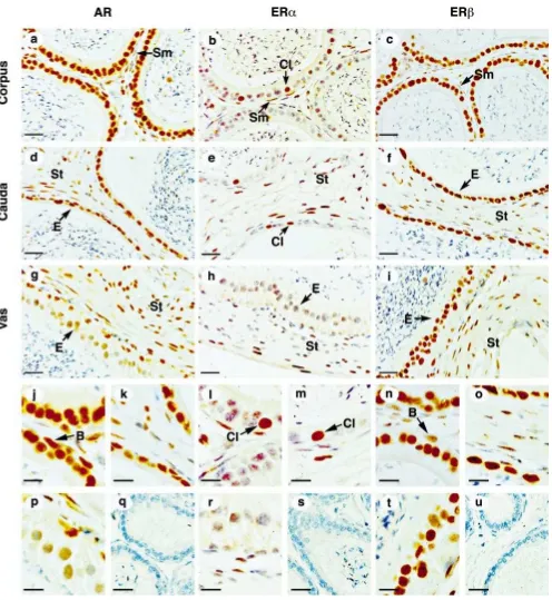
Corpus Epididymis
AR (Figure 2, a and j) and ERb(Figure 2, c and n) were expressed abundantly in all epithelial cells lining the cor-pus epididymal duct. In contrast, ERa was present only in clear cells of the epithelium and some peritubular cells of the stroma (Figure 2, b and l). In the stroma, AR and ERbwere positive in some cells but negative in others.
Cauda Epididymis
Epithelial cells stained intensely for AR (Figure 2, d and k) and ERb(Figure 2, f and o) but ERastaining (Figure 2, e and m) was identical to the corpus epididymis, where only clear cells and some peritubular smooth muscle cells were strongly positive. Many stromal cells were positive for all three receptors.
Vas Deferens
In the epithelium, AR was expressed weakly in epithelial cells but with moderate strength in basal cells (Figure 2, g and p). ERawas absent within the epithelium (Figure 2, h and r), whereas ERb was abundant in all epithelial cell types (Figure 2, i and t). In the stroma, AR was ex-pressed in smooth muscle and connective tissue cells, but ERawas found only in the outer layer of smooth muscle cells, whereas ERbwas abundant throughout the stroma.
Double Staining
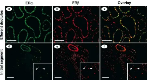
It was obvious from microscopic observation that both ERaand ERbwere colocalized in the same cells in var-ious regions of the male tract. Therefore, we selected to examine the efferent ductules and initial segment of epi-didymis using colocalization of fluorescent signals to de-termine whether the same cells express the 2 receptors. Both ERa (green) and ERb(red) were expressed in epi-thelial nuclei of the efferent ductules (Figure 3, a and b).
Colocalization of the 2 receptors in the same cell was detected by an orange to yellowish color, with variations in color intensities indicating differences in the proportion of the 2 receptors in an individual cell nucleus (Figure 3c). In the overlay view of the proximal efferent ductule epithelium (Figure 3c), some cells remained green, indi-cating expression of only ERa; some stained intensely yellow, indicating equivalent expression of ERaand ERb; and an occasional cell stained only red, indicating an ex-pression of only ERb.
A transition zone from efferent ductules to the initial segment of the epididymis can be viewed in Figure 3, d– f. The one tubule that stained intensely for both ERaand ERbbelongs to the common efferent duct that opens into the initial segment epididymis. This duct shows less var-iation in overlay staining (Figure 3f) than does the prox-imal ductules (Figure 3c), indicating nearly equal expres-sion of the 2 receptors in all epithelial cells. In the same photographs, a region of the initial segment is also noted. The only fluorescence detected for ERain the initial seg-ment was the apical and narrow cells (Figure 3d). ERb was expressed in all epithelial cell types (Figure 3e). The overlay shows considerable variation in staining of apical and narrow cells (Fig. 3f; note the unlabeled arrows), sim-ilar to what was observed with single receptor staining (Figure 1, h and i).
Antibody Competition
AR21 peptide competed away the nuclear staining given by the AR antibody PG21 reaction to efferent ductule (Figure 2q) and epididymal tissues (not shown), whereas the unrelated peptide AR462 was not able to ward off the specific nuclear staining (not shown). Recombinant hu-man ERaprotein competed away all nuclear staining giv-en by monoclonal antibody 6F11 reaction (Figure 2s), and peptide P40 also competed away all staining given by the ERbantibody S40 (Figure 2u).
Western Blot Analysis
Figure 1. Immunostaining of AR, ERa, and ERbin mouse testis (a–c, m, o, q), efferent ductules (d–f n, p, r), initial region of epididymis (g–i, s, u,
w), and caput epididymis (j–l, t, v, x). S indicates Sertoli cell; M, peritubular myoid cell; Ly, Leydig cell; C, ciliated cells; B, basal cell; Cl, clear cell;
Figure 2. Immunostaining of AR, ERa, and ERbin mouse corpus epididymis (a–c, j, l, n), cauda epididymis (d–f, k, m, o), and vas deferens (g–i, p,
r, t). Peptide competition of AR antibody (q), ERa(s), and ERb(u). B indicates basal cell; Cl, clear cell; E, epithelial cell; Sm, smooth muscle cell;
St, stromal cells. Bar525mm (a–i); bar512.5mm (j–u).
Discussion
This study provides specific cellular localization of an-drogen and estrogen receptors in the testis and its ex-current ducts in adult male mice. Based on previous studies in other species (Goyal et al, 1997b; Saunders et
Table 2. Androgen receptors in adult mouse testis
Location Cell Type Intensity of Stain
Interstitium Leydig cell
* All stages of spermatogenesis. † All germ cells were negative.
Testis
Although there is a substantial decrease in AR in the male mouse reproductive tract tissues from birth to adulthood (Gallon et al, 1989), androgens remain the primary steroid hormones for maintenance of male reproductive function in the adult (Ezer and Robaire, 2002), and the common presence of AR in the male would be expected. There is nearly universal agreement across species that in the adult testis, AR is present in Sertoli cells, peritubular cells, and Leydig cells (Sar et al, 1990; Bremner et al, 1994; Vorn-berger et al, 1994; Van Roijen et al, 1995; Goyal et al, 1997a,b; Suarez-Quian et al, 1999; Pelletier et al, 2000; Zhu et al, 2000). Expression of AR in Sertoli cells sup-ports previous resup-ports that androgens regulate Sertoli cell function and are essential for spermatogenesis (Sharpe, 1994). The present study found a stage-dependent ex-pression of AR in Sertoli cells, similar to other studies in rats and humans (Bremner et al, 1994; Vornberger et al, 1994; Suarez-Quian et al, 1999).
AR was not found in germ cells of mouse testis. How-ever, the presence of AR in testicular germ cells has been controversial, with most studies in other species having indicated a lack of AR, but some studies showing AR-positive spermatogonia (Kimura et al, 1993; Zhou et al, 1996) or stage-specific elongate spermatids (Vornberger et al, 1994). In support of our findings in the mouse, a recent study using spermatogonial stem cell transplant technology demonstrated that germ cells of the mouse testis do not require a functional AR to complete sper-matogenesis (Johnston et al, 2001). Therefore, if Sertoli cells in certain stages also express no AR or low concen-trations, it is likely that testosterone provides indirect stimulation of spermatogenesis in those stages through the AR-positive peritubular cells. AR expression in Sertoli cells appears to have more androgen dependency than in Leydig and peritubular cells (Zhu et al, 2000).
In the testes of adult mice, we detected ERain Leydig cells and peritubular myoid cells, whereas Sertoli and germ cells of the seminiferous epithelium were negative. This is consistent with a recent report that the presence
of ERain germ cells of the testis is not required for their development (Mahato et al, 2000). It is surprising that in testes of adult humans, macaques, marmosets, and goats, no ERaimmunoexpression was detected in any cell type (Goyal et al, 1997a; Makinen et al, 2001; Saunders et al, 2001). However, in another study of marmosets, ERawas reported in interstitial (Leydig) cells (Fisher et al, 1997). In cats and dogs (Nie et al, 2002) and rats (Sar and Welsch, 2000), ERa has also been detected in the inter-stitial (Leydig) cells but not in peritubular cells.
In the present study, ERb was expressed more exten-sively than ERafrom the testis to the vas deferens, both in cell types and in number of positive cells. Its expres-sion in the testis is controversial, with considerable var-iation across species and even within species. We found in mouse testis, using the S40 antibody, that ERb was expressed in Sertoli cells, spermatogonia, and spermato-cytes (except for cells in meiotic division). Spermatids and spermatozoa were negative. These results are similar to previous studies in several species (Enmark et al, 1997; Rosenfeld et al, 1998; Saunders et al, 1998, 2001; Pel-letier et al, 1999, 2000; van Pelt et al, 1999; Makinen et al, 2001; Nie et al, 2002), but contrasts with other reports. In one study of rats, ERbwas found only in Sertoli cells (Pelletier et al, 2000) and in a study of humans, Makinen et al (2001) found that ERbwas expressed only in germ cells. Rosenfeld (1998) found that in mice, ERbwas also expressed in elongated spermatids.
Efferent Ductules
tu-bules and the major role of ERa, rather than AR, in reg-ulation of efferent ductule function (Hess et al, 2002).
ERaexpression in efferent ductule epithelium has been consistent across all species studied, including rats, mice, roosters, dogs, cats, goats, monkeys, and humans (West and Brenner, 1990; Ergun et al, 1997; Fisher et al, 1997; Goyal et al, 1997a; Hess et al, 1997b; Kwon et al, 1997; Rosenfeld et al, 1998; Saunders et al, 2001; Nie et al, 2002). However, in some species, the ciliated cells were ERa-negative (West and Brenner, 1990; Ergun et al, 1997; Goyal et al, 1997a; Saunders et al, 2001), but in mice, all epithelial cells were ERa-positive in these duct-ules. ERa showed the highest intensity of reproductive tract staining in the efferent ductules, consistent with our previous studies showing that ERais responsible for reg-ulating fluid reabsorption in these ducts (Hess et al, 1997a) through the control of epithelial ion transporters (Lee et al, 2001; Zhou et al, 2001). Efferent ductules are responsible for reabsorbing more than 90% of the fluid entering from the rete testis (Clulow et al, 1998). This action of estrogen in the efferent ductules is now recog-nized as being essential for male fertility (Eddy et al, 1996; Hess et al, 2001b; Oliveira et al, 2001, 2002; Zhou et al, 2001).
The expression of ERbthroughout the excurrent ducts is ubiquitous even though it seems more predominant in epithelia than stroma. The expression profile of ERb in mouse tract is more similar to AR than it is to ERa. Due to its unclear physiological role in the male, further study is need to clarify ERbaction in the testis and its excurrent ducts (Krege et al, 1998; Couse et al, 1999; Dupont et al, 2000).
Epididymis
AR has been demonstrated by various techniques to be present in the epididymis of numerous species (Younes and Pierrepoint, 1981; Schleicher et al, 1984; Toney and Danzo, 1988; Gallon et al, 1989; Tekpetey et al, 1989; Sar et al, 1990; Cooke et al, 1991; Roselli et al, 1991; Goyal et al, 1997b; Ungefroren et al, 1997; You and Sar, 1998; Pelletier, 2000), and dependence of the epididymis on androgens for structure and function is well known (Ezer and Robaire, 2002). In mice, immunostaining for AR demonstrates a decrease in intensity from the efferent ductule epithelium to the vas deferens, with the caput, corpus, and cauda regions showing equivalent staining, but the vas exhibiting very low expression of AR. Some species variation is seen in the ram, in which there also appears to be regional differences in expression (Carreau et al, 1984; Tekpetey et al, 1989). In the present study, AR was expressed equally in principal and basal cells in mice, in contrast to that of rats (Zhu et al, 2000).
Although AR appears to be dominant in the epididymal epithelium, the aERKO male demonstrated epididymal
abnormalities in the absence of a functional ERa (Hess et al, 2000). It is interesting that the same cell types that were abnormal inaERKO clearly show ERa-positive im-munoreactivity in the present study, in particular in nar-row cells of the initial segment, apical cells of the caput, and in clear cells. Principal cells of the caput were also ERa-positive in mice, but they did not show gross ab-normalities in aERKO mice. These data are consistent with an earlier autoradiography study in mice (Schleicher et al, 1984), which showed much higher binding of 3 H-estradiol in the initial segment and caput epididymis than in the corpus through vas deferens, with greater binding in apical/narrow cells and in clear cells. What is not un-derstood is that ERb is abundant throughout the epidid-ymal epithelium of mice, and yet the autoradiographic data show little evidence of equivalent binding of estra-diol throughout the epididymal epithelium. Because ERa is absent in principal cells of the corpus, cauda, and vas, 3H-estradiol binding shown in these cells (Schleicher et
al, 1984) must represent the presence of ERb. In adult rats, inconsistent results for epididymal ERa have been reported, apparently due to the different antibodies ap-plied (Fisher et al, 1997; Hess et al, 1997b; Sar and Welsch, 2000; Atanassova et al, 2001). These data are consistent with messenger RNA hybridization studies in situ (Mowa and Iwanaga, 2001). Sar and Welsch (2000) reported all stroma to be positive but only some epithelial cells in the rat were positive. In contrast, the entire epi-didymis, both epithelium and stroma, were found to be negative in several species, including rats (Fisher et al, 1997; Atanassova et al, 2001), dogs (Nie et al, 2002), goats (Goyal et al, 1998) and marmoset monkeys (Fisher et al, 1997). Other studies have shown variable amounts of staining by the epididymal epithelium with the same antibodies (Saunders et al, 2001; Nie et al, 2002), which suggests there are major species difference for ERa ex-pression in the adult epididymis.
ERbwas expressed throughout the male mouse repro-ductive tract epithelium, similar to what has been reported in other species (Sar and Welsch, 2000; Atanassova et al, 2001; Saunders et al, 2001). There did not appear to be cell-specific expression of ERb; however, the epithelium showed more intense staining than did the stroma in all regions, except for the cauda and vas deferens.
Vas Deferens
Figure 3. Colocalization of ERaand ERbin mouse efferent ductules (a–c) and initial segment of epididymis (d–f). Immunostaining of ERa(green) and ERb(red) are both positive in the nucleus of efferent ductules, but only ERbin the connective tissue cells. The combined photo overlays (c and
f) show colocalization of the 2 receptors in the same cells (yellow). A transition area from efferent ductules to the head of epididymis (d–f) shows one
efferent ductule that is stained strongly positive for ERaand also some epithelial cells in initial segment. Most epithelial cells are positive for ERb. Unlabeled arrows in the inset photo enlargements point to narrow or apical cells in the initial segment of epididymis (d–f). Bar525mm (a–c); bar5 50mm (d–f).
Figure 4. Western blot analysis of AR, ERa, and ERbin mouse tissues. (a) AR. Mouse testis (1), mouse epididymis (2). (b) ERa. Mouse uterus (1), mouse epididymis (2). (c) ERb. Human recombinant protein (1), mouse epididymis (2). Arrow indicates molecular weight of dominant band in tissues for respective antibodies.
goat, rat (Goyal et al, 1997b), and human epithelium (Sar et al, 1990) expressed AR with intense immunostaining. In the mouse, there was abundant expression of AR in the stroma, which is consistent with autoradiographic studies (Weaker and Sheridan, 1983).
As observed in most species, (Goyal et al, 1997b; Hess et al, 1997b; Jefferson et al, 2000; Nie et al, 2002), the vas deferens epithelium was ERa-negative. Only the cat vas deferens shows ERa-positive staining (Nie et al, 2002). ERbis abundant in both the epithelium and stroma of the vas deferens, similar to all species examined (Jef-ferson et al, 2000; Nie et al, 2002).
Receptors in Stromal Tissue
for decreased expression of both ERaand ERbin stroma, going from the head of the epididymis through the vas. Even though ERb appears to be present in more cell types, ERamaintains much stronger intensity of staining in a cell-specific manner than does ERb. Therefore, these data suggest that further study is needed not only to de-termine the interactions between receptors and their spe-cific hormone ligands, but also to determine the interac-tions between epithelial and stromal cells in the epidid-ymal region.
Conclusions
This study demonstrates that the male mouse reproductive tract is substantially different from that reported in rats for the expression of ARs and ERs. This finding further supports the need to be cautious when extrapolating nu-clear steroid receptor data across species in the study of male reproductive tract biology. All epithelial and most stromal cells contained AR, except for the germ cells and some vas deferens cells. ERa was abundant in efferent ductules, similar to all other species, but its presence in specific cell types along the epididymis was novel, be-cause it differs from that seen in rats. ERb distribution was similar to that of AR, except that ERb alone was prominent in germ cells and vas deferens epithelium. Many cells expressed all three steroid receptors, including Leydig cells, peritubular myoid cells surrounding the seminiferous tubules, all epithelial cells from the efferent ductules, apical and narrow cells of the initial segment, and clear cells of the epididymis. The coexistence of mul-tiple receptors in the same cells raises important questions regarding steroid hormone interactions and receptor cross-talk in the control of male reproductive tract func-tion.
Acknowledgment
We appreciate the excellent laboratory assistance of Kay Carnes.
References
Atanassova N, McKinnell C, Williams K, Turner KJ, Fisher JS, Saunders PT, Millar MR, Sharpe RM. Age-, cell- and region-specific immu-noexpression of estrogen receptor alpha (but not estrogen receptor beta) during postnatal development of the epididymis and vas defer-ens of the rat and disruption of this pattern by neonatal treatment with diethylstilbestrol. Endocrinology. 2001;142:874–886.
Bremner WJ, Millar MR, Sharpe RM, Saunders PT. Immunohistochem-ical localization of androgen receptors in the rat testis: evidence for stage-dependent expression and regulation by androgens.
Endocri-nology. 1994;135:1227–1234.
Carreau S, Drosdowsky MA, Courot M. Androgen-binding proteins in sheep epididymis: characterization of a cytoplasmic androgen receptor in the ram epididymis. J Endocrinol. 1984;103:273–279.
Choi I, Ko C, Park-Sarge O-K, Nie R, Hess RA, Graves C, Katzenellen-bogen BS. Human estrogen receptor beta-specific monoclonal
anti-bodies: characterization and use in studies of estrogen receptor beta protein expression in reproductive tissues. Mol Cell Endocrinol. 2001; 181:139–150.
Clulow J, Jones RC, Hansen LA, Man SY. Fluid and electrolyte reab-sorption in the ductuli efferentes testis. J Reprod Fertil Suppl. 1998; 53:1–14.
Cooke PS, Buchanan DL, Lubahn DB, Cunha GR. Mechanism of estro-gen action: lessons from the estroestro-gen receptor-alpha knockout mouse.
Biol Reprod. 1998;59:470–475.
Cooke PS, Buchanan DL, Young P, Setiawan T, Brody J, Korach KS, Taylor J, Lubahn DB, Cunha GR. Stromal estrogen receptors mediate mitogenic effects of estradiol on uterine epithelium. Proc Natl Acad
Sci USA. 1997;94:6535–6540.
Cooke PS, Uchima F-DA, Fujii DK, Bern HA, Cunha GR. Restoration of normal morphology and estrogen responsiveness in cultured vag-inal and uterine epithelia transplanted with stroma. Proc Natl Acad
Sci USA. 1986;83:2109–2113.
Cooke PS, Young P, Cunha GR. Androgen receptor expression in devel-oping male reproductive organs. Endocrinology. 1991;128:2867– 2873.
Couse JF, Hewitt SC, Bunch DO, Sar M, Walker VR, Davis BJ, Korach KS. Postnatal sex reversal of the ovaries in mice lacking estrogen receptors alpha and beta. Science. 1999;286:2328–2331.
Couse JF, Korach KS. Estrogen receptor null mice: what have we learned and where will they lead us? Endocr Rev. 1999;20:358–417. Cunha G, Bigsby R, Cooke P, Sugimura Y. Stromal-epithelial interactions
in adult organs. Cell Differ. 1985;17:137–148.
Darne CH, Morel L, Claessens F, Manin M, Fabre S, Veyssiere G, Rom-bauts W, Jean CL. Ubiquitous transcription factors NF1 and Sp1 are involved in the androgen activation of the mouse vas deferens protein promoter. Mol Cell Endocrinol. 1997;132:13–23.
Dassouli A, Darne C, Fabre S, Manin M, Veyssiere G, Jean CI. Vas deferens epithelial cells in subculture: a model to study androgen regulation of gene expression. J Mol Endocrinol. 1995;15:129–141. Dupont S, Krust A, Gansmuller A, Dierich A, Chambon P, Mark M.
Effect of single and compound knockouts of estrogen receptorsa(ER a) andb(ERb) on mouse reproductive phenotypes. Development. 2000;127:4277–4291.
Eddy EM, Washburn TF, Bunch DO, Goulding EH, Gladen BC, Lubahn DB, Korach KS. Targeted disruption of the estrogen receptor gene in male mice causes alteration of spermatogenesis and infertility.
En-docrinology. 1996;137:4796–4805.
Enmark E, Pelto-Huikko M, Grandien K, Lagercrantz S, Lagercrantz J, Fried G, Nordenskjold M, Gustafsson JA. Human estrogen receptor beta-gene structure, chromosomal localization, and expression pattern.
J Clin Endocrinol Metab. 1997;82:4258–4265.
Ergun S, Ungefroren H, Holstein AF, Davidoff MS. Estrogen and pro-gesterone receptors and estrogen receptor-related antigen (ER-D5) in human epididymis. Mol Reprod Dev. 1997;47:448–455.
Ezer N, Robaire B. Androgenic regulation of the structure and functions of the epididymis. In: Robaire B, Hinton B, eds. The Epididymis:
From Molecules to Clinical Practice. New York: Kluwer Academic/
Plenum Publishers; 2002:297–316.
Fisher JS, Millar MR, Majdic G, Saunders PT, Fraser HM, Sharpe RM. Immunolocalisation of oestrogen receptor-alpha within the testis and excurrent ducts of the rat and marmoset monkey from perinatal life to adulthood. J Endocrinol. 1997;153:485–495.
Gallon C, Veyssiere G, Berger M, Jean-Faucher C, De Turckheim M, Jean C. Age-related changes in the concentration of cytosolic andro-gen receptors in the epididymis, vas deferens and seminal vesicle of maturing male mice. J Androl. 1989;10:188–194.
de-veloping testis and excurrent ducts of goats. Anat Rec. 1997a;249: 54–62.
Goyal HO, Bartol FF, Wiley AA, Khalil MK, Williams CS, Vig MM. Regulation of androgen and estrogen receptors in male excurrent ducts of the goat: an immunohistochemical study. Anat Rec. 1998;250:164– 171.
Goyal HO, Bartol FF, Wiley AA, Neff CW. Immunolocalization of re-ceptors for androgen and estrogen in male caprine reproductive tis-sues: unique distribution of estrogen receptors in efferent ductule ep-ithelium. Biol Reprod. 1997b;56:90–101.
Hess RA. Oestrogen in fluid transport and reabsorption in efferent ducts of the male reproductive tract. Rev Reprod. 2000;5:84–92.
Hess RA. The efferent ductules: structure and functions. In: Robaire B, Hinton B, eds. The Epididymis: From Molecules to Clinical Practice. New York: Kluwer Academic/Plenum Publishers; 2002:49–80. Hess RA, Bunick D, Bahr J. Oestrogen, its receptors and function in the
male reproductive tract—a review. Mol Cell Endocrinol. 2001a;178: 29–38.
Hess RA, Bunick D, Lee KH, Bahr J, Taylor JA, Korach KS, Lubahn DB. A role for oestrogens in the male reproductive system. Nature. 1997a;390:509–512.
Hess RA, Bunick D, Lubahn DB, Zhou Q, Bouma J. Morphologic chang-es in efferent ductulchang-es and epididymis in chang-estrogen receptor-alpha knockout mice. J Androl. 2000;21:107–121.
Hess RA, Gist DH, Bunick D, Lubahn DB, Farrell A, Bahr J, Cooke PS, Greene GL. Estrogen receptor (a& b) expression in the excurrent ducts of the adult male rat reproductive tract. J Androl. 1997b;18: 602–611.
Hess RA, Zhou Q, Nie R. The role of estrogens in the endocrine and paracrine regulation of the efferent ductules, epididymis and vas de-ferens. In: Robaire B, Hinton BT, eds. The Epididymis: From
Mole-cules to Clinical Practice. New York: Kluwer Academic/Plenum
Pub-lishers; 2002:317–338.
Hess RA, Zhou Q, Nie R, Oliveira C, Cho H, Nakai M, Carnes K. Es-trogens and epididymal function. Reproduction Fertility and
Devel-opment. 2001b;13:273–283.
Iguchi T, Uesugi Y, Sato T, Ohta Y, Takasugi N. Developmental pattern of estrogen receptor expression in male mouse genital organs. Mol
Androl. 1991;6:109–119.
Jefferson WN, Couse JF, Banks EP, Korach KS, Newbold R. Expression of estrogen receptor beta is developmentally regulated in reproductive tissues of male and female mice. Biol Reprod. 2000;62:310–317. Johnston DS, Russell LD, Friel PJ, Griswold MD. Murine germ cells do
not require functional androgen receptors to complete spermatogen-esis following spermatogonial stem cell transplantation.
Endocrinol-ogy. 2001;142:2405–2408.
Kimura N, Mizokami A, Oonuma T, Sasano H, Nagura H. Immunocy-tochemical localization of androgen receptor with polyclonal antibody in paraffin-embedded human tissues. J Histochem Cytochem. 1993; 41:671–678.
Krege JH, Hodgin JB, Couse JF, et al. Generation and reproductive phe-notypes of mice lacking estrogen receptor beta. Proc Natl Acad Sci
USA. 1998;95:15677–15682.
Kuiper G, Shughrue PJ, Merchenthaler I, Gustafsson JA. The estrogen receptor beta subtype: a novel mediator of estrogen action in neuro-endocrine systems. Front Neuroendocrinol. 1998;19:253–286. Kwon S, Hess RA, Bunick D, Kirby JD, Bahr JM. Estrogen receptors are
present in the epididymis of the rooster. J Androl. 1997;18:378–384. Lee KH, Finnigan-Bunick C, Bahr J, Bunick D. Estrogen regulation of ion transporter messenger RNA levels in mouse efferent ductules are mediated differentially through estrogen receptor (ER) alpha and ER-beta. Biol Reprod. 2001;65:1534–1541.
Mahato D, Goulding EH, Korach KS, Eddy EM. Spermatogenic cells do
not require estrogen receptor-alpha for development or function [see comments]. Endocrinology. 2000;141:1273–1276.
Makinen S, Makela S, Weihua Z, Warner M, Rosenlund B, Salmi S, Hovatta O, Gustafsson JK. Localization of oestrogen receptors alpha and beta in human testis. Mol Hum Reprod. 2001;7:497–503. McKinnell C, Atanassova N, Williams K, Fisher JS, Walker M, Turner
KJ, Saunders TK, Sharpe RM. Suppression of androgen action and the induction of gross abnormalities of the reproductive tract in male rats treated neonatally with diethylstilbestrol. J Androl. 2001;22:323– 338.
Mowa CN, Iwanaga T. Expression of estrogen receptor-alpha and -beta mRNAs in the male reproductive system of the rat as revealed by in situ hybridization. J Mol Endocrinol. 2001;26:165–174.
Nie R, Zhou Q, Jassim E, Saunders PTK, Hess RA. Differential expres-sion of estrogen receptorsa&bin the reproductive tract of the adult male dog and cat. Biol Reprod. 2002;66;1161–1168.
O’Donnell L, Robertson KM, Jones ME, Simpson ER. Estrogen and sper-matogenesis. Endocr Rev. 2001;22:289–318.
Oliveira CA, Carnes K, Franc¸a LR, Hess RA. Infertility and testicular atrophy in the antiestrogen-treated adult male rat. Biol Reprod. 2001; 65:913–920.
Oliveira CA, Zhou Q, Carnes K, et al. Estrogen receptor function in the adult male rat: short and long-term effects of the antiestrogen ICI 182,780 on the testis and efferent ductules, without changes in tes-tosterone. Endocrinology. 2002;143:2399–2409.
Pelletier G. Localization of androgen and estrogen receptors in rat and primate tissues. Histol Histopathol. 2000;15:1261–1270.
Pelletier G, Labrie C, Labrie F. Localization of oestrogen receptor alpha, oestrogen receptor beta and androgen receptors in the rat reproductive organs. J Endocrinol. 2000;165:359–370.
Pelletier G, Luu-The V, Charbonneau A, Labrie F. Cellular localization of estrogen receptor beta messenger ribonucleic acid in cynomolgus monkey reproductive organs. Biol Reprod. 1999;61:1249–1255. Prins G, Birch L, Greene G. Androgen receptor localization in different
cell types of the adult rat prostate. Endocrinology. 1991;129:3187– 3199.
Prins GS, Birch L, Couse JF, Choi I, Katzenellenbogen B, Korach KS. Estrogen imprinting of the developing prostate gland is mediated through stromal estrogen receptor alpha: studies with alphaERKO and betaERKO mice. Cancer Res. 2001;61:6089–6097.
Roselli CE, West NB, Brenner RM. Androgen receptor and 5 alpha-re-ductase activity in the ductuli efferentes and epididymis of adult rhe-sus macaques. Biol Reprod. 1991;44:739–745.
Rosenfeld CS, Ganjam VK, Taylor JA, Yuan X, Stiehr JR, Hardy MP, Lubahn DB. Transcription and translation of estrogen receptor-beta in the male reproductive tract of estrogen receptor-alpha knock-out and wild-type mice. Endocrinology. 1998;139:2982–2987.
Sar M, Lubahn DB, French FS, Wilson EM. Immunohistochemical lo-calization of the androgen receptor in rat and human tissues.
Endo-crinology. 1990;127:3180–3186.
Sar M, Welsch F. Oestrogen receptor alpha and beta in rat prostate and epididymis. Andrologia. 2000;32:295–301.
Saunders PT, Fisher JS, Sharpe RM, Millar MR. Expression of oestrogen receptor beta (ER beta) occurs in multiple cell types, including some germ cells, in the rat testis. J Endocrinol. 1998;156:R13–17. Saunders PT, Millar MR, Williams K, et al. Differential expression of
estrogen receptor-alpha and -beta and androgen receptor in the ovaries of marmosets and humans. Biol Reprod. 2000;63:1098–1105. Saunders PT, Sharpe RM, Williams K, Macpherson S, Urquart H, Irvine
DS, Millar MR. Differential expression of oestrogen receptor alpha and beta proteins in the testes and male reproductive system of human and non-human primates. Mol Hum Reprod. 2001;7:227–236. Schindelmeiser J, Kutzner M, Bergmann M, Aumuller G, Heck H,
and androgen receptor levels of the epididymis and the ductus defer-ens of Phodopus sungorus. Andrologia. 1988;20:507–515.
Schleicher G, Drews U, Stumpf WE, Sar M. Differential distribution of dihydrotestosterone and estradiol binding sites in the epididymis of the mouse. An autoradiographic study. Histochemistry. 1984;81:139– 147.
Sharpe R. Regulation of spermatogenesis. In: Knobil E, Neill J, eds. The
Physiology of Reproduction. Vol 3, 2nd ed. New York: Raven Press;
1994:1363–1434.
Sharpe RM. The roles of oestrogen in the male. Trends in Endocrinology
and Metabolism. 1998;9:371–377.
Suarez-Quian CA, Martinez-Garcia F, Nistal M, Regadera J. Androgen receptor distribution in adult human testis. J Clin Endocrinol Metab. 1999;84:350–358.
Tekpetey FR, Veeramachaneni DN, Amann RP. Localization of androgen receptors in ram epididymal principal cells. J Reprod Fertil. 1989;87: 311–319.
Toney TW, Danzo BJ. Developmental changes in and hormonal regulation of estrogen and androgen receptors present in the rabbit epididymis.
Biol Reprod. 1988;39:818–828.
Ungefroren H, Ivell R, Ergun S. Region-specific expression of the andro-gen receptor in the human epididymis. Mol Hum Reprod. 1997;3:933– 940.
van Pelt AM, de Rooij DG, van der Burg B, van der Saag PT, Gustafsson JA, Kuiper GG. Ontogeny of estrogen receptor-beta expression in rat testis. Endocrinology. 1999;140:478–483.
Van Roijen JH, Van Assen S, Van Der Kwast TH, De Rooij DG, Boersma
WJ, Vreeburg JT, Weber RF. Androgen receptor immunoexpression in the testes of subfertile men. J Androl. 1995;16:510–516.
Vornberger W, Prins G, Musto NA, Suarez-Quian SA. Androgen receptor distribution in rat testis: new implications for androgen regulation of spermatogenesis. Endocrinology. 1994;134:2307–2316.
Weaker FJ, Sheridan PJ. Autoradiographic localization of 3H-dihydrotes-terone in the reproductive organs of baboons. Acta Anat. 1983;115: 244–251.
West NB, Brenner RM. Estrogen receptor in the ductuli efferentes, epi-didymis, and testis of rhesus and cynomolgus macaques. Biol Reprod. 1990;42:533–538.
You L, Sar M. Androgen receptor expression in the testes and epididy-mides of prenatal and postnatal Sprague-Dawley rats. Endocrine. 1998;9:253–261.
Younes MA, Pierrepoint CG. Androgen steroid-receptor binding in the canine epididymis. Prostate. 1981;2:133–142.
Zhou Q, Clarke L, Nie R, et al. Estrogen action and male fertility: roles of the sodium/hydrogen exchanger-3 and fluid reabsorption in repro-ductive tract function. Proc Natl Acad Sci USA. 2001;98:14132– 14137.
Zhou X, Kudo A, Kawakami H, Hirano H. Immunohistochemical local-ization of androgen receptor in mouse testicular germ cells during fetal and postnatal development. Anat Rec. 1996;245:509–518. Zhu LJ, Hardy MP, Inigo IV, Huhtaniemi I, Bardin CW, Moo-Young AJ.
Effects of androgen on androgen receptor expression in rat testicular and epididymal cells: a quantitative immunohistochemical study. Biol