VOL. 6, NO. 3, pp. 205 - 209, September, 2016 Submitted August 2016; Revised August 2016; Accepted September 2016
Baculovirus Surface Display Using Infuenza Neuraminidase (NA) Transmembrane Anchor
Irisa Trianti1, Saengchai Akeprathumchai1, Phenjun Mekvichitsaeng2, Kanokwan Poomputsa1*
1Biotechnology Division, School of Bioresources and Technology, King Mongkuts’s University of Technology Thonburi
(Bangkhuntien), Bangkok, Thailand
2Pilot Plant Development and Training Institute, King Mongkuts’s University of Technology Thonburi (Bangkhuntien), Bangkok,
Thailand
ABSTRACT
Baculovirus surface display has been employed as an excellent tools for presentation of foreign peptides and proteins on virus surface with native conformation, functions and immunogenicity. A baculovirus major envelope protein, gp64, or a capsid protein, vp39 are generally used as fusion partners for displaying of polypeptides on the surface of virions. Alternatively, a membrane anchoring domain of vesicular stomatitis virus G protein (VSV-G) can also be used. In this study, an influenza neuraminidase (NA) was proposed as a new membrane anchor for the display of Angiotensin II (AngII), DRVYIHPFHL, peptides. The AngII peptides were inserted into NA by replacing NA amino acid number 60-67 with AngII, and then integrated into a baculovirus genome. A recombinant baculovirus expressing the NA fusion-AngII peptides was generated from infected insect cells. Those peptides were found to express and translocated on the membrane of the baculovirus infected insect cell (Sf9 cell) as detected by immunocytochemistry using anti-AngII monoclonal antibody. Upon budding of the recombinant baculovirus progenies through the insect cells membrane, the recombinant NA-AngII peptides was acquired to envelopes of the new baculovirus progenies. The conformation of NA on baculovirus surface was not affected by the deletion, as the 55 kDa band of NA can be detected from Western Blotting analysis by specific anti-NA monoclonal antibody. In addition, the same protein was also found by anti-AngII antibody indicating that the AngII peptides had been successfully fused with the recombinant NA. Interestingly, electron microscopy analysis demonstrated that not only the recombinant baculovirus displaying AngII peptides were generated by infected insect cells, but also the NA virus-like-particle displaying AngII peptides.
Keywords: Baculovirus surface display, Neuraminidase, AngII peptides, Virus like particles
Baculovirus, an insect virus has been widely used as an expression system [1]. Baculovirus is able to trans-duce a broad range of mammalian and avian cells, therefore, it also potentially be used as gene delivery vector [2]. Interestingly, foreign protein can be manip-ulated to be displayed on baculovirus surface. It has been reported that baculovirus surface display is poten-tially be used as an efficient vaccine, such as vaccine against classical swine fewer virus [3], malaria [4] and human enterovirus 71 (EV71) [5].
The display of antigen on baculovirus surface can
be facilitated by fusion of antigen with gp64 protein which is the major envelope protein of Baculovirus. Gp64 comprised of amino acid signal peptide domain, carboxyl proximal transmembrane domain and cyto-plasmic tail domain. After gp64 expression, it will translocate to host cell membrane that control by sig-nal peptide domain. During virus budding, the virion will take up proteins from host cell membrane to gen-erate envelope protein, consequently, the gp64 will be incorporated in baculovirus envelope [6]. However, low number of gp64 fusion protein can be incorporated into viral membrane due to competition of fusion gp64 INTRODUCTION
*Corresponding author: Kanokwan Poomputsa
Biotechnology Division, School of Bioresources and Technology King Mongkuts’s University of Technology Thonburi (Bangkhuntien) 49 Soi Thian Thale 25, Bang Khun Thian, Bangkok 10150, Thailand E-mail address: [email protected]
How to cite:
and wild type gp64 to be uptake by virion during virus budding [7].
Thus, some other transmembrane proteins such as vesicular somatic virus glycoprotein (VSVG) has been used as an alternative. The VSVG membrane anchor approach provides higher level of display of GFP pro-tein on baculovirus in comparison with that of gp64 fusion protein base [8]. In addition, Kolpe et al. (2012) reported that VP1 of human enterovirus 71 together with signal peptide and transmembrane domain of in-fluenza neuraminidase (NA) can be displayed on bac-ulovirus surface [5]. NA signal peptide and transmem-brane domain fusion protein seems to have a non-polar virus distribution, unlike gp64 fusion protein that lim-ited to the pole of baculovirus [9].
In this study, a modified full length NA fused with AngII peptide was shown to be displayed on bac-ulovirus surface as fusion peptide. Furthermore, NA virus like particle (VLP) was also produced with AngII displayed on its surface.
Cells and virus
The Spodoptera frugiperda (Sf-9) insect cells line (ATCC, USA) were grown as suspension culture in TMN-FH medium (Gibco, USA) supplemented with 10% heat inactivated fetal bovine serum (Hyclone, USA). Recombinant baculoviruses were amplified in Sf-9 cells according to Bac to Bac manual (Invitrogen, USA). The virus titter was determined by end point di-lution assay [10].
Generation of recombinant baculovirus displaying NA-AngII
Recombinant baculovirus was generated according to Bac to Bac system protocol (Invitrogen, USA). A re-combinant donor plasmid pFASTBac_ie1_na-angII was constructed by insertion of White Spot Syndrome Virus ie1 promoter (IE1) and na_angII into pFAST-BacHT A, baculovirus transfer vector with polyhedron promoter (Invitrogen, USA). Firstly, the pFASTBacHT-A polyhedrin promoter was removed and replaced with a PCR amplified ie1 promoter of WSSV. WSSV IE1 promoter was amplified by using IE1 specific primer, IE1F-5’-GTGCGTACGTAGGCTGTTTGAATCAT-GTTAAGG-3’ and IE1 R-5’ATTAGGCGCC TATCG-GACCGCTTGAGTGGAGAGAGAGA-3’,
pBACsurf_IE1was used as template.
AngII was inserted into influenza Neuraminidase (NA) at amino acid number 60 by overlapping PCR using pGEM-NA vector as template. After second
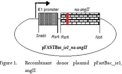
Figure 1. Recombinant donor plasmid pFastBac_ie1_na-angII
round PCR, the full length NA-AngII with size of 1380 bp was generated.
The amplified PCR products were then clones into SnaBI and RsrII restriction enzyme into pFASTBac_ie1. The pFASTBac_ie1_na-angII recom-binant donor plamid (figure 1) was then integrated into a baculovirus DNA (bacmid) in DH10BacTM through site-specific transposition. The positive DH10Bac E. coli colonies were selected by blue/white colony selection. The recombinant bacmid with ie1 promoter and na_angII was then extracted from white colony and was transfected into Sf-9 cells by using cell-fectin II (Invitrogen, USA) to generate recombinant baculovirus, rBV_IE1_NA-AngII.
rBV_IE1_NA-AngII production
rBV_IE1_NA-AngII was produced by infecting into Sf9 cells at multiplicity of infection (MOI) of 0.1 and harvested after 5 days post infections. The virus was collected by overlaying onto 30% sucrose and ul-tracentrifugation at 26,500 rpm for 1.5 hour. The virus pellet was resuspended in PBS.
NA-AngII gene expression
To verify the expression of NA-AngII in insect cells, total RNA from infected Sf-9 cells was extracted by TRIZOL reagent (Invitrogen, USA) and cDNA was prepared according to manufacture protocol (Thermo scientific, USA). Twenty nanogram of synthesized cDNA was used as template for DNA amplification us-ing primers specific to NA sequences and AngII.
Immuno-fluorescent analysis
Sf-9 cells were seeded in 12-well plate at cell density of 1 × 10⁶
cells per well and infected with rBV_IE1_NA-AngII at MOI 0.1 for 48 hour at 27°C. The culture medium was removed and infected cells were washed 3 times with PBS. The anti-NA and/or anti-AngII antibodies at a dilution of 1 : 1000 was used as primary antibody and a goat anti-rabbit IgG
conju-JTLS | J. Trop. Life. Science 207 Volume 6 | Number 3 | September | 2016 MATERIALS AND METHODS
gated with green fluorescence at dilution of 1 : 5000 were used as secondary antibody. The green fluores-cence on the infected cells membrane were detected us-ing fluorescence microscopy IX71 (Olympus, Japan)
Western blot analysis
The concentrated rBV_IE1_NA-AngII was sub-jected to 12% SDS-PAGE (Amersham, UK) and trans-ferred to nitrocelullose membrane (Bio-Rad, USA). The anti-NA and/or anti-AngII monoclonal antibodies at a dilution of 1:500 was used as primary antibody and goat anti-rabbit IgG conjugated with horseradish per-oxidase (HRP) at dilution of 1:5000 was used as sec-ondary antibody. For signal detection, the protein band was developed by 3,3 ,5,5 -Tetramethylbenzidine′ ′ (TMB) liquid substrate (Sigma, USA).
Transmission electron microscope analysis
Carbon coated grids (EMS, USA) were floated on concentrated rBV_IE1_NA-AngII solution for 30 min, washed with 5 drops of deionized water and negatively stained with 2% uranyl acetate solution (Sigma, USA) and examined under transmission electron microscope HT7700 (Hitachi, Japan).
Construction of recombinant baculovirus displaying NA-AngII
AngII peptides, an eight amino acid peptide that re-sponsible in controlling blood pressure in renin an-giotensin system [11] was chosen as model peptides to be displayed by fusion with influenza neuraminidase (NA) and displayed on baculovirus surface. The shuttle promoter that active in both insect and mammalian cells, IE1 [12], was chosen to control NA-AngII gene expression. The NA fragment used for fusion with AngII consists of a signal peptides, a trans-membrane region, a hypervariable stalk and a globular do-main to facilitate the display of AngII on baculovirus surface [13].
AngII peptide was inserted into the NA protein be-tween amino acid residues from 60 to 67. Deletion of these amino acids at this region did not not disrupt NA conformation, in addition, the AngII peptide was expected to be located on the exposed location [14].
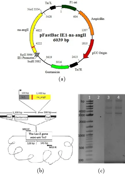
Figure 2a and 2b show introduction of NA-AngII into baculovirus genome. Firstly, the recombinant transfer vector containing ie1 promoter and NA-AngII was constructed and transformed into DH10Bac E. coli containing baculovirus genome (bacmid). By site spe-cific transposition, the NA-AngII under the control of
(a)
(b) (c)
Figure 2. (a) Recombinant transfer vector, (b) Transposition of ie1-na-angII into baculovirus genome (bacmid) (c) PCR analysis of recombinant bacmid using M13 (LacZ) specific primers. Lane 1: 2 long DNA ladder, 2: Negative control of PCR product using M13 primer and 3 and 4: PCR product of recombinant bacmid with ie1-na-angII insertion using M13 primer.
Figure 3. 9 cell at 72 days in culture (a) and transfected Sf-9 cells at 72 hours (b)
ie1 promoter was transferred to bacmid according to Bac to Bac system (Invitrogen, USA) at LacZ. Thus, re-combinants baculoviru DH10Bac colonies become white when cultured on the LB containing X-gal (sub-strate to produce a blue product). The white colonies were then selected for extraction of the recombinant bacmid DNA.
Figure 4. Reverse transcriptase PCR using NA specific primer (Lane 1-4) and NA, AngII specific primer (Lane 5-7) for detection of NA-AngII expression in infected cell with rBV_IE1_NA-AngII. Lane 1: 2log DNA ladder, 2 and 5: negative control without template, 3 and 6: pFASTBac_ie1_NA-AngII was used as template for positive control, 4 and 7: synthesized cDNA from infected cell was used as template
Figure 5. NA-AngII fusion protein on infected Sf-9 cell mem-brane. Cells were infected with wild type bac-ulovirus, AcMNPV and recombinant baculovirus rBV_IE1_NA-AngII. 48 hours after infections, the infected cells were analyzed by immuno-flourescent using anti-NA specific antibody (A) and anti-AngII specific antibody (B), followed secondary antibody conjugated with green fluorescent.
The recombinant bacmid NA-AngII was transfected into Sf-9 insect cells using CellFECTIN II reagent (In-vitrogen, USA). The recombinant baculovirus was then harvested from transfected cell culture medium when cytopathic effect of the Sf-9 insect cell was observed at 72 hour post transfection (Figure 3).
Expression of NA-AngII fusion gene
Reverse transcriptase PCR was used to verify NA-AngII expression in infected Sf-9 cell with recombinant baculovirus, rBV_IE1_NA-AngII. Figure 4 shows a specific band of PCR product at 1380 bp correspond-ing to the transcription of NA-AngII, when the NA specific primers were used. In addition, specific PCR product at 201 bp, which represents N-terminal of NA (59 amino acids) with additional eight amino acids of AngII could also be detected when the NA specific for-ward primer and AngII specific reverse primer were used.
Characterization of rBV_IE1_NA-AngII
In order to confirm the translocation of NA_AngII to Sf-9 infected cell membrane, the immune-fluorescent analysis was performed. The Sf-9 cells were infected with rBV_IE1_NA-AngII or wild type baculovirus, AcMNPV (as control) for 48 h. The green fluorescent were observed from infected cell with rBV-NA_AngII detected by using anti-NA antibody and anti-AngII specific monoclonal antibody, while there is no fluores-cent signal can be observed from infected cells. This in-dicated that the NA_AngII has been expressed and translocated on the infected cell membrane, before virus budding (Figure 5).
Western blot analysis showed that NA can be de-tected on baculovirus surface, as the 55 kDa band of NA can be detected by anti-NA monoclonal antibody. The same size of protein was also detected by anti-AngII antibody indicating that the anti-AngII peptides had been successfully fused with the recombinant NA and was displayed on baculovirus surface on the exposed position (Figure 6).
Electron micrograph of rBV_IE1_NA-AngII
Transmission electron microscope was performed to observe the budded virus rBV_IE1_NA-AngII that accumulated in infected cell culture medium. TEM pic-ture shows rod shapes rBV_NA_AngII budded out from infected cell and it is expected to display NA_AngII on baculovirus surface. Interestingly, there are also NA-AngII-Virus Like Particles (VLP) can be observed in infected cell culture supernatant (Figure 7).
Figure 6. Western Blot analysis of recombinant BV_ IE1_NA-AngII against (a) anti-NA monoclonal an-tibody and (b) anti-AngII monoclonal anan-tibody (Ar-row indicate the NA-AngII band). Lane 1: Wild type AcMNPV, 2: rBV_IE1_NA-AngII
Figure 7. rBV- NA_AngII particles under transmission elec-tron microscope. White arrow indicate rBV-NA_AngII particle and black arrow indicated NA_AngII VLP
This result agree with Lai, et al (2010) that reported NA play key role in virus budding and it could facili-tate the formation of NA-VLP which morphologically similar to influenza virion [15].
AngII peptides has been successfully fused with in-fluenza neuraminidase and was located on the exposed position and the recombinant baculovirus displaying NA-AngII antigen on the surface of virus particle was successfully constructed
Irisa Trianti received financial support from Ministry of National Education, Republic of Indonesia through Directorate General of Higher Education (DGHE) postgraduate scholarship and technical support for research from Animal Cell Culture Laboratory, KMUTT.
1. Van Oers MMV (2011) Opportunities and challenges for the baculovirus expression system. Journal of Invertebrate
Pathology 107:3-15.
2. Kataoka C, Kaname Y, Taguwa S et al (2012) Baculovirus GP-64 mediated entry into mammalian cells. J Virol 86(5): 2610-2610.
3. Xu XG, Tong DW, Chiou MT et al (2009) Baculovirus surface display of NS3 nonstructural protein of classical swine fever virus. Journal of Virological Methods 159: 259-264.
4. Yoshida S, Araki H, Yokomine T (2010) Baculovirus-based nasal drop vaccine confers complete protection against malaria by natural boosting of vaccine-induced antibodies in mice. Infection and Immunity 78(2): 595-602.
5. Kolpe AB, Kiener TK, Grotenbreg GM, Kwang J (2012) Display of enterovirus 71 VP1 on baculovirus as a type II transmembrane protein elicits protective B and T cell responses in immunized mice. Virus Research 168: 64-72. 6. Rohrmann G (2013) Baculovirus molecular biology. 3rd
edition. Bethesda (MD): National Library of Medicine (US).
7. Blom CO, Airenne KJ, Grabherr (2003) Technique reviews baculovirus display strategy: emerging tools for eukaryotic libraries and gene delivery. Briefing in Functional Genomics and Proteomics 2(3): 244-253. 8. Chapple SDJ, Jones IM (2002) Non-polar distribution of
green fluorescent protein on the surface of Autographa californica nucleopolyhedrovirus using a heterologous membrane anchor. Journal of Biotechnology 95: 269-275. 9. Borg J, Nevsten P, Wallenberg R et al (2004) Amino
terminal anchored surface display in insect cells and budded baculovirus using the amino-terminal end of neuraminidase. Journal of Biotechnology 114: 21-30 10. O’Reilly DR, Miller LK, Luckow VA (1992) Baculovirus
expression vector a laboratory manual. New York: WH Freeman and Company.
11. Maurer P, Bachmann MF (2009) Immunization against angiotensins for the treatment of hypertension. Clinical Immunology 134: 89-95
12. Madhan S, Prabakaran M, Kwang J (2010) Baculovirus as vaccine vectors. Current Gene Therapy 10: 201-213. 13. Wohlbold TJ, Krammer F (2014) In the shadow of
hemagglutinin: a growing interest in influenza viral neuraminidase and its role. Viruses 6: 2465-2494. 14. Castrucci MR, Bilsel P and Kawaoka Y (1992)
Attenuation of influenza A virus by insertion of a foreign epitope into the neuraminidase. Journal of Virology 66(8): 4647-4653.
15. Lai JCC, Chan WWL, Kien F et al (2010) Formation of virus-like particle from human cell line exclusively expressing influenza neuraminidase. Journal of General Virology 91: 2322-2330
ACKNOWLEDGMENT