Original Article
Periodontal disease influences the recovery processes in the skeletal muscles in
trained mices
BÁRBARA CAPITANIO DE SOUZA1, MARCELO EKMAN RIBAS1, ANDRÉ LUIZ LOPES2, BRUNO COSTA TEIXEIRA2, MARCELO LAZZARON LAMERS1,3
1Dentistry School, Federal University of Rio Grande do Sul (UFRGS), Porto Alegre, BRAZIL.
2 Laboratory of Exercise Research (LAPEX), College of Physical Education (ESEF), Federal University of Rio
Grande do Sul (UFRGS), Porto Alegre, BRAZIL.
Objective: The goal of this study was to analyze the role of periodontal disease associated with physical exercise on inflammatory mediators and muscle hypertrophy. Materials and Methods: Wistar rats were divided into four experimental groups (n=6): sedentary and healthy (SH), trained and healthy (TH), sedentary with PD (SP) and trained with PD (TP). Animals were submitted to 30 days of PD induction (SP and TP) followed by an 8 week long treadmill exercise protocol (TH and TP). Blood samples were analyzed for interleukin serum levels and leukocyte count, while the tibialis anterior and gastrocnemius muscle underwent histomorphologic analysis. Results: Trained animals with periodontal disease showed a significant decrease on muscle fibers perimeter and anaerobic endurance throughout the exercise protocol. It was also observed that PD increases the IL-6, IL-10 and TNF-α serum levels and the neutrophils and eosinophils blood count, while the association between PD-physical exercises exacerbated the PD changes on leukocyte count. Conclusion: These data suggest that the association between the pro-inflammatory state induced by PD and the physical exercise load might play an important role on muscle hypertrophy.
Key words: Interleukins, leukocytes, tibialis, gastrocnemius, treadmill training.
Introduction
Periodontal disease (PD) is an infectious and inflammatory condition that affects hard and soft tissues surrounding the teeth (Liu et al., 2009). Microbial challenge may generate an imbalance in the disease process, modifying the size and severity of the inflammatory immune response which can be modulated by environmental and genetic factors and systemic individual (Liu et al., 2010; Ebersole et al., 2008). Epidemiological data suggest that PD may be associated with other systemic pathological conditions probably due to exacerbation of host immune response (Liu et al., 2009; Ebersole et al., 2010; Keelan et al., 2010) probably due to modulation of serum cytokines levels as IL-6 (Interleukin 6), TNF-α (tumor necrosis factor alpha) and IL-10 (interleukin 10) (Liu et al., 2010; Keelan et al., 2010; Baek and Choi, 2012). Regular physical activity has been shown to have a protective role against several chronic diseases, including reducing the risk of developing periodontitis (Al-Zahrani et al., 2005; Sanders et al., 2009). However, different types and loads of exercise might have an ambiguous role, since it may lead to depression of immunity, increase on stress hormones, especially cortisol, and changes in the concentrations of interleukins such as IL-6 and IL-10 (Keast et al., 1988; Walsh et al., 2011). During physical training, muscle damage may occur due to mechanical overload imposed mainly by the action of eccentric exercise and muscle exhaustiveness, causing a local inflammatory response and migration of myeloid cells into the damaged tissue (Malaguti et al., 2013). It was already demonstrated that exhaustive exercise causes oxidative stress, inflammatory response, and structural damage to muscle cells, which is evidenced by an increase in the plasma activity of cytosolic enzymes as lactate dehydrogenase (LDH) (Malaguti et al., 2009).
--- --- 573 Taken together, these data indicate that changes on systemic inflammatory profile induced by odontogenic infections might have an indirectly relationship with muscle catabolic processes during the according to the exercise protocol (Tidball and Villalta, 2010). The goal of this study was to analyze the association between periodontal disease and physical exercise on serum inflammatory mediators and muscle hypertrophy.
Material and Methods Ethics Statement
The experimental protocol followed the Brazilian Law of Procedure for the Use of Animal for Science and it was approved by the Research Ethics Committee of Federal University of Rio Grande do Sul (approval #19728). All surgery was performed under anesthesia, and all efforts were made to minimize suffering.
Samples Distribution
It was used 60 days old male Wistar rats, weighing on average 250g. During the experiment, the animals were kept in collective polypropylene cages, getting balanced diet for rodents and water ad libitum and kept in a photoperiodic cycle of 12 hours light/dark. The animals were randomly divided into four experimental groups by stratified randomization (n=6): Sedentary and Healthy (SH), Sedentary with PD (SP), Trained and Healthy (TH) and Trained with PD (TP). The animals were weighed, over the weeks of training.
Induction of Periodontal Disease
The induction of PD was performed by the ligature technique (Abe and Hajishengallis, 2013) performed by a trained operator. The animals were sedated with xylazine (IP, 1 mg/kg) and anesthetized with sodium pentobarbital (IP, 15 mg/kg). After anesthesia, it was placed a strand of sterile 4.0 suture silk in the gingival sulcus around the molar teeth (maxilla second molars and mandible first molars). This procedure was performed on groups SP and TP 30 days prior to the physical training protocol.
Exercise Protocol
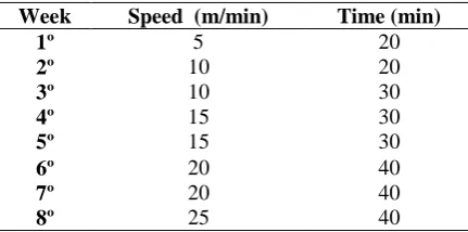
The treadmill training protocol was adapted from the study protocol for verification of the anaerobic threshold in rats (De Araújo et al., 2009), and lasted for five days a week during eight weeks with an increase on speed on every week. During the exercise protocol, the speed received 5m/min gradual increases every five minutes until the value for the week. The treadmill speed and the arrangement of duration of exercise were adapted accordingly (Table 1). To analyze anaerobic endurance, blood lactate levels were measured during the training protocol and blood samples (25 µl) were dispensed directly onto a lactimeter tape collector (Accutrend® Roche, Basel, Switzerland).
Animals were subjected to daily training with gradual increase in the intensity and time of realization.
Blood Collection, cytokine measurement and leukocyte count
It was realized three blood samples collection from the tail vein at different time-points: t0 = before the beginning of the training; t1 = at the end of the fourth week of the exercise protocol; t2 = at the end of the eighth week of the exercise protocol. Blood samples (1 ml) were centrifuged (4°C, 3.200g, 10 min) and the serum stored at -80oC. Quantification of inflammatory markers (IL-6, IL-10 and TNF-α) levels were measured by ELISA (enzyme - linked immunosorbent assay) (BD Biosciense, San Diego, CA). The blood analysis was performed by changing absorbance at a wavelength of 450nm with correction for 560nm using an ELISA reader (XMark Absorbance Microplate Spectrophotometer, Bio-Rad, Hercules, CA).
---Muscle Morphological Analysis
At the end of the exercise protocol, rats were sacrificed and the tibialis anterior and gastrocnemius muscle were collected, fixed in a Methacarn solution (60% methanol, 10% acetic acid and chloroform 30%, 4 h, 4 °C),
dehydrated, cleared in xileno, embedded in paraffin (Oxford Paraplast®, St. Louis, MO., USA), cut in microtome (5µM) and submitted to hematoxylin-eosin (HE) staining. A blinded researcher randomly selected
three slices for each muscle and, for each slice, it was obtained 400x microscopic digital images (Olympus Optical Co.) from regions containing cross section of muscle fibers. Images of at least 200 fibers for each animal
were analyzed using the software Image J® 1.45, a public domain software developed by W. Rasband (National Institute of Mental Health, National Institutes of Health, Bethesda, MD, USA). It was applied a digital mask
consisting of parallel lines where it was measured the distance between the lines (d) and the number of intersections between the fibers and the lines (i) (Supplementary Figure 1). The values obtained were applied in
the formula B = π/2xdxi, where B represents the muscle fiber circumference (Battlehner, 2003; Morath et al, 2013).
Supplemental Fig. 1. System testing parallel lines for determining the mean circumference of muscle fibers. B = π/2xdxi formula was used to calculate the perimeter corresponds to the perimeter where b, d is the distance between the lines i is the number of intersections between the fibers and the lines. Hematoxylin-eosin staining.
400X magnification. Statistical Analysis
Data were analyzed using SPSS (Statistical Package for Social Sciences) version 17.0 for Windows. Normality test was performed to the data found. Results are expressed as mean ± standard deviation and percentages. To perform the comparison of means between groups in different time points analyzed, it was used the One-Way ANOVA test followed by the post-hoc Tukey test with a significance level of 0.05.
Results
Periodontal disease impairs physiologic parameters on trained mice.
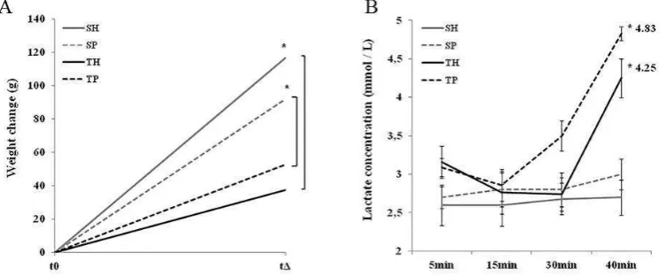
All animals showed an increase in weight during the study period, and the animals of the sedentary groups showed the greatest weigth variations when compared to trained animals (Figure 1A). Periodontal disease did not influence the weight variation, when compared to their respective experimental group (sedentary or trained). Sedentary rats showed a basal blood lactate level, which was not affected by periodontal disease (Figure 1B). After 8 weeks of training, the trained animals showed an increase in the lactate generation during the exercise period, which was more significant in the group with periodontal disease. This data suggests that periodontal disease acted as a negative factor for the treadmill training performance.
--- --- 575 without periodontal disease, SP: sedentary with periodontal disease, TH: trained without periodontal disease, TP:
trained with periodontal disease. Test One-Way ANOVA and post-hoc Tukey test (* p <0.05, n = 6).
The analysis of the muscle fiber perimeter showed that treadmill exercise led to a hypertrophy of the gastrocnemius and the tibialis anterior muscle (8.99%, p<0.05 and 31.9%, p<0.000, respectively, Figure 2A), when compared to the sedentary group. The induction of periodontal disease per se did not alter the muscle fiber
size, but the association between exercise and PD was responsible for a significant decrease in the perimeter of the fibers of the gastrocnemius muscle (29%, p<0.001, Figure 2E) when compared to all experimental groups (Figure 2B-D). For the tibialis anterior muscle, there was no significant differences between groups SH and SP (Figure F and G), but periodontal disease (Figure 2I) impaired muscle hypertrophy when compared to the trained
group (Figure 2H). Histological analysis showed no changes on the local leukocyte profile on both muscles.
Fig. 2. Periodontal disease alters muscle hypertrophy. (A) Morphometric analysis of gastrocnemius and tibialis anterior muscle. Hematoxilin and eosin staining of gastronecmius (B-E) and tibialis anterior (F-I) of sedentary and without periodontal disease (SH, B and F), sedentary with periodontal disease (SP, C and G), trained without
periodontal disease (TH, d and h) and trained with periodontal disease group (TP, E and I). Bars indicate standard deviation. Test One-Way ANOVA and post-hoc Tukey test (* p <0.05, ** p <0.001 and *** p <0.000,
n = 6). 400X magnification.
Periodontal diseases promotes changes on leukocyte count and serum cytokine levels of trained and sedentary rats
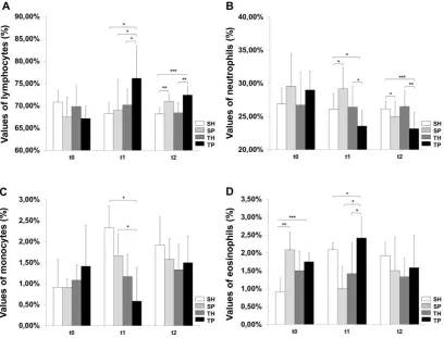
Sedentary animals without periodontal disease (SH) showed a predominance in circulating lymphocytes (70.83%+/-2.66%, t0, Figure 3A), followed by neutrophills (26.91%+/-2.31%, t0, Figure 3B), monocytes (0.92%+/-0.66%, t0, Figure 3C), eosinophills (0.92%+/-0.37%, t0, Figure 3D) and basophills (data not shown), with small variations during the timepoints used in this study. The levels of basophills showed no difference among experimental groups. Trained animals without periodontal disease (TH) showed a leukocyte count similar to the SH group. The sedentary group with periodontal disease (SP) showed a high variation on leukocyte count at each timepoint, suggesting adaptative changes during the time course of the pathology. When compared to sedentary animals without PD, it was observed that, in early stages of PD there was an increase on eosinophills at 30 days (2.08%+/-0.49%, p<0.001, t0, Figure 3D) and on neutrophills at 60 days (29.17%+/-2.94%, p<0.05, t1, Figure 3B). After 90 days of PD there was an increase on lymphocyte (71%+/-1.55%, p<0.001, t2, Figure 3A), suggesting a switch from an acute to a chronic inflammatory profile in the pathology course along this study.
---after 30 days of exercise (76.17%+/-7.2%, p<0.05, t1, Figure 3A) and 60 days (+72.42%+/-1.9%, p<0.000, t2, Figure 3A) of treadmill training, which was accompanied by a decrease on neutrophills (23.58%+/-2.39%, p<0.05 and 23.17%+/-2.48%, p<0.000, t1 and t2 respectivelly, Figure 3B). The monocyte count was also diminished after 30 days of exercise (0.58%+/-0.8%, p<0.05, t1, Figure 3C).
Fig. 3. Changes in leukocyte counts triggered by physical exercise and periodontal disease. Blood smear leukocyte count of lymphocytes (A), neutrophils (B), monocytes (C) and eosinophills (D) in the beginning (t0), 4
weeks (t1) or 8 weeks (t2) of physical exercise. SH: sedentary rats without periodontal disease, SP: sedentary with periodontal disease, TH: trained without periodontal disease, TP: trained with periodontal disease. Test
One-Way ANOVA and post-hoc Tukey test (* p <0.05, ** p <0.001 and *** p <0.000, n = 6).
The analysis of plasma interleukin levels showed that the training protocol alone induced a small increase on IL-6 (10.55%+/-7.6%, p<0.05 and 11.76%+/-7.6%, p<0.001, t1 and t2, respectively, Figure 4A) and IL-10 (37.9%+/-19.3%, p<0.05, t2, Figure 4C) levels when compared to sedentary animals, with no changes on TNF-α levels (2.17%+/-3.5%, t2, Figure 4B). Periodontal disease per se induced an increase on IL-6 (11.8%+/-7.24%, p<0.05) and TNF-α levels (12%+/-10.9%, p<0.05, Figure 4B) after 30 days of PD induction, which remained elevated at 60 and 90 days when compared to the sedentary group. After 90 days of PD it was also observed an increase on serum levels of the anti-inflamatory IL-10 (37.3%+/-19.9%, p<0.05, t2, Figure 4C).
--- --- 577 Fig. 4. Periodontal disease alters the interleukin serum levels. Serum levels of IL-6 (A), TNF-α (B) and IL-10
(C) was measured by ELISA in the beginning (t0), 4 weeks (t1) or 8 weeks (t2) of physical exercise. Data is expressed as percentage of sedentary rats without periodontal disease serum levels. SP: sedentary with periodontal disease, TH: trained without periodontal disease, TP: trained with periodontal disease. Test One-
Way ANOVA and post-hoc Tukey test (* p <0.05, ** p <0.001 and *** p <0.000, n=6).
Discussion
Our most important result was able to demonstrate that the treadmill training induced an increase in muscle fiber perimeter on both muscles as expected (Macaluso et al., 2013) and the periodontal disease process influenced negatively muscle hypertrophy.
In this study, we demonstrated that animals with PD that underwent treadmill exercise showed higher levels of blood lactate, a tissue hypoxia marker used as a reference for training intensity, when compared to the trained healthy group. Tissue hypoxia may be due to the association between heterogeneous distribution of microvascular blood flow, low systemic blood flow (ischemic hypoxia) and failure in cellular metabolism (Thomas et al, 2012; Gatterer et al., 2012). Loo et al. (2012) also demonstrated that PD might induce increase on lactate levels, but using different experimental protocol. These data suggest that PD can affect metabolic processes involving skeletal muscle adaptation to exercise load.
---data show that the treadmill training induced an increase in muscle fiber perimeter on both muscles as expected (Macaluso et al., 2013) and the periodontal disease process influenced negatively muscle hypertrophy. We showed that in our result what the type II muscle fibers were more susceptible to the effects of periodontal disease, since the gastrocnemius was the most affected. To our knowledge, this is the first report of the selective action of periodontal disease on skeletal muscle hypertrophy according to the muscle fiber type.
Several studies suggest that there is a crosstalk between muscle physiology and the immune system. It was already showed that moderate physical exercise (< 60% VO2max) might be related to an increased response
of defense mechanisms, while a more intense and prolonged exercise (> 65 % VO2max) or the overtraining is
related to impair the immune system (Keast et al., 1988; Brines et al, 1996; Suzuki et al., 2003). The model used in our study indicates a moderate training, which did not cause dramatically changes on leukocyte count of trained animals. However, it was observed an increase on IL-10, an anti-inflammatory cytokine, suggesting a protective role of physical exercise (Hirose et al., 2004; Thomas, 2007).
The systemic inflammatory condition may influence the size of muscle fibers, as well as its performance (Thomas, 2007; Marklund et al., 2013). For example, Tidball and Villalta (2010) showed that changes in eosinophils activity might influence negatively the hypertrophy of skeletal muscle. Other studies emphasize the importance of proper interaction of myeloid cells and inflammatory mediators for normal muscle growth (Marklund et al., 2013). We showed that in early stages of periodontal disease there was a fluctuation on circulating levels of neutrophils and eosinophils, while the lymphocytes count was affected only after 90 days, suggesting a switch on the host immunological responsiveness to PD from a destructive to a restorative condition overtime. In the other hand, we showed that after 30 days of the association between physical exercise and PD there was an increase on lymphocytes and eosinophils count and a decrease on neutrophils and monocytes. These data support the evidence of the role of physical exercise on the immune system and it also suggests that odontogenic infections might indirectly influence muscle hypertrophy due to changes on leukocytes count.
Muscle metabolism is susceptible to various pathological conditions mediated by cytokines (Cannon, 1993; Clark, 2007). IL-10, for instance, reduces the expression of proinflammatory cytokines and regulates the macrophage phenotype in injured muscle, through reduction of class 1 macrophage (M1) – involved on muscle cell lysis, and increase on class 2 macrophage (M2), involved on proliferation of satellite cells (Marklund et al., 2013; Guttridge et al., 2000; Coletti et al., 2002). In our study, all conditions increased the IL-10 serum levels after 8 weeks of exercise. These data are correlated with the values of monocytes, which were reduced after 4 week of training, and presented a small increase after the 8th week. This suggests that it might have some loss in the hability of tissue repair process after muscle injury (Villalta et al., 2009; Deng et al., 2012).
The increased production of pro inflammatory cytokines such as IL-6 and TNF-α is probably the most common cause of depletion of the muscle tissue. Studies in animals and humans have shown that an increase in serum IL-6 levels inhibits the anabolic effect of IGF-1 (Insulin-like growth factor 1), and that high concentrations of IL-6 and low concentrations of IGF-1 contribute to aggravation of sarcopenia (Muñoz-Cánoves et al., 2013). IL-6 may also induce the loss of muscle mass by modulating the secretion of anabolic hormones (Pedersen et al., 2004; Febbraio et al., 2004). It was also shown that TNF-α stimulates the proteolysis of myosin heavy chain and it is involved in the pathogenesis of muscle (Teles et al., 2009; Hodgetts et al., 2006; Clark, 2007). Prolonged exposure to TNF-α is also known to block cell differentiation and muscle regeneration (Clark, 2007; Perniconi et al., 2008) and it can also induce alterations of muscle proteins independently of protein loss, which results in diminished force production (Li et al., 1998; Li et al., 2000; Noh et al., 2013). It is possible that in physiological conditions this cytokines might be differentially expressed in the muscle microenvironment, leading to different response of muscle fibers to exercise load in accordance with the intensity and duration of activity. Under normal health conditions, exercise treadmill decreases TNF-α and IL-6 plasma levels and increases IL-10 (Nunes et al., 2013). In our study, the treadmill protocol alone induced an increase of IL-6, but not of TNF-α Increased levels of IL-6 alone may indicate a regulation of TNF-α, preventing the loss of muscle mass (Igaz et al., 2000).
The chronic systemic proinflammatory state could result in skeletal muscle atrophy (Li et al., 2000). We observed that PD per se induced an increase on TNF-α and IL-6 (Noh et al., 2013) and the association between PD and treadmill exercise did not change these values, suggesting that PD might indirectly interfere with muscle responsiveness to exercise load due to the local presence of TNF-α. It is possible that this association prevented not only the muscle metabolism but it also induced a catabolic process on muscle fiber in response to the exercise load. It appears that this effect is more pronounced on type 2 muscle fibers, since the gastrocnemius muscle showed a decrease on fiber hypertrophy due to PD-physical exercise association. In fact, there are several reports of the synergic effects of the IL-6 and TNF-α association on muscle sarcopenia (De Benedetti et al., 1997; Ferrucci et al., 2002; Visser et al., 2002).
Conclusions
---Al-Zahrani, M.S., Borawski, E.A., & Bissada, N.F. (2005). Periodontitis and three health-enhancing behaviors: maintaining normal weight, engaging in recommended level of exercise, and consuming a high-quality diet. J Periodontol, 76(8), 1362-1366.
Baek, K.J., Choi, Y., & Ji, S. (2012). Gingival fibroblasts from periodontitis patients exhibit inflammatory characteristics in vitro. Arch Oral Biol, 58(10), 1282-1292.
Baldwin, K.M., & Haddad, F. (2001). Effects of different activity and inactivity paradigms on myosin heavy chain gene expression in striated muscle. J Appl Physiol, 90(1), 345-357.
Battlehner, C.N., Caldini, E.G., Pereira, J.C., Luque, E.H., & Montes, G.S. (2003). How to measure the increase in elastic system fibres in the lamina propria of the uterine cervix of pregnant rats. J Anat, 203(4), 405– 418.
Brines, R., Hoffman-Goetz, L., & Pedersen, B.K. (1996). Can you exercise to make your immune system fitter? Immunol Today, 17(6), 252-254.
Cannon, J.G. (1993). Exercise and resistance to infection. J Appl Physiol, 74(3), 973-981.
Clark, I.A. (2007). How TNF was recognized as a key mechanism of disease. Cytokine Growth Factor Rev, 18(3-4), 335-343.
Coletti D, Yang E, Marazzi G, & Sassoon D (2002). TNFalpha inhibits skeletal myogenesis through a PW1-dependent pathway by recruitment of caspase pathways. EMBO J, 21(4),631-642.
De Araújo, M.B., Gobatto, F.B.M., Voltarelli, F.A., Ribeiro, C., Mota, C.S.A, Gobatto, C.A., & De Mello, M.A.R. (2009). Running training effects in different intensities on the aerobic capacity and lactate production by the muscle of wistar rats. Rev Bras Med Esporte 15(5), 365-369.
De Benedetti, F., Alonzi, T., Moretta, A., Lazzaro, D., Costa, P., Poli, V., Martini, A., Ciliberto, G., & Fattori, E. (1997). Interleukin 6 causes growth impairment in transgenic mice through a decrease in insulin-like growth factor-I. A model for stunted growth in children with chronic inflammation. J Clin Invest, 99(4), 643–650.
Delp, M.D., & Duan, C. (1996). Composition and size of type I, IIA, IID/X, and IIB fibers and citrate synthase activity of rat muscle. J Appl Physiol, 80(1), 261-270.
Deng, B., Wehling-Henricks, M., Villalta, S.A., Wang, Y., & Tidball, J.G. (2012). IL-10 triggers changes in macrophage phenotype that promote muscle growth and regeneration. J Immunol, 189(7), 3669-3680. Ebersole, J.L., Steffen, M.J., Reynolds, M.A., Branch-Mays, G.L., Dawson, D.R., Novak, K.F., Gunsolley,
J.C., Mattison, J.A., Ingram, D.K., & Novak, M.J. (2008). Differential Gender Effects of a Reduced Calorie Diet on Systemic Inflammatory and Immune Parameters in Nonhuman Primates. J Periodontal Res, 43(5),500-507.
Ebersole, J.L., Stevens, J., Steffen, M.J., Dawson Iii, D., & Novak, M.J. (2010). Systemic endotoxin levels in chronic indolent periodontal infections. J Periodontal Res, 45(1), 1-7.
Febbraio, M.A., Hiscock, N., Sacchetti, M., Fischer, C.P., & Pedersen, B.K. (2004). Interleukin-6 is a novel factor mediating glucose homeostasis during skeletal muscle contraction. Diabetes, 53(7), 1643-1648. Ferrucci, L., Penninx, B.W., Volpato, S., Harris, T.B., Bandeen-Roche, K., Balfour, J., Leveille, S.G., Fried,
L.P., & Md JM. (2002). Change in muscle strength explains accelerated decline of physical function in older women with high interleukin-6 serum levels. J Am Geriatr Soc, 50(12), 1947-1954.
Foster, W.H., Tidball, J.G., & Wang, Y. (2012). p38γ activity is required for maintenance of slow skeletal muscle size. Muscle Nerve, 45(2), 266-273.
Gatterer, H., Greilberger, J., Philippe, M., Faulhaber, M., Djukic, R., & Burtscher, M. (2012). Short-term supplementation with alpha-ketoglutaric acid and 5-hydroxymethylfurfural does not prevent the hypoxia induced decrease of exercise performance despite attenuation of oxidative stress. Int J Sports Med, 34(1), 1-7.
---Hirose, L., Nosaka, K., Newton, M., Laveder, A., Kano, M., Peake, J., & Suzuki, K. (2004). Changes in
inflammatory mediators following eccentric exercise of the elbow flexors. Exerc Immunol Rev, 10:75-90.
Hodgetts, S., Radley, H., Davies, M., & Grounds, M.D. (2006). Reduced necrosis of dystrophic muscle by depletion of host neutrophils, or blocking TNF alpha function with Etanercept in mdx mice. Neuromuscul Disord, 16(9-10), 591-602.
Igaz, P., Horváth, A., Horváth, B., Szalai, C., Pállinger, E., Rajnavölgyi, E., Tóth, S., Rose-John, S. &, Falus, A. (2000). Soluble interleukin-6 receptor (sIL-6R) makes IL-6R negative T cell line respond to IL-6 it inhibits TNF production. Immunol Lett, 71(3), 143-148.
Keast, D., Cameron, K., & Morton, A.R (1988). Exercise and the immune response. Sports Med, 5(4), 248-267. Keelan, J.A., Wong, P.M., Bird, P.S., & Mitchell, M.D. (2010). Innate inflammatory responses of human
decidual cells to periodontopathic bacteria. Am J Obstet Gynecol, 202(5), 1-11.
Krogh-Madsen, R., Plomgaard, P., Moller, K.., Mittendorfer, B., & Pedersen, B.K. (2006). Influence of TNF-alpha and IL-6 infusions on insulin sensitivity and expression of IL-18 in humans. Am J Physiol Endocrinol Metab, 291(1), E108-114.
Li, X., Moody, M.R., Engel, D., Walker, S., Clubb, F.J. Jr, Sivasubramanian, N., Mann, D.L., & Reid, M.B. (2000). Cardiac-specific overexpression of tumor necrosis factor-alpha causes oxidative stress and contractile dysfunction in mouse diaphragm. Circulation, 102(14), 1690-1696.
Li, Y.P., & Reid, M.B. (2000). NF-kappaB mediates the protein loss induced by TNF-alpha in differentiated skeletal muscle myotubes. Am J Physiol Regul Integr Comp Physiol, 279(4), R1165–R1170.
Li, Y.P., Schwartz, R.J., Waddell, I.D., Holloway, B.R., & Reid, M.B. (1998). Skeletal muscle myocytes undergo protein loss and reactive oxygen-mediated NF-kappaB activation in response to tumor necrosis factor alpha. FASEB J, 12(10), 871-880.
Liu, K.Z., Xiang, X.M., Man, A., Sowa, M.G., Cholakis, N., Ghiabi, E.,Singer, D.L., & Scott, D.A. (2009). In vivo determination of multiple indices of periodontal inflammation by optical spectroscopy. J Periodontal Res, 44(1), 117-124.
Liu, L., Li, C., Cai, C., Xiang, J., & Cao, Z. (2010). Cyclophilin A (CypA) is associated with the inflammatory infiltration and alveolar bone destruction in an experimental periodontitis. Biochem Biophys Res Commun, 391(1), 1000-1006.
Loo, W.Y., Yue, Y., Fan, C.B., Bai, L., Dou, Y., Wang, M., Liang, H., Cheung, M.N., Chow, L.W.C, Li, J., Tian, Y. & Qing, L. (2012). Comparing serum levels of cardiac biomarkers in cancer patients receiving chemotherapy and subjects with chronic periodontitis. J Transl Med, 10(suppl 1):S5.
Macaluso, F., Brooks, N.E., Niesler, C.U., & Myburgh, K.H. (2013). Satellite cell pool expansion is affected by skeletal muscle characteristics. Muscle Nerve, 48(1), 109-116.
Malaguti, M., Angeloni, C., Garatachea, N., Baldini, M., Leoncini, E., Collado, P.S., Teti, G., Falconi, M., Gonzalez-Gallego, J., & Hrelia, S. (2009). Sulforaphane treatment protects skeletal muscle against damage induced by exhaustive exercise in rats. J Appl Physiol, 107(4), 1028-1036.
Malaguti, M., Angeloni, C., & Hrelia, S. (2013). Polyphenols in exercise performance and prevention of exercise-induced muscle damage. Oxid Med Cell Longey, 2013. Doi: 10.1155/2013/825928.
Marklund, P., Mattsson, C.M., Wåhlin-Larsson, B., Ponsot, E., Lindvall, B., Lindvall, L., Ekblom, B., & Kadi, F. (2013). Extensive inflammatory cell infiltration in human skeletal muscle in response to an ultraendurance exercise bout in experienced athletes. J Appl Physiol, 114(1), 66-72.
Morath, V., Keuper, M., & Rodriguez-Franco, M. (2013). Semi-automatic determination of cell surface areas used in systems biology. Front Biosci, 5:533-545.
Muñoz-Cánoves, P., Scheele, C., Pedersen, B.K.., & Serrano, A.L. (2013). Interleukin-6 myokine signaling in skeletal muscle: a double-edged sword? FEBS J, 280(17), 4131-4148.
Noh, M.K., Jung, M., Kim, S.H., Lee, S.R., Park, K.H., Kim, D.H., Kim, H.H., & Park, Y.G. (2013). Assessment of IL-6, IL-8 and TNF-α levels in the gingival tissue of patients with periodontitis.
Exp Ther Med, 6(3), 847-851.
Nunes, R.B., Alves, J.P., Kessler, L.P., & Dal Lago, P. (2013). Aerobic exercise improves the inflammatory profile correlated with cardiac remodeling and function in chronic heart failure rats. Clinics, 68(6), 876-882.
Pedersen, B.K., Steensberg, A., Fischer, C., Keller, C., Keller, P., Plomgaard, P., Wolsk-Petersen, E., & Febbraio, M. (2004). The metabolic role of IL-6 produce during exercise: is IL-6 an exercise factor? Proc Nutr Soc, 63(2), 263-267.
Perniconi, B., Albertini, M.C., Teodori, L., Belli, L., & Rocchi, M. (2008). A meta-analysis on a therapeutic dilemma: to exercise or not to exercise in cachexia. Basic Appl Myol, 18:105-120.
Pette, D. (1998). Training effects on the contractile apparatus. Acta Physiol Scand, 162(3), 367-376.
--- --- 581 Roubenoff, R. (1997). Inflammatory and hormonal mediators of cachexia. J Nutr, 127(suppl 5), 1014S-1016S. Sanders, A.E., Slade, G.D., Fitzsimmons, T.R., & Bartold, P.M. (2009). Physical activity, inflammatory
biomarkers in gingival crevicular fluid and periodontitis. J Clin Periodontol, 36(5), 388-395.
Staron, R.S., Kraemer, W.J., Hikida, R.S., Fry, A.C., Murray, J.D., & Campos, G.E. (1999). Fiber type composition of four hindlimb muscles of adult Fisher 344 rats. Histochem Cell Biol, 111(2), 117-123. Suzuki, K., Nakaji, S., Kurakake, S., Totsuka, M., Sato, K., Kuriyama, T., Fujimoto, H., Shibusawa,
K., Machida, K., & Sugawara, K. (2003). Exhaustive exercise and type-1/type-2 cytokine balance with special focus on interleukin-12 p40/p70. Exerc Immunol Rev 9:48-55.
Tefferi, A., Hanson, C.A., & Inwards, D.J. (2005). How to Interpret and Pursue an Abnormal Complete Blood Cell Count in Adults. Mayo Clin Proc, 80(7), 923-936.
Teles, R.P., Likhari, V., Socransky, S.S., & Haffajee, A.D. (2009). Salivary Cytokine Levels in Chronic Periodontitis and Periodontally Healthy Subjects. A cross-sectional Study. J Periodontal Res, 44(3), 411-417.
Thomas, C., Bishop, D.J., Lambert, K.., Mercier, J., & Brooks, G.A. (2012). Effects of acute and chronic exercise on sarcolemmal MCT1 and MCT4 contents in human skeletal muscles: current status. Am J Physiol Regul Integr Comp Physiol, 302(1), 1-14.
Thomas, D.R (2007). Loss of skeletal muscle mass in aging: examining the relationship of starvation, sarcopenia and cachexia. Clin nutr, 26(4), 389-399.
Tidball, J.G., & Villalta, S.A. (2010). Regulatory interactions between muscle and the immune system during muscle regeneration. Am J Physiol Regul Integr Comp Physiol, 298(5), 1173-1187.
Villalta, S.A., Nguyen, H.X., Deng, B., Gotoh, T., & Tidball, J.G. (2009). Shifts in macrophage phenotypes and macrophage competition for arginine metabolism affect the severity of muscle pathology in muscular dystrophy. Hum Mol Genet, 18(3), 482-496.
Villalta, S.A., Rinaldi, C., Deng, B., Liu, G., Fedor, B., & Tidball, J.G. (2011). Interleukin-10 reduces the pathology of mdx muscular dystrophy by deactivating M1 macrophages and modulating macrophage phenotype. Hum Mol Genet, 20(4), 790-805.
Visser, M., Pahor, M., Taaffe, D.R., Goodpaster, B.H., Simonsick, E.M., Newman, A.B., Nevitt, M., & Harris, T.B. (2002). Relationship of interleukin-6 and tumor necrosis factor-alpha with muscle mass and muscle strength in elderly men and women: the Health ABC Study. J Gerontol A Biol Sci Med Sci, 57(5), M326–M332.