Genetic Diversity of
Trypanosoma evansi
in Buffalo based on Internal Transcribed
Spacer (ITS) Regions
Sintawee KHUCHAREONTAWORN1), Phirom SINGHAPHAN1), Nareerat VISESHAKUL2) and Kosum CHANSIRI1)*
1)Department of Biochemistry, Faculty of Medicine, Srinakharinwirot University, Sukhumvit 23, Bangkok 10110 and 2)Department of
Parasitology, Faculty of Veterinary Medicine, Chulalongkorn University, Patumwan Road, Bangkok 10330 Thailand
(Received 8 June 2006/Accepted 16 January 2007)
ABSTRACT. The nucleotide sequences of 18S rDNA and internal transcribed spacer (ITS) regions were used for studying the relationships of Trypanosoma evansi isolate from a buffalo. The sequences were analyzed and compared to 18S rDNA and the ITS regions of the other Trypanosoma spp. Maximum likelihood phylogenetic trees were constructed using Leishmania major as the outgroup. The tree of 18S rDNA indicated that T. evansi (buffalo B18) isolate was closely related to those of Taiwan and T. brucei stock. The ITS tree showed the genetic diversity among 32 clones of T. evansi (B18) within a single host. This data will be useful for epidemiological and dynamic studies for designing the rational control programs of the disease.
KEYWORDS: internal transcribed spacers (ITS), phylogenetic, small subunit rDNA, Trypanosoma evansi.
J. Vet. Med. Sci. 69(5): 487–493, 2007
Trypanosoma spp. consists of a large group of obligated flagellate blood parasites that infect members of every ver-tebrate class [20]. Members of this genus can be transmitted by various vectors such as blood-sucking invertebrates; pri-marily arthropods (for species infecting mammals and birds) and primarily hirudinean annelids (for species infect-ing poikilothermic vertebrates). In Thailand, where T. evansi is the only pathogenic trypanosome, it causes consid-erable loss in productivity in domestic animals such as horses, beef cattle, dairy cattle, and buffalos [5, 8, 31]. Ani-mals which recover from an acute episode of disease are often clinically normal and T. evansi isolates can be obtained from them.
In Thailand, buffaloes are one of the most common hosts of T. evansi, and are infected via tabanid flies. It has been reported that there are 45 species of tabanids comprising three genera: Tabanus, Haematopota, and Crysops.
Tabanus rubidus was found most frequently, distributed in every part of the country and present in 25 provinces. Clin-ical signs in animals infected with T. evansi are variable, depending on the animal species. Chronic cases occur mostly in infected buffaloes and cattle, which sometimes do not show any clinical signs [31]. However, abortion at late pregnancy or early parturition caused by T. evansi is known to occur in buffaloes [27]. In addition, stress factors such as fasciolosis, combined with seasonal malnutrition during the dry season, lower the resistance of the animals, causing exacerbation of clinical surra [26].
Generally, classification of trypanosomes has been based on a morphological description of bloodstream trypomastig-otes and morphometric parameters. However, identification by means of such descriptions is often very difficult because of extreme morphological variation. Consequently, there is
much confusion in the literature [10, 11, 24].
Molecular characterization has produced additional infor-mation on the affinities of trypanosomes. Some specific genes have been sequenced, aligned, and analyzed in order to study the phylogenetic relationships among morphologi-cally indistinguishable trypanosomes [19]. Evolutionary events have been addressed by studying the ribosomal RNA (rRNA) genes and their associated spacer regions, collec-tively called ribosomal DNA [32]. Techniques such as the analysis of small or large subunit ribosomal RNA sequences [23, 35]. Restriction Fragment Length Polymorphisms (RFLPs) in DNA [6, 29, 49], ribosomal DNA restriction [7], the polymorphisms of PCR amplification products [4, 12], and molecular karyotypes have all been successfully used in distinguishing between morphologically similar protozoa [44, 45]. Nevertheless, the aforementioned methods have been unsuccessful in determining the relationships within trypanosome species.
The 18S rDNA gene has been studied because it is among the slowest evolving sequence found extensively throughout living organisms, and has therefore been very useful for examining ancient evolutionary events. The first molecular phylogenetic studies based on comparisons of genes encod-ing mitochondrial and nuclear ribosomal RNA (rRNA) showed trypanosomes to be a monophyly [14, 28, 38–41] as did studies based on protein-coding genes [1, 3, 15–17, 36, 47]. Doubt has now been cast on this consensus by a re-analysis of SSU rRNA gene sequences, but the previous SSU rRNA gene trees did not adequately prove monophyly of trypanosomes because they either included an inadequate number and selection of taxa, or were rooted inappropri-ately. In recent SSU rRNA gene trees, trypanosomes and trypanosomatids appear to be a paraphyly [21, 22].
The internal transcribed spacers (ITS) are versatile genetic markers and have been used for phylogenetic analy-sis, evaluation of the evolutionary process, and for the
mination of taxonomic identities [34]. The ITS are located between the repeating array of nuclear 18S, 5.8S, and 28S ribosomal RNA genes; a locus that has 100–200 copies per genome [18]. In 1999, the phylogenetic analysis of ITS sequences was used for studying the polymorphism of T. rangeli strains isolated from different hosts and geographic areas (southern Brazil, Central America, and northern South America) [13].
Previously, the intra-species genetic variation of Trypa-nosoma have been reported in T. brucei [2], T. congolense
[30], and T. evansi [33, 46] in different hosts basically revealed by PCR based method. Herein, we investigated the diversity of T. evansi in a single host (buffalo) using the nucleotide data of 18S rDNA, 5.8S rDNA, and ITS regions. Phylogenetic trees were constructed and compared to those of other trypanosomes reported in GenBank. The data will be useful for epidemiological and dynamic studies for designing the rational control programs of the disease.
MATERIALS AND METHODS
Parasite collection: T. evansi were collected during 1999 from the infected blood samples of a buffalo namely B18 in the Ladkrabang district, Singburi province, Thailand. It was previously screened using the thin blood smear technique. Blood were preserved in the phosphate saline glucose buffer ( P S G ) , p H 8 . 0 ( 5 0 m M N a2H P O4. H2O , 2 m M
NaH2PO4.2H2O, 36 mM NaCl and 1.5% glucose) and stored
in the liquid nitrogen tank (–196°C) until use.
Parasite infected blood preservation and isolation: Infected blood in the liquid nitrogen tank was thawed at room temperature and 0.1 ml was injected to a mouse using 1 ml Tuberculin syringe and needle gauge No 27. Para-sitemia was daily checked by wet blood smear, starting from the third day after injection. At the highest parasitemia (108
cells/ml) which was usually in the 3rd -5th day of injection, blood was withdrawn from euthanized mouse by cardiac punctured. An anion exchange column (DE 52 DEAE cel-lulose) was used to purify parasite from the horde blood cells according to the method of described by Chao [9]. The eluent with infect protozoa was collected and proceeding to parasite lysis and DNA extraction.
DNA extraction and purification: Pellet of parasites was resuspended in PSG buffer in the presence of SDS (final concentration was 2%) and proteinase K (final concentra-tion was 1 mg/ml). The solution was incubated at 42°C for 14 hr. Parasite DNA was extracted by conventional phenol/ chloroform which responsed for deproteinization of the aqueous solution containing the desired nucleic acid. The purified DNAs were precipitated by the addition of 2
vol-umes of cold absolute ethanol. The pellet was dried, dis-solved in sterile distilled water and kept at 4°C until use.
PCR amplification: PCR amplifications of 18S rDNA gene and ITS spacer region were carried out using their spe-cific primers (Table 1). All reactions were performed in 25 µl volume containing 100–200 ng of genomic DNA. The 18S rDNA reaction contained 1X PCR buffer, 1.7 mM of each primer, 200 mM of dNTP, 1.5 mM MgCl2 and 3 units
of Taq DNA polymerase. Sterile distilled water was used to make volume to 25 µl. The PCR amplification was per-formed using Peltier Thermal Cycler (MJ Research, PTC-200) for 30 cycles. Each cycle consisted of denaturation at 94°C for 1 min, annealing at 37°C for 1 min and extension at 72°C for 1 min. The PCR amplification of ITS regions contained 1 × PCR buffer, 2 mM of each primer, 400 mM of dNTP, 6 mM MgCl2 and 1.5 units of Taq DNA
poly-merase. Sterile distilled water was used to make volume to 25 µl. A PCR amplification was performed using Peltier Thermal Cycler (MJ Research, PTC-200) for 30 cycles. Each cycle consisted of denaturation at 94°C for 1 min, annealing at 60°C for 1 min and extension at 72°C for 1 min. PCR products were analyzed using electrophoresis in 1.2% agarose gel at 110 volts approximately 35 min prior to stain-ing in ethidium bromide solution and observation.
Cloning and DNA sequencing: The PCR fragments were eluted from the gels, purified using TOPO XL PCR cloning kit (Invitrogen). The procedure of purification was per-formed according to the instruction manual provided by the company. The entire amplified both of 18S rDNA and ITS regions were purified and cloned using the procedures as specified by the manufacturer. After checking the cloned products by colony PCR amplification, 32 clones of ITS region were selected for further DNA sequencing using the Big Dye Terminator Cycle Sequencing procedure. The 300
ng of sample was used for PCR amplification using 25 cycles of denaturation at 96°C for 10 sec, annealing at 50°C for 4 sec and extension at 60°C for 4 min. The sample was dissolved with dye and formamide solution prior to DNA sequencing. The nucleotide sequence data were analyzed using software of ABI PRISM Model 377 version 3.4. The alignment of the sequences was achieved by using Clustal X software Version 1.83 (multiple sequence alignment) pro-gram [43] and refined using the manual method. Leishma-niamajor and Trypanosoma congolense were used as the outgroup to root the 18S rDNA and ITS trees, respectively. Blocks of sequence data containing 2,349 characters for 18S rDNA and 1,125 characters for ITS regions were used for the tree analysis and were indicated by nucleotide position. The phylogenetic trees were constructed by PAUP program version 4.0 [42]. The cladograms were created by using
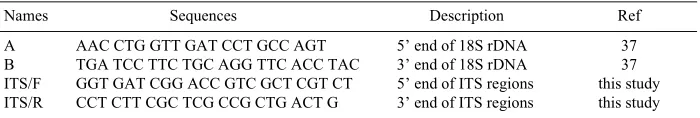
Table 1. Oligonucletides used for PCR amplification
Names Sequences Description Ref
A AAC CTG GTT GAT CCT GCC AGT 5’ end of 18S rDNA 37
B TGA TCC TTC TGC AGG TTC ACC TAC 3’ end of 18S rDNA 37
ITS/F GGT GAT CGG ACC GTC GCT CGT CT 5’ end of ITS regions this study
maximum likelihood and maximum parsimony methods for 18S rDNA gene and ITS region, respectively. Bootstrap values were replicated 100 times and computed by PAUP. The T. evansi sequences were compared to those of other trypanosomes reported in Genbank as indicated in Table 2.
RESULTS
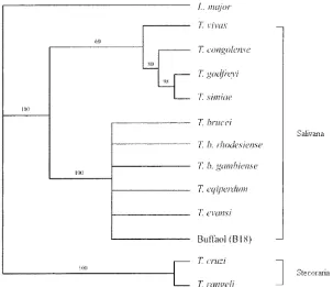
This study used the molecular phylogenetics of 18S rDNA to identify the species of trypanosome isolated from a buffalo in Thailand. The PCR product of 18S rDNA was approximately 2.1 kb in size. The phylogenetic tree of 18S rDNA sequences from different Trypanosoma spp. stocks confirmed the phylogenetic position of T. evansi in the Try-panosomatids family and suggested that T. evansi stocks from buffalo in Thailand belong to a single genus receiving highly significant support when using Leishmania major as the outgroup (Fig. 1). According to the tree, Salivaria, except for T. vivax, formed a cluster which was supported by a highly significant internal branch falling outside the highly significantly supported cluster of Stercoraria and outgroup. The tree also revealed that T. evansi strain B18 was
homol-ogous to T. evansi, T. brucei, T. b. gambiense, T. b. rhode-siense, and T. eqiperdum stock submitted to GenBank. This larger cluster was supported by a highly significant internal branch.
Sequence analysis of the ITS region containing ITS-1, 5.8S rDNA, and ITS-2 from 32 clones of T. evansi B18 iso-lates was performed. The 5.8S rDNA sequences showed no significant differences among 32 clones of T. evansi against
T. brucei, whereas the ITS-1 and ITS-2 genes exhibited sin-gle nucleotide polymorphisms (SNPs) characterized by base alterations, insertions, or deletion. Along with probable insertion/deletion events, several perfect and imperfect mic-rosatellite repeats with variable motifs (TA, AT, GG, GT, TT, and CTT) and length were mainly observed in the sequences. A comparative analysis of the sequence align-ments carried out by ClustalX (Version 1.83) revealed that 180 and 312 variable sites were observed in 1 and ITS-2 regions, respectively.
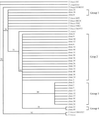
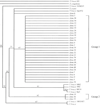
The phylogenetic tree based on ITS-1 and ITS-2 regions (Figs. 2 and 3) of 32 clones from T. evansi B18 stock showed that most of them were separated from T. brucei
when T. congolense was used as the outgroup. However,
Table 2. GenBank accession numbers of 18S rDNA and ITS regions of Trypanosomaspp. and other blood parasites
Parasites names specific Host/Lab host GenBank accession numbers
Leishmania major AC005806.3
118S rDNA T. brucei AL929605.1
T. b. gambiense AJ009141.1
T. b. rhodesiense AJ009142.1
T. congolense Rattus norvegicus U22319.1
T. cruzi AJ009148.1
T. eqiperdum AJ009153.1
T. evansi D89527.1
T. evansi Buffalo (B18) AY904050
T. godfreyi AJ009155.1
T. rangeli AJ012416.1
T. simiae U22320.1
T. vivax Rattus norvegicus U22316.1
T. congolense Rattus norvegicus U22319.1
ITS region T. evansi D89527.1
T. evansi Buffalo (B18) Buffalo AY904050
T. evansi clone No.2–10 Buffalo DQ472679-DQ472687
T. evansi clone No.12–21 Buffalo DQ472688-DQ472697
T. evansi clone No.24 Buffalo DQ472698
T. evansi clone No.26 Buffalo DQ472699
T. evansi clone No.30–34 Buffalo DQ472700-DQ472704
T. evansi clone No.36 Buffalo DQ472705
T. evansi clone No.38–40 Buffalo DQ472706-DQ472708
T. evansi clone No.48–49 Buffalo DQ472709-DQ472710
T. brucei Homo sapiens X05682.1
T. brucei NW2 Homo sapiens AF306776.1
T. brucei TH2 Homo sapiens AF306777.1
T. brucei Da1972 Pig AF306774.1
T. brucei B8/18 Glossina palpalis AF306772.1
T. brucei KP2 Lion AF306773.1
T. brucei STIB215 Lion AF306771.1
T. brucei H3 AF306770.1
some of them were classified within the same clade as T. brucei. Based on the ITS-1 tree (Fig. 2), the data revealed that these clones were diverse and separated into two groups, group 1 and group 2, which consisted of 29 and 3 taxa, respectively. Referring to the ITS-2 tree (Fig. 3), the clones were separated into four groups containing 3, 18, 9, and 2 taxa, respectively. The phylogram showed intermedi-ate significant bootstrap supported clusters and the variation of nucleotides in the ITS-2 region showed the variation of T. evansi in buffalo.
DISCUSSION
The nucleotide sequence alignment of 18S rDNA from T. evansi showed a sufficiently high degree of homology to demonstrate that nucleotide misincorporation may occur while using regular Taq DNA polymerase in PCR; the con-sistency of these polymorphisms was assessed by separate analysis of each clone sequence. Nucleotide alterations on the consensus sequences of all clones were also observed. The 18S rDNA data clearly indicated that T. evansi B18 stock was relatively straightforward to include under the T. evansi clade.
Analysis of the ITS regions revealed that mononucleotide insertion/deletion or transition and transversion occurred,
whereas the 5.8S rDNA was completely conserved among all of selected clones. This indicated that the ITS regions were more informative than the 5.8S rDNA. When phylo-genetic trees were separately constructed using the nucle-otide data of either 1 or 2, it was clear that the ITS-2 tree was more informative than the ITS-1 tree. However, both ITS regions of T. evansi B18 isolate revealed intra-spe-cies genetic diversity; at least 2–4 groups within one host were observed. Previously, the genetic diversity of T. evansi has been reported using MGE-, SSR-, RAPD-PCR profiling, and expression-site-associated genes (ESAG 6) [25, 48]. These studies indicated that there are at least 3–7 different patterns of T. evansi with different hosts. How-ever, the data showed no genetic diversity of T. evansi
within a single host.
Thus, it is proposed that the ITS-2 nucleotide sequence data represent a useful genetic marker for classification within the group of T. evansi which can be applied in epide-miological and dynamic studies for designing the rational control programs of the parasites.
ACKNOWLEDGEMENTS. We would like to thank Dr. Nopporn Sarataphan, National Institute of animal health, Ministry of Agriculture, for providing the T. evansi B18 stock.
REFERENCES
1. Adj’e, C. A., Operdoes, F. R. and Michels, P. A. 1998. Molec-ular analysis of phosphoglycerate kinase in Trypanoplasma borreli and the evolution of this enzyme in kinetoplastida.
Gene217: 91–99.
2. Agbo, E. E., Majiwa, P. A., Claassen, H. J. and te Pas, M. F. 2002. Molecular variation of Trypanosoma brucei subspecies as revealed by AFLP fingerprinting. Parasitology124: 349– 358.
3. Alvarez, F., Cortinas, M. N. and Musto, H. 1996. The analysis
of protein coding genes suggests monophyly of Trypanosoma.
Mol. Phylogenet. Evol.5: 333–343.
4. Artama, W. T., Agey, M. W. and Donelson, J. E. 1992. DNA comparisons of Trypanosoma evansi (Indonesia) and Trypano-soma brucei spp. Parasitology104: 67–74.
5. Boonyawong, T., Chantaraprateep, P. and Muangyai, M. 1975. Surra in horse. Thai J. Vet. Med.5: 665–672.
6. Borst, P., Fase-Fowler, F. and Gibson, W. C. 1981. Quantita-tion of genetic differences between Trypanosoma brucei gam-biense, rhodesiense and brucei by restriction enzyme analysis of kinetoplast DNA. Mol. Biochem. Parasitol.3: 117–131. Fig. 2. A maximum parsimony methods of ITS-1 among 32 clones of T. evansi B18 isolates and T. brucei,
7. Camargo, E. P., Sbravate, C., Teixeira, M. M., Uliana, S. R., Soares, M. B., Affonso, H. T. and Floeter-Winter, L. 1992. Ribosomal DNA restriction analysis and synthetic oligonucle-otide probing in the identification of genera of lower trypano-somatids. J. Parasitol.78: 40–48.
8. Chaichanapunpol, I., Sirirangkamanont, T., Trongwongsa, L. and Suvanwasi, P. 1985. Observations on trypanosomiasis in a native bull. Kasetsart Vet.6: 1–9.
9. Chao, D. and Dusanic, D. G. 1984. Comparative studies of the isolation of metacyclic trypomastigotes of Trypanosoma cruzi
by DEAE ion exchange chromatography. Zhonghua Min Guo Wei Sheng Wu Ji Mian Yi Xue Za Zhi17: 146–152.
10. Desser, S. S. 2001. The blood parasites of anurans from Costa Rica with reflections on the taxonomy of their trypanosomes. J.
Parasitol.87: 152–160.
11. Diamond, L. S. 1965. Studies in the morphology, biology and taxonomy of the trypanosomes of Anura. Wildlife Dis.44: 1–77. 12. Dirie, M. F., Murphy, N. B. and Gardiner, P. R. 1993. DNA fin-gerprinting of Trypanosoma vivax isolates rapidly identifies intraspecific relationships. J. Eukaryot. Microbiol.40: 132–134. 13. Grisard, E. C., Campbell, D. A. and Romanha, A. J. 1999. Mini-exon gene sequence polymorphism among Trypanosoma rangeli strains isolated from distinct geographical regions.
Parasitology118: 375–382.
14. Haag, J., O'hUigin, C. and Overath, P. 1998. The molecular phylogeny of tryanosomes: evidence for an early divergence of the salivaria. Mol. Biochem. Parasitol.91: 37–49.
15. Hannaert, V., Blaauw, M., Kohl, L., Allert, S., Opperdoes, F. Fig. 3. A maximum parsimony of ITS-2 among 32 clones of T. evansi B18 isolated and T. brucei, outgroup with T.
R. and Michels, P. A. 1992. Molecular analysis of the cytosolic and glycosomal glyceraldehyde-3-phosphate dehydrogenase in
Leishmania mexicana. Mol. Biochem. Parasitol. 55: 115–126. 16. Hannaert, V., Operdoes, F. R. and Michels, P. A. 1998.
Com-parison and evolutionary analysis of the glycosomal glyceral-d e h y glyceral-d e - 3 - p h o s p h a t e glyceral-d e h y glyceral-d r o g e n a s e f r o m glyceral-d i f f e r e n t Kinetoplastida. J. Mol. Evol. 47: 728–738.
17. Hashimoto, T., Nakamura, Y., Kamaishi, T., Adachi, J., Naka-mura, F., Okamoto, K. and Hasegawa, M. 1995. Phylogenetic place of kinetoplastid protozoa inferred from a protein phylog-eny of elongation factor 1 alpha. Mol. Biochem. Parasitol.70: 181–185.
18. Hernandez, R., Martinez-Calvillo, S., Hernandez-Rivas, R. and Gomez, E. 993. Trypanosoma cruzi ribosomal RNA genes: a review. Biol. Res.26: 109–114.
19. Hillis, D. M. and Moritz, C. 1990. An overview of applications of molecular systematics, pp. 502–515. In: Molecular System-atics (Hillis, D. M. and Moitz, C. E. eds.), Sinauer Associates, Sunderland.
20. Hoare, C. A. 1972. The trypanosomes of mammals, pp. 749.
In: A Zoological Monograph. Blackwell Scientific Publication, Oxford.
21. Hughes, A. L. and Piontkivska, H. 2003. Molecular phyloge-netics of Trypanosomatidae: contrasting results from 18S rRNA and protein phylogenies. Kinetoplastid Biol. Dis.2: 15. 22. Hughes, A. L. and Piontkivska, H. 2003. Phylogeny of
Trypa-nosomatidae and Bodonidae (Kinetoplastida) based on 18S rRNA: evidence for paraphyly of Trypanosoma and six other genera. Mol. Biol. Evol.20: 644–652.
23. Johnson, A. M. and Baverstock, P. R. 1989. Rapid ribosomal RNA sequencing and the phylogenetic analysis of protists.
Parasitol. Today5: 102–105.
24. Lebedeff, W. 1910. pp. 397–436. Ueber Trypanosoma rotato-rium Gruby, Festschr. 60., Richard Hertwigs (Munchen), Geburts.
25. Li, F. J., Gasser, R. B., Zheng, J. Y., Claes, F., Zhu, X. Q. and Lun, Z. R. 2005. Application of multiple DNA fingerprinting techniques to study the genetic relationships among three members of the subgenus Trypanozoon (Protozoa: Trypanoso-matidae). Mol .Cell Probes.19: 400–407.
26. Lohr, K. F., Pholpark, S., Srikitjakarn, L., Betterman, G. and Staa, C. 1985. Trypanosoma evansi infection in buffaloes in Northeast Thailand. Field investigation, Trop. Anim. Health. Prod17: 121–125.
27. Lohr, K. F., Pholpark, S., Upatum, N., Leidl, K. and Staak, C. 1986. Trypanosoma evansi infection, a frequent cause of abor-tion in buffaloes. Trop. Anim. Hlth. Prod 18: 103–108. 28. Lukes, J., Jirku, M., Dolezel, D., Kral’ova, I., Hollar, L. and
Maslov, D. A. 1997. Analysis of ribosomal RNA genes sug-gests that trypanosomes are monophyletic. J. Mol. Evol.44: 521–527.
29. Lun, Z. R., Allingham, R., Brun, R. and Lanham, S. M. 1992. The isoenzyme characteristics of Trypanosoma evansi and Try-panosoma equiperdum isolated from domestic stocks in China.
Ann. Trop. Med. Parasitol.86: 333–340.
30. Majiwa, P. A., Hamers, R., Van Meirvenne, N. and Matthys-sens, G. 1986. Evidence for genetic diversity in Trypanosoma (Nannomonas) congolense. Parasitology93 (Pt 2): 291–304. 31. Mathias, E. and Muangyai, M. 1980. Trypanosoma evansi
infection in a swamp buffalo calf. Thai. J. Vet. Med.10: 47–54. 32. Mindell, D. P. and Honeycutt, R. L. 1989. Variability in tran-scribed regions of ribosomal DNA and early divergences in birds. Auk106: 539–548.
33. Omanwar, S., Rao, J. R., Singh, R. K. and Butchaiah, G. 2001. DNA polymorphism in Trypanosoma evansi isolates defined by randomly amplified polymorphic DNA-PCR. Vet. Rec. 148: 244–246.
34. Powers, T. O., Todd, T. C., Burnell, A. M., Murray, P. C. B., Fleming, C. C., Szalanki, A. L., Adams, B. A. and Harris, T. S. 1997. The internal transcribed spacer region as a taxonomic marker for nematodes. J. Nematol.29: 441–450.
35. Qu, L. H., Yu, X. O. and Lun, Z. R. 1990. Nucleotide sequence of the 5’ terminal region of L-rRNA from Trypanosoma evansi
and T. brucei and comparative analysis to D2 domain. Chin. Sci. Bull. 35: 1096–1098.
36. Simpson, A. G. B., Lukes, J. and Roger, A. J. 2002. The evolu-tionary histoly of kinetoplastids and their kinetoplasts. Mol. Biol. Evol. 19: 2071–2083.
37. Sogin, M. L. 1990. Amplification of ribosomal RNA genes for molecular evolution studies. pp. 307–314. In: PCR Protocols. A guide to methods and applications (Innis, M. A., Gelfand, D. H., Sninsky, J. J. and White, T. J. eds.), Academic Press, New York. 38. Stevens, J., Noyes, H. and Gibson, W. 1998. The evolution of trypanosomes infecting humans and primates. Mem. Inst. Oswaldo. Cruz. 93: 669–676.
39. Stevens, J. R. and Gibson, W. 1999. The molecular evolution of trypanosomes. Parasitol. Today15: 432–437.
40. Stevens, J. R., Noyes, H. A., Dover, G. A. and Gibson, W. C. 1999. The ancient and divergent origins of the human patho-genic trypanosomes, Trypanosoma brucei and T. cruzi. Parasi-tology118 (Pt 1): 107–116.
41. Stevens, J. R., Noyes, H. A., Schofield, C. J. and Gibson, W. 2001. The molecular evolution of Trypan osomatidae. Adv. Parasitol.48: 1–56.
42. Swofford, D. L. 2001. PAUP*: Phylogenetic Analysis Using Par-simony (and Other Methods). Sinauer Associates, Sunderland, MA.
43. Thompson, J. D., Gibson, T. J., Plewniak, F., Jeanmougin, F. and Hoiggins, D. G. 1997. The CLUSTALX windows inter-face: flexible strategies for multiple sequence alignment aided by quality analysis tools. Nucleic Acids Res 25.
44. Waitumbi, J. N., Murphy, N. B. and Peregrine, A. S. 1994. Genotype and drug-resistance phenotype of Trypanosoma evansi isolated from camels in northern Kenya. Ann. Trop. Med. Parasitol.88: 677–683.
45. Waitumbi, J. N. and Young, J. R. 1994. Electrophoretic karyo-typing is a sensitive epidemiological tool for studying Trypa-nosoma evansi infections. Vet. Parasitol. 52: 47–56.
46. Watanapokasin, Y., Tananyutthawongese, C., Uthaisang, W., Chansiri, K., Boonmatit, C. and Sarataphan, N. 1998. Intra-species differentiation of Trypanosoma evansi by DNA finger-printing with arbitrary primered polymerase chain reaction.
Vet. Parasitol. 78: 259–264.
47. Wiemer, E. A., Hannaert, V., van den, I. P. R., Van Roy, J., Opperdoes, F. R. and Michels, P. A. 1995. Molecular analysis of glyceraldehyde-3-phosphatedehydrogenase in Trypano-plasma borelli: an evolutionary scenario of subcellular com-partmentation in kinetoplastida. J. Mol. Evol.40: 443–454. 48. Witola, W. H., Sarataphan, N., Inoue, N., Ohashi, K. and
Onuma, M. 2005. Genetic variability in ESAG6 genes among
Trypanosoma evansi isolates and in comparison to other Try-panozoon members. Acta Tropica93: 63–73.