20
13
E
DI
TIO
N
GUIDELINES FOR THE MANAGEMENT OF
COMMON CHILDHOOD ILLNESSES
Second edition
POCKET BOOK
OF
ANTIMICROBIAL DRUGS FOR COMMON CONDITIONS
Please fill the blanks with your country’s most recent updated treatment guidelines. Page numbers refer to where generic guidance is found in the Pocket Book.
Condition Drug Dose
Dysentery (p. 144) HIV treatment (p. 233)
drug 2 drug 3 Malaria, non severe (p. 164–5)
drug 2 Malaria, severe (p. 158) Mastoiditis (p. 182)
drug 2 Meningitis (p. 169)
drug 2 Osteomyelitis (p. 187)
drug 2 Otitis media, acute (p. 183) Pneumonia, non-severe (p. 86) Pneumonia, severe (p. 82)
drug 2 Sepsis, neonatal (p. 55)
drug 2 Sepsis, older child (p. 180)
drug 2 Severe acute malnutrition,
uncomplicated (p. 207) complicated (p. 207) drug 2 drug 3 Tuberculosis (p.116-7)
drug 2 drug 3 drug 4 Typhoid fever (p. 181)
POCKET BOOK
OF
Hospital care
for children
GUIDELINES FOR THE MANAGEMENT OF
COMMON CHILDHOOD ILLNESSES
WHO Library Cataloguing-in-Publication Data :
Pocket book of hospital care for children: guidelines for the management of common childhood illnesses – 2nd ed.
1.Pediatrics. 2.Child care. 3.Child, Hospitalized. 4.Child health services. 5.Guideline. I.World Health Organization.
ISBN 978 92 4 154837 3 (NLM classifi cation: WS 29)
© World Health Organization 2013
All rights reserved. Publications of the World Health Organization are available on the WHO web site (www.who.int) or can be purchased from WHO Press, World Health Organization, 20 Avenue Appia, 1211 Geneva 27, Switzerland (tel.: +41 22 791 3264; fax: +41 22 791 4857; e-mail: [email protected]). Requests for permission to reproduce or translate WHO publications – whether for sale or for non-commercial distribution – should be addressed to WHO Press through the WHO web site (www. who.int/about/licensing/copyright_form/en/index.html).
The designations employed and the presentation of the material in this publication do not imply the expression of any opinion whatsoever on the part of the World Health Organization concerning the legal status of any country, territory, city or area or of its authorities, or concerning the delimitation of its frontiers or boundaries. Dotted lines on maps represent approximate border lines for which there may not yet be full agreement.
The mention of specifi c companies or of certain manufacturers’ products does not imply that they are endorsed or recommended by the World Health Organization in preference to others of a similar nature that are not mentioned. Errors and omissions excepted, the names of proprietary products are distinguished by initial capital letters.
All reasonable precautions have been taken by the World Health Organization to verify the information contained in this publication. However, the published material is being distributed without warranty of any kind, either expressed or implied. The responsibility for the interpretation and use of the material lies with the reader. In no event shall the World Health Organization be liable for damages arising from its use. Designed by minimum graphics
iii
Co ntents
Preface xv
Acknowledgements xviii Abbreviations xxi
Chart 1: Stages in the management of a sick child admitted
to hospital: key elements xxii
1. TRIAGE AND EMERGENCY CONDITIONS 1
1.1 Triage 2
1.2 Summary of steps in emergency triage assessment and treatment 3 1.3 Assessment of emergency and priority signs 4
Triage of all sick children 5
How to manage a choking infant or child 7 How to manage the airway in a child with obstructed breathing 9
How to give oxygen 11
How to position the unconscious child 12
Give IV fl uids for shock in a child without severe acute malnutrition 13 Give IV fl uids for shock in a child with severe acute malnutrition 14
Give diazepam rectally 15
Give IV glucose 16
Treat severe dehydration in an emergency setting 17 1.4 Emergency treatment for a child with severe malnutrition 19 1.5 Diagnostic considerations for children with emergency conditions 20 1.5.1 Child presenting with an airway or severe breathing problem 20
1.5.2 Child presenting with shock 21
1.5.3 Child presenting with lethargy, unconsciousness
or convulsions 23
1.6 Common poisoning 26
iv
1.6.3 Principles for inhaled poisons 29
1.6.4 Specifi c poisons 29
Corrosive compounds 29
Petroleum compounds 30
Organophosphorus and carbamate compounds 30
Paracetamol 31
Aspirin and other salicylates 31
Iron 32
Morphine and other opiates 32
Carbon monoxide 33
1.6.5 Prevention of poisoning 33
1.7 Drowning 33
1.8 Electrocution 34
1.9 Common causes of envenoming 34
1.9.1 Snake bite 34
1.9.2 Scorpion sting 37
1.9.3 Other sources of envenoming 38
1.10 Trauma and injuries 38
1.10.1 Primary survey or initial assessment 38
1.10.2 Secondary survey 39
2. DIAGNOSTIC APPROACHES TO THE SICK CHILD 41
2.1 Relationship to the IMCI approach and stages of hospital care 41
2.2 Taking history 42
2.3 Approach to the sick child and clinical examination 43
2.4 Laboratory investigations 43
2.5 Differential diagnoses 44
3. PROBLEMS OF THE NEONATE AND YOUNG INFANT 45
3.1 Essential newborn care at delivery 46
3.2 Neonatal resuscitation 46
3.2.1 Post resuscitation care 50
3.2.2 Cessation of resuscitation 50
3.3 Routine care for all newborns after delivery 50
3.4 Prevention of neonatal infections 51
v 3.5 Management of the infant with hypoxic ischaemic encephalopathy 51 3.6 Danger signs in newborns and young infants 52
3.7 Convulsions or fi ts 53
3.8 Serious bacterial infection 54
3.9 Meningitis 55
3.10 Supportive care for sick neonates 56
3.10.1 Thermal environment 56
3.10.2 Fluid management 57
3.10.3 Oxygen therapy 58
3.10.4 High fever 58
3.11 Preterm and low-birth-weight infants 58 3.11.1 Infants with a birth weight of 2.0–2.5 kg
(35–36 weeks’ gestation) 58
3.11.2 Infants with a birth weight < 2.0 kg
(< 35 weeks’ gestation) 59
3.11.3 Common problems of low-birth-weight infants 61 3.11.4 Discharge and follow-up of low-birth-weight infants 63
3.12 Other common neonatal problems 64
3.12.1 Jaundice 64
3.12.2 Conjunctivitis 66
3.12.3 Congential malformations 67
3.13 Infants of mothers with infectious diseases 67
3.13.1 Congenital syphilis 67
3.13.2 Infants of mothers with tuberculosis 68 3.13.3 Infants of mothers with HIV infection 68 3.14 Doses of common drugs for neonates and low-birth-weight
infants 69
4. COUGH OR DIFFICULTY IN BREATHING 75
4.1 Child presenting with cough 76
4.2 Pneumonia 80
4.2.1 Severe pneumonia 80
4.2.2 Pneumonia 86
4.3 Complications of pneumonia 88
4.3.1 Pleural effusion and empyema 88
vi
4.3.2 Lung abscess 89
4.3.3 Pneumothorax 90
4.4 Cough or cold 90
4.5 Conditions presenting with wheeze 91
4.5.1 Bronchiolitis 94
4.5.2 Asthma 96
4.5.3 Wheeze with cough or cold 101
4.6 Conditions presenting with stridor 102
4.6.1 Viral croup 102
4.6.2 Diphtheria 105
4.6.3 Epiglottitis 107
4.6.4 Anaphylaxis 108
4.7 Conditions presenting with chronic cough 109
4.7.1 Pertussis 111
4.7.2 Tuberculosis 115
4.7.3 Foreign body inhalation 119
4.8 Heart failure 120
4.9 Rheumatic heart disease 122
5. DIARRHOEA 125
5.1 Child presenting with diarrhoea 126
5.2 Acute diarrhoea 127
5.2.1 Severe dehydration 129
5.2.2 Some dehydration 132
5.2.3 No dehydration 134
5.3 Persistent diarrhoea 137
5.3.1 Severe persistent diarrhoea 137
5.3.2 Persistent diarrhoea (non-severe) 142
5.4 Dysentery 143
6. FEVER 149
6.1 Child presenting with fever 150
6.1.1 Fever lasting 7 days or less 150
6.1.2 Fever lasting longer than 7 days 153
vii
6.2 Malaria 156
6.2.1 Severe malaria 156
6.2.2 Uncomplicated malaria 163
6.3 Meningitis 167
6.3.1 Bacterial meningitis 167
6.3.2 Meningococcal epidemics 170
6.3.3 Tuberculous meningitis 171
6.3.4 Cryptococcal meningitis 172
6.4 Measles 174
6.4.1 Severe complicated measles 175
6.4.2 Non-severe measles 178
6.5 Septicaemia 179
6.6 Typhoid fever 180
6.7 Ear infections 182
6.7.1 Mastoiditis 182
6.7.2 Acute otitis media 183
6.7.3 Chronic otitis media 184
6.8 Urinary tract infection 184
6.9 Septic arthritis or osteomyelitis 186
6.10 Dengue 188
6.10.1 Severe dengue 188
6.11 Rheumatic fever 193
7. SEVERE ACUTE MALNUTRITION 197
7.1 Severe acute malnutrition 198
7.2 Initial assessment of a child with severe acute malnutrition 198
7.3 Organization of care 200
7.4 General management 200
7.4.1 Hypoglycaemia 201
7.4.2 Hypothermia 202
7.4.3 Dehydration 203
7.4.4 Electrolyte imbalance 206
7.4.5 Infection 207
7.4.6 Micronutrient defi ciencies 208
viii
7.4.7 Initial re-feeding 209
7.4.8 Catch-up growth feeding 210
7.4.9 Sensory stimulation 215
7.4.10 Severe acute malnutrition in infants aged < 6 months 216
7.5 Treatment of associated conditions 217
7.5.1 Eye problems 217
7.5.2 Severe anaemia 218
7.5.3 Skin lesions in kwashiorkor 218
7.5.4 Continuing diarrhoea 219
7.5.5 Tuberculosis 219
7.6 Discharge and follow-up 219
7.6.1 Discharge to outpatient care 219
7.6.2 Discharge from nutritional treatment 220
7.6.3 Follow up 221
7.7 Monitoring the quality of care 221
7.7.1 Mortality audit 221
7.7.2 Weight gain during rehabilitation 222
8. CHILDREN WITH HIV/AIDS 225
8.1 Sick child with suspected or confi rmed HIV infection 226
8.1.1 Clinical diagnosis 226
8.1.2 HIV counselling 228
8.1.3 Testing and diagnosis of HIV infection 229
8.1.4 Clinical staging 230
8.2 Antiretroviral therapy 232
8.2.1 Antiretroviral drugs 233
8.2.2 When to start antiretroviral therapy 235
8.2.3 Side-effects and monitoring 235
8.2.4 When to change treatment 238
8.3 Supportive care for HIV-positive children 240
8.3.1 Vaccination 240
8.3.2 Co-trimoxazole prophylaxis 241
8.3.3 Nutrition 243
ix 8.4 Management of HIV-related conditions 243
8.4.1 Tuberculosis 243
8.4.2 Pneumocystis jiroveci pneumonia 244 8.4.3 Lymphoid interstitial pneumonitis 245
8.4.4 Fungal infections 246
8.4.5 Kaposi sarcoma 246
8.5 Prevention of mother-to-child HIV transmission, and infant feeding 247 8.5.1 Prevention of mother-to-child HIV transmission 247 8.5.2 Infant feeding in the context of HIV infection 248
8.6 Follow-up 249
8.6.1 Discharge from hospital 249
8.6.2 Referral 249
8.6.3 Clinical follow-up 250
8.7 Palliative and end-of-life care 250
8.7.1 Pain control 250
8.7.2 Management of anorexia, nausea and vomiting 252 8.7.3 Prevention and treatment of pressure sores 252
8.7.4 Care of the mouth 252
8.7.5 Airway management 252
8.7.6 Psychosocial support 253
9. COMMON SURGICAL PROBLEMS 255
9.1 Care before, during and after surgery 256
9.1.1 Preoperative care 256
9.1.2 Intraoperative care 258
9.1.3 Postoperative care 260
9.2 Congenital anomalies 264
9.2.1 Cleft lip and palate 264
9.2.2 Bowel obstruction 265
9.2.3 Abdominal wall defects 266
9.2.4 Myelomeningocoele 267
9.2.5 Congenital dislocation of the hip 267 9.2.6 Talipes equinovarus (club foot) 268
x
9.3 Injuries 269
9.3.1 Burns 269
9.3.2 Head injuries 272
9.3.3 Chest injuries 273
9.3.4 Abdominal injuries 275
9.3.5 Fractures 275
9.3.6 Principles of wound care 279
9.4 Abdominal problems 281
9.4.1 Abdominal pain 281
9.4.2 Appendicitis 282
9.4.3 Bowel obstruction after the neonatal period 283
9.4.4 Intussusception 284
9.4.5 Umbilical hernia 285
9.4.6 Inguinal hernia 285
9.4.7 Incarcerated hernia 286
9.4.8 Testicular torsion 286
9.4.9 Rectal prolapse 287
9.5 Infections requiring surgery 287
9.5.1 Abscess 287
9.5.2 Osteomyelitis 288
9.5.3 Septic arthritis 289
9.5.4 Pyomyositis 291
10. SUPPORTIVE CARE 293
10.1 Nutritional management 294
10.1.1 Supporting breastfeeding 294
10.1.2 Nutritional management of sick children 299
10.2 Fluid management 304
10.3 Management of fever 305
10.4 Pain control 306
10.5 Management of anaemia 307
10.6 Blood transfusion 308
10.6.1 Storage of blood 308
10.6.2 Problems in blood transfusion 308
xi 10.6.3 Indications for blood transfusion 309
10.6.4 Giving a blood transfusion 309
10.6.5 Transfusion reactions 310
10.7 Oxygen therapy 312
10.8 Toys and play therapy 315
11. MONITORING THE CHILD’S PROGRESS 319
11.1 Monitoring procedures 319
11.2 Monitoring chart 320
11.3 Audit of paediatric care 320
12. COUNSELLING AND DISCHARGE FROM HOSPITAL 321
12.1 Timing of discharge from hospital 321
12.2 Counselling 322
12.3 Nutrition counselling 323
12.4 Home treatment 324
12.5 Checking the mother’s health 324
12.6 Checking immunization status 325
12.7 Communicating with the fi rst-level health worker 325
12.8 Providing follow-up care 327
BIBLIOGRAPHY 329 ANNEXES
Annex 1. Practical procedures 333
A1.1 Giving injections 335
A1.1.1 Intramuscular 336
A1.1.2 Subcutaneous 336
A1.1.3 Intradermal 336
A1.2 Giving parenteral fl uids 338
A1.2.1 Insertion of an indwelling intravenous
cannula in a peripheral vein 338
A1.2.2 Intraosseous infusion 340
A1.2.3 Central vein cannulation 342
A1.2.4 Venous cut-down 343
A1.2.5 Umbilical vein catheterization 344 A1.3 Insertion of a nasogastric tube 345
A1.4 Lumbar puncture 346
xii
A1.5 Insertion of a chest drain 348
A1.6 Supra-pubic aspiration 350
A1.7 Measuring blood glucose 350
Annex 2. Drug dosages and regimens 353
Annex 3. Equipment sizes 375
Annex 4. Intravenous fl uids 377
A4.1 Choice of intravenous fl uids 378
Annex 5. Assessing nutritional status 379
A5.1 Calculating a child’s weight-for-age 379 A5.2 Calculating a child’s weight-for-length or height 386
Annex 6. Job aids and charts 403
INDEX 405 CHARTS
Chart 1. Stages in the management of a sick child admitted to
hospital: key elements xxii
Chart 2. Triage of all sick children 5
Chart 3. How to manage a choking infant or child 7 Chart 4. How to manage the airways in a child with obstructed
breathing (or who has just stopped breathing) 9
Chart 5. How to give oxygen 11
Chart 6. How to position an unconscious child 12 Chart 7. How to give intravenous fl uids rapidly to a child in shock
without severe malnutrition 13
Chart 8. How to give intravenous fl uids to a child in shock with
severe malnutrition 14
Chart 9. How to give diazepam rectally 15
Chart 10. How to give glucose intravenously 16 Chart 11. How to treat severe dehydration in an emergency after
initial management of shock 17
Chart 12. Neonatal resuscitation 47
Chart 13. Diarrhoea treatment plan C: Treat severe dehydration quickly 130 Chart 14. Diarrhoea treatment plan B: Treat some dehydration with
oral rehydration salts 135
Chart 15. Diarrhoea treatment plan A: Treat diarrhoea at home 138 Chart 16. Feeding recommendations during sickness and health 302
xiii TABLES
Table 1. Differential diagnosis in a child presenting with
an airways or severe breathing problem 21 Table 2. Differential diagnosis in a child presenting with shock 22 Table 3. Differential diagnosis in a child presenting with lethargy,
unconsciousness or convulsions 24
Table 4. Differential diagnosis in a young infant (< 2 months) presenting with lethargy, unconsciousness or convulsions 25 Table 5. Poisoning: amount of activated charcoal per dose 28 Table 6. Differential diagnosis in a child presenting with cough or
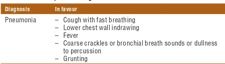
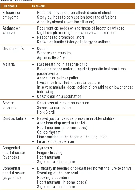
diffi culty in breathing 77
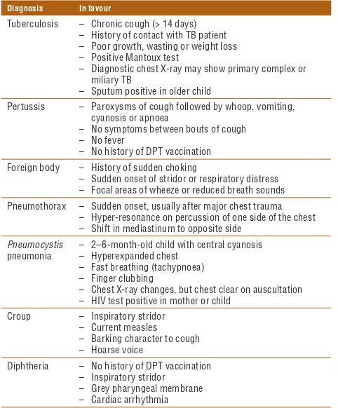
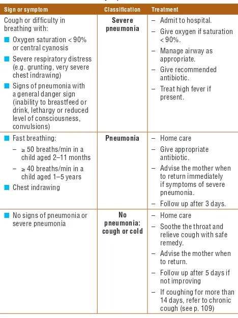
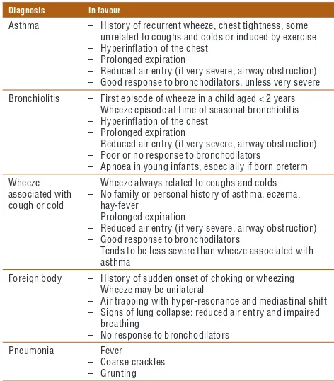
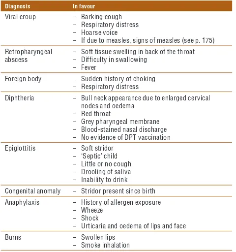
Table 7. Classifi cation of the severity of pneumonia 81 Table 8. Differential diagnosis in a child presenting with wheeze 93 Table 9. Differential diagnosis in a child presenting with stridor 103 Table 10. Differential diagnosis in a child presenting with
chronic cough 110
Table 11. Differential diagnosis in a child presenting with diarrhoea 127 Table 12. Classifi cation of the severity of dehydration in children
with diarrhoea 128
Table 13. Administration of intravenous fl uids to a severely
dehydrated child 130
Table 14. First diet for persistent diarrhoea: a starch-based,
reduced-milk (low-lactose) diet 141
Table 15. Second diet for persistent diarrhoea: a reduced-starch (cereal) no-milk (lactose-free) diet 141 Table 16. Differential diagnosis of fever without localizing signs 151 Table 17. Differential diagnosis of fever with localized signs 152 Table 18. Differential diagnosis of fever with rash 153 Table 19. Additional differential diagnosis of fever lasting longer
than 7 days 155
Table 20 WHO criteria for the diagnosis of rheumatic fever
(based on the revised Jones criteria) 194 Table 21. Time frame for the management of a child with
severe acute malnutrition 201
Table 22. Volumes of F-75 per feed for malnourished children
(approximately 130 ml/kg per day) 211 Table 23. WHO paediatric clinical staging system for HIV infection 231 Table 24. Classes of antiretroviral drugs recommended for use
in children 234
xiv
Table 25. First-line treatment regimens for children 234 Table 26. Common side-effects of antiretroviral drugs 236 Table 27. Recommended second-line treatment regimens
for children 240
Table 28. Endotracheal tube size by age 259 Table 29. Blood volume of children by age 260 Table 30. Normal pulse rate and blood pressure in children 261 Table 31. Examples of local adaptations of feeding
recommendations on the mother’s card in Bolivia, Indonesia, Nepal, South Africa and the
United Republic of Tanzania 303
Table 32. Maintenance fl uid requirements 304 Table 33. Primary vaccination schedule for infants recommended
in the Expanded Programme on Immunization 326 Table A2.1 Drug dosage by surface area (m2) of the child 354 Table A5.1.1 Weight-for-age from birth to 5 years: Boys 379 Table A5.1.2 Weight-for-age from birth to 5 years: Girls 381 Table A5.2.1 Weight-for-length from birth to 2 years: Boys 386 Table A5.2.2 Weight-for-length from birth to 2 years: Girls 391 Table A5.2.3 Weight-for-height from 2 to 5 years: Boys 395 Table A5.2.4 Weight-for-height from 2 to 5 years: Girls 399
xv
Preface
This is the second edition of the World Health Organization (WHO) Pocket book of hospital care for children, which was fi rst published in 2005. It is a compila-tion of the updated WHO guidelines for the management of common childhood illnesses at the fi rst-referral level in low-resource countries. It presents relevant, up-to-date, evidence-based clinical guidelines that can be used by clinicians in their daily work in hospitals with basic laboratory facilities and inexpensive medicines. The guidelines focus on inpatient management of children who are severely ill with conditions that are major causes of childhood mortality, such as neonatal illness, pneumonia, diarrhoea, fever (mainly malaria, meningitis and septicaemia), severe acute malnutrition and HIV/AIDS. It also includes guidance on common surgical problems, appropriate supportive care and monitoring of patients on the ward.
The Pocket book is part of a series of tools for improving the quality of care for severely ill children and is consistent with the Integrated Management of Childhood Illness (IMCI) guidelines for outpatient management of sick chil-dren. It is for use by doctors, senior nurses and other senior health workers who are responsible for the care of young children at the fi rst referral level in developing countries.
The fi rst edition of the Pocket book was reviewed by a WHO guidelines steering committee, which identifi ed those chapters that required updating, comprising: • revisions to align the Pocket book with recently published, WHO-approved
guidelines; and
• priorities for which new information had become available, which was col-lated, analysed and synthesized before updating.
xvi
All the changes were reviewed by external clinical experts and were approved by the WHO Guidelines Review Committee. A web version of the Pocket book
will be updated regularly as new evidence with clinical implications emerges. Printed editions will be published every 5 years if there are substantial new changes. Users are therefore advised to check the WHO web site regularly for
Pocket book updates (http://www.who.int/maternal_child_adolescent/en/). The main changes in the second edition are listed below.
Chapters unchanged from the fi rst edition of the Pocket book (2005): Chapters with only editorial changes or reorganization but with no major update of previous information:
• Chapter 1. Triage and emergency conditions • Chapter 2. Diagnostic approaches to the sick child • Chapter 5. Diarrhoea
• Chapter 9. Common surgical problems • Chapter 11. Monitoring the child’s progress • Chapter 12. Counselling and discharge from hospital • Annexes 1, 3 and 6
Chapters substantially changed from the fi rst edition of the
Pocket book (2005):
Chapters with substantial changes to clinical guidance or which have been restructured are:
• Chapter 3. Problems of the neonate and young infant • Chapter 4. Cough or diffi culty in breathing • Chapter 6. Fever
xvii Additional sections or subsections in this second edition
Several sections of some chapters were added or substantially expanded in response to demand from users:
• Chapter 1, section 1.10. Trauma and injuries • Chapter 3, section 3.7. Convulsions or fi ts
• Chapter 3, section 3.11.3. Respiratory distress syndrome • Chapter 4, section 4.6.3. Epiglottitis
• Chapter 4, section 4.6.4. Anaphylaxis • Chapter 4, section 4.9. Rheumatic heart disease • Chapter 6, section 6.11. Rheumatic fever
• Chapter 8, section 8.5. Prevention of mother to child HIV transmission, and infant feeding
xviii
Acknowledgements
WHO expresses its gratitude to the following members of the group that up-dated the guidelines, people who made original contributions, and reviewers, institutions and consultants for their contributions to updating the Pocket book of hospital care for children.
Guideline development group
WHO thanks the members of the guideline development group who reviewed most of the evidence and made recommendations for updating the Pocket book
and also those who reviewed the chapters: Dr Fizan Abdullah, Johns Hopkins University School of Medicine, USA; Shinjini Bhatnagar, All India Institute of Medical Sciences, India; Bridget Wills, Clinical Research Unit, University of Oxford Centre for Tropical Diseases, Viet Nam; Harry Campbell, University of Edinburgh Medical School, United Kingdom; Leonila Dans, University of Philippines, Philippines; Trevor Duke, Centre for International Child Health, University of Melbourne, Australia; Michael English, University of Nairobi and Kenya Medical Research Institute, Kenya; Andy Gray, University of KwaZulu-Natal, South Africa; Sandra Grisi, São Paulo University, Brazil; Stuart Macleod, University of British Columbia, Canada; Hilda Mujuru, University of Zimbabwe, Zimbabwe; Susan Niermeyer, University of Colorado, USA; Jesca Nsungwa, Ministry of Health, Uganda; Vinod Paul, All India Institute of Medical Sci-ences, India; Haroon Saloojee, Witwatersrand University, South Africa; Mathu Santosham, Johns Hopkins School of Public Health, USA; Giorgio Tamburlini, Institute of Child Health, Italy; and Anita Zaidi, Aga Khan University, Pakistan. Special gratitude is owed to Rhona MacDonald, Maternal Child Health Advo-cacy International, who incorporated the changes and prepared the fi rst draft.
Original contributors and external reviewers
WHO coordinated the international contributions for the 2005 edition of the
xix Dr Giorgio Tamburlini (Italy), Dr Bridget Wills (Viet Nam) and Fabienne Jäger (Switzerland).
WHO wishes to acknowledge the following for comments and contributions made at various stages of the Pocket book updating: Sabrina Bakeere-Kitaka, Makerere Medical School, Uganda; Zulfi qar Bhutta, Aga Khan University, Pakistan; Stephen W. Bickler, University of California-San Diego, USA; Uday Bodhankar, Commonwealth Association for Health and Disability, United Kingdom; Adegoke Falade, College of Medicine, University of Ibadan, Nigeria; Jeremy Farrar, Centre for Tropical Medicine, Ho Chi Minh City, Viet Nam; Julian Kelly, Royal Children’s Hospital, Centre for International Child Health, Melbourne, Australia; Carolyn Maclennan, Flinders University, Australia; Rhona MacDonald, David Southall and Barbara Phillips, Maternal Child Health Advo-cacy International; Amha Mekasha, Addis Ababa University, Ethiopia; Elizabeth Molyneux, College of Medicine, Malawi; Maria Asuncion Silvestre, University of the Philippines, Manila, Philippines, Joan Skinner, Victoria University of Wellington, New Zealand and Andrew Steer, Royal Children’s Hospital, Centre for International Child Health, Melbourne, Australia.
Valuable input was provided by several WHO clusters and the departments of Family, Women’s and Children’s Health, Health Systems and Services, HIV/AIDS, Tuberculosis, Neglected Tropical Diseases, Noncommunicable Diseases, and Mental Health. We particularly acknowledge the WHO staff who participated as members of the Guidelines Steering Committee or who contributed to and reviewed various draft chapters: Desta Teshome, WHO Regional Offi ce for Africa; Meena Cherian, Essential Health Technologies; Tarun Dua, Mental Health and Substance Abuse; Lisa Nelson, Martina Penazzato, and Sandra Gove, HIV/ AIDS; Malgorzata Grzemska, Stop TB; Emmalita Manalac, WHO Regional Of-fi ce for the Western PaciOf-fi c; Peter Olumese, Global Malaria Programme; Ma del Carmen Casanovas, Zita Weise Prinzo and Chantal Gegout, Nutrition for Health and Development; Susan Hill and Clive Ondari, Essential Medicines and Pharmaceutical Policies; Raman Velayudhan, Neglected Tropical Diseases; and Martin Weber, WHO Country Offi ce, Indonesia.
Special thanks to Rami Subhi at the Centre for International Child Health in Australia, who helped in collating the evidence for recommendations for updating the Pocket book.
xx Institutions
We are grateful to the following institutions for providing input and support during the review of the Pocket book: Centre for International Child Health, University of Melbourne, Australia; University of Edinburgh, Scotland; Kenya Medical Research Institute, Kenya; Asociación Colaboración Cochrane Iber-oamericana, Spain; Aga Khan University, Pakistan; Institute of Child Health Burlo Garofolo, Italy; University of Malawi, Malawi; Capital Institute of Pae-diatrics, China; University of Western Australia, Australia; and Instituto de Medicina Integral Professor Fernando Figueira, Brazil.
xxi
Abbreviations
AIDS acquired immunodefi ciency syndrome ART antiretroviral therapy
AVPU alert, responding to voice, responding to pain, unconscious (simple consciousness scale)
BCG bacille Calmette-Guérin CSF cerebrospinal fl uid DPT diphtheria, pertussis, tetanus
EVF erythrocyte volume fraction (haematocrit) Hb haemoglobin
HIV human immunodefi ciency virus IM intramuscular (injection), intramuscularly IMCI Integrated Management of Childhood Illness IV intravenous (injection), intravenously MDR multidrug-resistant
NNRTI non-nucleoside reverse transcriptase inhibitor NRTI nucleoside reverse transcriptase inhibitor NSAID non-steroidal anti-infl ammatory drug ORS oral rehydration salt(s)
PCP Pneumocystis carinii pneumonia ReSoMal rehydration solution for malnutrition SD standard deviation
TB tuberculosis
WHO World Health Organization
Symbols
xxii
Chart 1. Stages in the management of a sick child
admitted to hospital: key elements
TRIAGE
(present) • Check for emergency signs
(absent)
• Check for priority signs or conditions
Give emergency
treatment until stable
HISTORY AND EXAMINATION
(including assessment of vaccination status, nutritional status and feeding) • Check children with emergency and priority conditions fi rst.
Laboratory and other investigations, if required
List and consider differential diagnoses
Select main diagnoses (and secondary diagnoses)
Plan and begin inpatient treatment (including supportive care)
Monitor for signs of — improvement — complications — failure of treatment.
Plan and begin outpatient
treatment.
Arrange follow-up, if required.
(not improving or new problem) (improving)
Reassess for causes of failure of
treatment.
Revise treatment.
Continue treatment.
Plan discharge.
Discharge home.
Arrange continuing care or
follow-up at hospital or in
1
1.
T
R
IA
G
E
CHAPTER 1
T riage and emergency
conditions
1.1 Triage 2
1.2 Summary of steps in emergency triage assessment and treatment 3
1.3 Assessment of emergency and priority signs 4
Triage of all sick children 5
How to manage a choking infant or child 7
How to manage the airway in a child with obstructed breathing 9
How to give oxygen 11
How to position the unconscious child 12
Give IV fl uids for shock in a child without severe acute malnutrition 13 Give IV fl uids for shock in a child with severe acute malnutrition 14
Give diazepam rectally 15
Give IV glucose 16
Treat severe dehydration in an emergency setting 17 1.4 Emergency treatment for a child with severe malnutrition 19 1.5 Diagnostic considerations for children with emergency conditions 20
1.5.1 Child presenting with an airway or severe breathing
problem 20
1.5.2 Child presenting with shock 21
1.5.3 Child presenting with lethargy, unconsciousness
or convulsions 23
1.6 Common poisoning 26
1.6.1 Principles for ingested poisons 27
1.6.2 Principles for poisons in contact with skin or eyes 29
1.6.3 Principles for inhaled poisons 29
1.6.4 Specifi c poisons 29
Corrosive compounds 29
Petroleum compounds 30
Organophosphorus and carbamate compounds 30
Paracetamol 31
Aspirin and other salicylates 31
Iron 32
Morphine and other opiates 32
Carbon monoxide 33
2
1.
T
R
IA
G
E
1.1 Triage
Triage is the process of rapidly screening sick children soon after their arrival in hospital, in order to identify:
– those with emergency signs, who require immediate emergency treatment; – those with priority signs, who should be given priority in the queue so that
they can be assessed and treated without delay; and
– non-urgent cases, who have neither emergency nor priority signs. Emergency signs include:
■obstructed or absent breathing
■severe respiratory distress
■central cyanosis
■signs of shock (cold hands, capillary refi ll time longer than 3 s, high heart rate with weak pulse, and low or unmeasurable blood pressure)
■coma (or seriously reduced level of consciousness)
■convulsions
■signs of severe dehydration in a child with diarrhoea (lethargy, sunken eyes, very slow return after pinching the skin or any two of these).
Children with these signs require immediate emergency treatment to avert death.
The priority signs (see p. 6) identify children who are at higher risk of dying. These children should be assessed without unnecessary delay. If a child has one or more emergency signs, don’t spend time looking for priority signs. TRIAGE
1.7 Drowning 33
1.8 Electrocution 34
1.9 Common causes of envenoming 34
1.9.1 Snake bite 34
1.9.2 Scorpion sting 37
1.9.3 Other sources of envenoming 38
1.10 Trauma and injuries 38
1.10.1 Primary survey or initial assessment 38
3
1.
T
R
IA
G
E
1.2
Summary of steps in emergency triage assessment
and treatment
Steps in emergency triage assessment and treatment are summarized in the charts on pp. 5–17.
First check for emergency signs in three steps:
• Step 1. Check whether there is any airway or breathing problem; start im-mediate treatment to restore breathing. Manage the airway and give oxygen. • Step 2. Quickly check whether the child is in shock or has diarrhoea with severe dehydration. Give oxygen and start IV fl uid resuscitation. In trauma, if there is external bleeding, compress the wound to stop further blood loss. • Step 3. Quickly determine whether the child is unconscious or convulsing. Give IV glucose for hypoglycaemia and/or an anti-convulsant for convulsing. If emergency signs are found:
• Call for help from an experienced health professional if available, but do not delay starting treatment. Stay calm and work with other health workers who may be required to give the treatment, because a very sick child may need several treatments at once. The most experienced health professional should continue assessing the child (see Chapter 2, p. 41), to identify all underlying problems and prepare a treatment plan.
• Carry out emergency investigations (blood glucose, blood smear, haemoglo-bin [Hb]). Send blood for typing and cross-matching if the child is in shock, appears to be severely anaemic or is bleeding signifi cantly.
• After giving emergency treatment, proceed immediately to assessing, diagnosing and treating the underlying problem.
Tables of common differential diagnoses for emergency signs are provided from p. 21 onwards.
If no emergency signs are found, check for priority signs:
■Tiny infant: any sick child aged < 2 months
■Temperature: child is very hot
■Trauma or other urgent surgical condition
■Pallor (severe)
■Poisoning (history of)
■Pain (severe)
■Respiratory distress
■Restless, continuously irritable or lethargic
4
1.
T
R
IA
G
E
■Referral (urgent)
■Malnutrition: visible severe wasting
■Oedema of both feet
■Burns (major)
The above can be remembered from the mnemonic 3TPR MOB.
These children need prompt assessment (no waiting in the queue) to determine what further treatment is needed. Move a child with any priority sign to the front of the queue to be assessed next. If a child has trauma or other surgical problems, get surgical help where available.
1.3
Assessment of emergency and priority signs
■Assess the airway and breathing (A, B)Does the child’s breathing appear to be obstructed? Look at the chest wall movement, and listen to breath sounds to determine whether there is poor air movement during breathing. Stridor indicates obstruction.
Is there central cyanosis? Determine whether there is bluish or purplish dis-coloration of the tongue and the inside of the mouth.
Is the child breathing? Look and listen to determine whether the child is breathing.
Is there severe respiratory distress? The breathing is very laboured, fast or gasping, with chest indrawing, nasal fl aring, grunting or the use of auxiliary muscles for breathing (head nodding). Child is unable to feed because of respiratory distress and tires easily.
■Assess circulation (for shock) (C)
Children in shock who require bolus fl uid resuscitation are lethargic and have cold skin, prolonged capillary refi ll, fast weak pulse and hypotension.
Check whether the child’s hand is cold. If so, determine whether the child is in shock.
Check whether the capillary refi ll time is longer than 3 s. Apply pressure to whiten the nail of the thumb or the big toe for 5 s. Determine the time from the moment of release until total recovery of the pink colour.
If capillary refi ll is longer than 3 s, check the pulse. Is it weak and fast? If the radial pulse is strong and not obviously fast, the child is not in shock. If you cannot feel the radial pulse of an infant (< 1 year old), feel the brachial pulse or, if the infant is lying down, the femoral pulse. If you cannot feel the radial pulse of a child, feel the carotid.
5
Chart 2. Triage of all sick children
Emergency signs:If any sign is positive, call for help, assess and resuscitate, give treatment(s), draw blood for emergency laboratory investigations (glucose, malaria smear, Hb)
CHART 2. TRIAGE OF ALL SICK CHILDREN
TREAT
Do not move neck if a cervical spine injury is possible, but open the airway.
ASSESS
■ Severe respiratory distress
If no foreign body aspirated
왘 Manage airway (Chart 4) giving fl uids rapidly (Chart 7).
If peripheral IV cannot be inserted, insert an intraosseous or external jugular line (see pp. 340, 342).
If not lethargic or unconscious:
왘 Give glucose orally or by nasogastric tube. 왘 Proceed immediately to full
6
E CHART 2. TRIAGE OF ALL SICK CHILDREN
Chart 2. Triage of all sick children
Emergency signs:If any sign is positive: call for help, assess and resuscitate, give treatment(s), draw blood for emergency laboratory investigations (glucose, malaria smear, Hb)
PRIORITY SIGNS
These children need prompt assessment and treatment
ASSESS TREAT
Do not move neck if you suspect cervical spine injury, but open the airway.
Coma/ 왘 If convulsing, give diazepam rectally
(Chart 9)
왘 Position the unconscious child (if head or neck trauma is suspected, stabilize the neck fi rst) (Chart 6). 왘 Give IV glucose (Chart 10).
왘 Make sure the child is warm. If no severe malnutrition: 왘 Insert an IV line and begin giving
fl uids rapidly following Chart 11 and diarrhoea treatment plan C in hospital (Chart 13, p. 131).
If severe malnutrition: 왘 Do not insert an IV line. 왘 Proceed immediately to full
assessment and treatment (see section 1.4, p. 19). ■ Trauma or other urgent surgical
condition ■ Pallor (severe) ■ Poisoning (history of) ■ Pain (severe) ■ Respiratory distress
■ Restless, continuously irritable, or lethargic ■ Referral (urgent)
■ Malnutrition: visible severe wasting ■ Oedema of both feet or face ■ Burns (major)
Note: If a child has trauma or other surgical problems, get surgical help or follow surgical guidelines.
NON-URGENT
7
1.
T
R
IA
G
E
CHART 3. HOW TO MANAGE A CHOKING INFANT
Chart 3. How to manage a choking infant
Chest thrusts
왘 Lay the infant on your arm or thigh in a head-down position.
왘 Give fi ve blows to the middle of the infant’s back with the heel of the hand. 왘 If obstruction persists, turn
the infant over and give fi ve chest thrusts with two fi ngers on the lower half of the sternum.
왘 If obstruction persists, check infant’s mouth for any obstruction that can be removed.
왘 If necessary, repeat sequence with back slaps.
8
1.
T
R
IA
G
E CHART 3. HOW TO MANAGE A CHOKING CHILD
Chart 3. How to manage a choking child (> 1 year of age)
Heimlich manoeuvre for a choking older child
Administer back blows to clear airway obstruction in a choking child. 왘 Give fi ve blows to the middle of the
child’s back with the heel of the hand, with the child sitting, kneeling or lying.
왘 If the obstruction persists, go behind the child and pass your arms around the child’s body; form a fi st with one hand immediately below the child’s sternum; place the other hand over the fi st and pull upwards into the abdomen (see diagram); repeat this Heimlich manoeuvre fi ve times.
왘 If the obstruction persists, check the child’s mouth for any obstruction that can be removed.
왘 If necessary, repeat this sequence with back blows.
9
CHART 4. HOW TO MANAGE THE AIRWAY IN A CHILD
Chart 4. How to manage the airway in a child with
obstructed breathing (or who has just stopped
breathing)
A: When no neck trauma is suspected
■ OLDER CHILD
Look, listen and feel for breathing
Child conscious 1. Inspect mouth and
remove foreign body, if present.
2. Clear secretions from the throat.
keep it tilted and lift chin to open airway.
2. Inspect mouth and remove foreign body if present and easily visible.
3. Clear secretions from the throat. 4. Check the airway
by looking for chest movements, listening for breath sounds and feeling for breath (see diagram).
■ INFANT
Neutral position to open the airway in an infant
10
1.
T
R
IA
G
E CHART 4. HOW TO MANAGE THE AIRWAY IN A CHILD
Chart 4. How to manage the airway in a child with
obstructed breathing (or who has just stopped
breathing)
B: When neck trauma or cervical spine injury is suspected: jaw thrust
Use jaw thrust if airway are still not open. Place the fourth and fi fth fi ngers behind the angle of the jaw and move it upwards so that the bottom of the jaw is thrust forwards, at 90° to the body
1. Stabilize the neck as shown in Chart 6, and open the airway. 2. Inspect mouth and remove foreign body, if present.
3. Clear secretions from throat under direct vision.
4. Check the airway by looking for chest movements, listening for breath sounds and feeling for breath.
11
1.
T
R
IA
G
E
Chart 5. How to give oxygen
CHART 5. HOW TO GIVE OXYGEN
Give oxygen through nasal prongs or a nasal catheter.
■ NASAL PRONGS
왘 Place the prongs just inside the nostrils and secure with tape.
■NASAL CATHETER
왘 Use an 8 French gauge size tube
왘 Measure the distance from the side of the nostril to the inner eyebrow margin with the catheter.
왘 Insert the catheter as shown in the diagram.
왘 Secure with tape.
12
1.
T
R
IA
G
E
Chart 6. How to position an unconscious child
CHART 6. HOW TO POSITION AN UNCONSCIOUS CHILD■If neck trauma is suspected:
왘 Stabilize the child’s neck and keep the child lying on the back.
왘 Tape the child’s forehead and chin to the sides of a fi rm board to secure this position.
왘 Prevent the neck from moving by supporting the child’s head (e.g. using litre bags of IV fl uid on each side). 왘 If the child is vomiting,
turn on the side, keeping the head in line with the body.
■If neck trauma is not suspected:
왘 Turn the child on the side to reduce risk of aspiration.
왘 Keep the neck slightly extended, and stabilize by placing cheek on one hand.
13
Chart 7. How to give intravenous fl uids to a child in
shock without severe malnutrition
왘 Check that the child is not severely malnourished, as the fl uid volume and rate are different. (Shock with severe malnutrition, see Chart 8.) 왘 Insert an IV line (and draw blood for emergency laboratory
investigations).
왘 Attach Ringer’s lactate or normal saline; make sure the infusion is running well.
왘 Infuse 20 ml/kg as rapidly as possible.
Age (weight) Volume of Ringer’s lactate or normal saline solution (20 ml/kg)
Reassess the child after the appropriate volume has run in. Reassess
after fi rst infusion:
• If no improvement, repeat 10–20 ml/kg as rapidly as possible.
• If no improvement with signs of dehydration (as in profuse diarrhoea or cholera), repeat 20 ml/kg of Ringer’s lactate or normal saline.
• If no improvement, with suspected septic shock, repeat 20 ml/kg and consider adrenaline or dopamine if available (see Annex 2, p. 353).
• If no improvement, see disease-specifi c treatment guidelines. You should have established a provisional diagnosis by now.
After improvement at any stage (pulse volume increases, heart rate slows, blood pressure increases by 10% or normalizes, faster capillary refi ll < 2 s), go to Chart 11, p. 17.
Note:In children with suspected malaria or anaemia with shock, rapid fl uid infusion must be administered cautiously, or blood transfusion should be given in severe anaemia instead.
14
1.
T
R
IA
G
E
Chart 8. How to give intravenous fl uids to a child in
shock with severe malnutrition
Give this treatment only if the child has signs of shock (usually there will also be a reduced level of consciousness, i.e. lethargy or loss of consciousness): 왘 Insert an IV line (and draw blood for emergency laboratory investigations). 왘 Weigh the child (or estimate the weight) to calculate the volume of fl uid to be given. 왘 Give IV fl uid at 15 ml/kg over 1 h. Use one of the following solutions according to
availability:
– Ringer’s lactate with 5% glucose (dextrose);
– Half-strength Darrow’s solution with 5% glucose (dextrose); – 0.45% NaCl plus 5% glucose (dextrose).
Weight Give over 1 h (15 ml/kg)Volume of IV fl uid Weight Give over 1 h (15 ml/kg)Volume of IV fl uid
4 kg 60 ml 12 kg 180 ml
6 kg 90 ml 14 kg 210 ml
8 kg 120 ml 16 kg 240 ml
10 kg 150 ml 18 kg 270 ml
왘 Measure the pulse rate and volume and breathing rate at the start and every 5–10 min.
If there are signs of improvement (pulse rate falls, pulse volume increases or respiratory rate falls) and no evidence of pulmonary oedema
– repeat IV infusion at 15 ml/kg over 1 h; then
– switch to oral or nasogastric rehydration with ReSoMal at 10 ml/kg per h up to 10 h (see p. 204);
– initiate re-feeding with starter F-75 (see p. 209).
If the child fails to improve after two IV boluses of 15 ml/kg,
– give maintenance IV fl uid (4 ml/kg per h) while waiting for blood;
– when blood is available, transfuse fresh whole blood at 10 ml/kg slowly over 3 h (use packed cells if the child is in cardiac failure); then
– initiate re-feeding with starter F-75 (see p. 209); – start IV antibiotic treatment (see p. 207).
If the child deteriorates during IV rehydration (breathing rate increases by 5/min and pulse rate increases by 15/min, liver enlarges, fi ne crackles throughout lung fi elds, jugular venous pressure increases, galloping heart rhythm develops), stop the infusion, because IV fl uid can worsen the child’s condition by inducing pulmonary oedema.
15
Chart 9. How to give diazepam rectally
■ Give diazepam rectally:
왘 Draw up the dose from an ampoule of diazepam into a tuberculin (1-ml) syringe. Base the dose on the weight of the child, when possible. Then remove the needle.
왘 Insert the syringe 4–5 cm into the rectum, and inject the diazepam solution.
왘 Hold the buttocks together for a few minutes.
Age (weight)
a Use phenobarbital (200 mg/ml solution) at a dose of 20 mg/kg to control convulsions
in infants < 2 weeks of age:
Weight 2 kg – initial dose, 0.2 ml; repeat 0.1 ml after 30 min If convulsions Weight 3 kg – initial dose, 0.3 ml; repeat 0.15 ml after 30 min continue
If convulsions continue after 10 min, give a second dose of diazepam (or give diazepam IV at 0.05 ml/kg = 0.25 mg/kg if IV infusion is running).
Do not give more than two doses of diazepam.
If convulsions continue after another 10 min, suspect status epilepticus:
왘 Give phenobarbital IM or IV at 15 mg/kg over 15 min; or
왘 Phenytoin at 15–18 mg/kg IV (through a different line from diazepam) over 60 min. Ensure a very good IV line, as the drug is caustic and will cause local damage if it extravasates.
■ If high fever:
왘 Undress the child to reduce the fever.
왘 Do not give any oral medication until the convulsion has been controlled (danger of aspiration).
왘 After convulsions stop and child is able to take orally, give paracetamol or ibuprofen.
Warning: Always have a working bag and mask of appropriate size available in case the patient stops breathing, especially when diazepam is given.
16
Chart 10. How to give glucose intravenously
왘 Insert an IV line, and draw blood for emergency laboratoryinvestigations.
왘 Check blood glucose with a glucose monitoring stick. If the level is < 2.5 mmol/litre (45 mg/dl) in a well-nourished or < 3 mmol/litre (54 mg/dl) in a severely malnourished child or if blood glucose cannot be measured as no stick test is available, treat as for hypoglycaemia: 왘 Give 5 ml/kg of 10% glucose solution rapidly by IV injection
Age (weight)
Volume of 10% glucose solution as bolus (5 ml/kg)
If the child is unable to feed without danger of aspiration, give: – milk or sugar solution via a nasogastric tube (to make sugar solution,
dissolve four level teaspoons of sugar (20 g) in a 200-ml cup of clean water), or
– IV fl uids containing 5–10% glucose (dextrose) (see Annex 4, p. 377)
Note: 50% glucose solution is the same as 50% dextrose solution.
If only 50% glucose solution is available: dilute one part 50% glucose solution in four parts sterile water, or dilute one part 50% glucose solution in nine parts 5% glucose solution. For example, 10 ml 50% solution with 90 ml 5% solution gives 100 ml of approximately a 10% solution.
Note: To use blood glucose stick tests, refer to instructions on box. Generally, the strip must be stored in its box at 2–3 °C, avoiding sunlight or high humidity. A drop of blood should be placed on the strip (it should cover all the reagent area). After 60 s, the blood should be washed off gently with drops of cold water and the colour compared with the key on the bottle or on the blood glucose reader. (The exact procedure varies for different strips.)
Note: Sublingual sugar may be used as an immediate ‘fi rst aid’ measure in managing hypoglycaemia if IV access is impossible or delayed. Place one level teaspoonful of sugar moistened with water under the tongue every 10–20 min.
17
1.
T
R
IA
G
E
Chart 11. How to treat severe dehydration in an
emergency after initial management of shock
For children with severe dehydration but without shock, refer to diarrhoea treatment plan C, p. 131.If the child is in shock, fi rst follow the instructions in Charts 7 and 8 (pp. 13 and 14). Switch to the chart below when the child’s pulse becomes slower or capillary refi ll is faster.
왘 Give 70 ml/kg of Ringer’s lactate (Hartmann’s) solution (or, if not available, normal saline) over 5 h to infants (aged < 12 months) and over 2.5 h to children (aged 12 months to 5 years).
Total volume IV fl uid (volume per hour) Weight Age < 12 months
Give over 5 h
Age 12 months to 5 years Give over 2.5 h
< 4 kg 200 ml (40 ml/h) –
4–6 kg 350 ml (70 ml/h) –
6–10 kg 550 ml (110 ml/h) 550 ml (220 ml/h)
10–14 kg 850 ml (170 ml/h) 850 ml (340 ml/h)
14–19 kg – 1200 ml (480 ml/h)
Reassess the child every 1–2 h. If the hydration status is not improving, give the IV drip more rapidly.
Also give oral rehydration salt (ORS) solution (about 5 ml/kg per h) as soon as the child can drink, usually after 3–4 h (in infants) or 1–2 h (in children).
Weight Volume of ORS solution per hour
< 4 kg 15 ml
4–6 kg 25 ml
6–10 kg 40 ml
10–14 kg 60 ml
14–19 kg 85 ml
Reassess after 6 h for infants and after 3 h for children. Classify dehydration. Then choose the appropriate plan A, B or C (pp. 138, 135, 131) to continue treatment.
If possible, observe the child for at least 6 h after rehydration to be sure that the mother can maintain hydration by giving the child ORS solution by mouth.
18
1.
T
R
IA
G
E
If the room is very cold, rely on the pulse to determine whether the child is in shock.
Check whether the systolic blood pressure is low for the child’s age (see Table below). Shock may be present with normal blood pressure, but very low blood pressure means the child is in shock.
Normal blood pressure ranges in infants and children
Age Systolic blood pressure
Premature 55–75
0–3 months 65–85
3–6 months 70–90
6–12 months 80–100
1–3 years 90–105
3–6 years 95–110
■Assess for coma or convulsions or other abnormal mental status (C)
Is the child in coma? Check the level of consciousness on the ‘AVPU’ scale: A alert,
V responds to voice, P responds to pain, U unconscious.
If the child is not awake and alert, try to rouse the child by talking or shaking the arm. If the child is not alert but responds to voice, he or she is lethargic. If there is no response, ask the mother whether the child has been abnormally sleepy or diffi cult to wake. Determine whether the child responds to pain or is unresponsive to a painful stimulus. If this is the case, the child is in coma (unconscious) and needs emergency treatment.
Is the child convulsing? Are there spasmodic repeated movements in an unresponsive child?
■Assess the child for severe dehydration if he or she has diarrhoea
Does the child have sunken eyes? Ask the mother if the child’s eyes are more sunken than usual.
Does a skin pinch go back very slowly (longer than 2 s)? Pinch the skin of the abdomen halfway between the umbilicus and the side for 1 s, then release and observe.
19
1.
T
R
IA
G
E
■Assess for priority signs
While assessing the child for emergency signs, you will have noted several possible priority signs:
Is there any respiratory distress (not severe)?
Is the child lethargic or continuously irritable or restless?
This was noted when you assessed for coma. Note the other priority signs (see p. 6).
1.4
Emergency treatment for a child with
severe malnutrition
During triage, all children with severe malnutrition will be identifi ed as having priority signs, which means that they require prompt assessment and treatment. A few children with severe malnutrition will be found during triage assessment to have emergency signs.
Those with emergency signs for ‘airway and breathing’ or ‘coma or convulsions’ should receive emergency treatment accordingly (see charts on pp. 5–17). • Those with signs of severe dehydration but not in shock should not be
rehy-drated with IV fl uids, because severe dehydration is diffi cult to diagnose in severe malnutrition and is often misdiagnosed. Giving IV fl uids puts these children at risk of over-hydration and death from heart failure. Therefore, these children should be rehydrated orally with the special rehydration solu-tion for severe malnutrisolu-tion (ReSoMal). See Chapter 7 (p. 204).
• In severe malnutrition, individual emergency signs of shock may be pre-sent even when there is no shock. Malnourished children with many signs of shock: lethargy, reduced level of consciousness, cold skin, prolonged capillary refi ll and fast weak pulse, should receive additional fl uids for shock as above.
• Treatment of a malnourished child for shock differs from that for a well-nourished child, because shock from dehydration and sepsis are likely to coexist, and these are diffi cult to differentiate on clinical grounds alone, and because children with severe malnutrition may not cope with large amounts of water and salt. The amount of fl uid given should be guided by the child’s response. Avoid over-hydration. Monitor the pulse and breathing at the start and every 5–10 min to check whether they are improving. Note that the type of IV fl uid differs for severe malnutrition, and the infusion rate is slower.
All severely malnourished children require prompt assessment and treatment to deal with serious problems such as hypoglycaemia, hypothermia, severe
20
infection, severe anaemia and potentially blinding eye problems. It is equally important to take prompt action to prevent some of these problems, if they were not present at the time of admission to hospital.
1.5
Diagnostic considerations for children with
emergency conditions
The following text provides guidance for approaches to the diagnosis and dif-ferential diagnosis of presenting conditions for which emergency treatment has been given. After you have stabilized the child and provided emergency treatment, determine the underlying cause of the problem, in order to provide specifi c curative treatment. The following lists and tables are complemented by the tables in the disease-specifi c chapters.
1.5.1 Child presenting with an airway or severe breathing problem
History
• Onset of symptoms: slow or sudden • Previous similar episodes • Upper respiratory tract infection • Cough and duration in days • History of choking
• Present since birth or acquired
• Vaccination history: diphtheria, pertussis, tetanus (DPT), measles • Known HIV infection
• Family history of asthma
Examination
• Cough and quality of cough • Cyanosis
• Respiratory distress • Grunting
• Stridor, abnormal breath sounds • Nasal fl aring
• Swelling of the neck • Crepitations
21
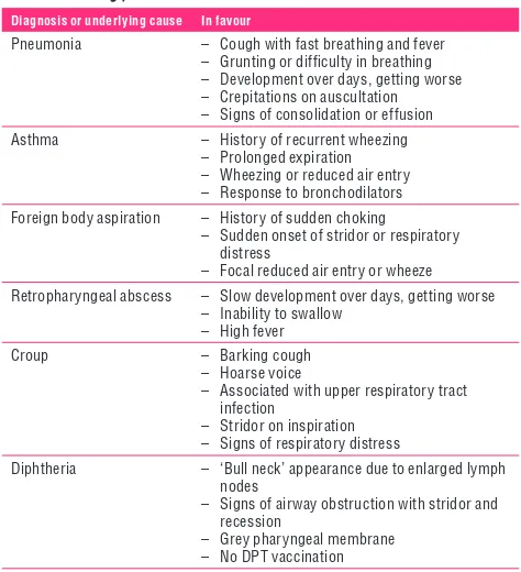
Table 1. Differential diagnosis in a child presenting with an airway or severe breathing problem
Diagnosis or underlying cause In favour
Pneumonia – Cough with fast breathing and fever – Grunting or diffi culty in breathing – Development over days, getting worse – Crepitations on auscultation – Signs of consolidation or effusion
Asthma – History of recurrent wheezing
– Prolonged expiration – Wheezing or reduced air entry – Response to bronchodilators
Foreign body aspiration – History of sudden choking
– Sudden onset of stridor or respiratory distress
– Focal reduced air entry or wheeze
Retropharyngeal abscess – Slow development over days, getting worse – Inability to swallow
– High fever
Croup – Barking cough
– Hoarse voice
– Associated with upper respiratory tract infection
– Stridor on inspiration – Signs of respiratory distress
Diphtheria – ‘Bull neck’ appearance due to enlarged lymph nodes
– Signs of airway obstruction with stridor and recession
– Grey pharyngeal membrane – No DPT vaccination
1.5.2 Child presenting with shock
History
• Acute or sudden onset • Trauma
• Bleeding
• History of congenital or rheumatic heart disease • History of diarrhoea
• Any febrile illness
22 • Cold or warm extremities
• Neck veins (elevated jugular venous pressure) • Pulse volume and rate
• Blood pressure • Liver size increased • Petaechiae • Purpura
CHILD PRESENTING WITH SHOCK
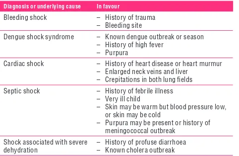
Table 2. Differential diagnosis in a child presenting with shock
Children with shock are lethargic, have fast breathing, cold skin, prolonged capillary refi ll, fast weak pulse and may have low blood pressure as a late sign. To help make a specifi c diagnosis of the cause of shock, look for the signs below.
Diagnosis or underlying cause In favour
Bleeding shock – History of trauma – Bleeding site
Dengue shock syndrome – Known dengue outbreak or season – History of high fever
– Purpura
Cardiac shock – History of heart disease or heart murmur – Enlarged neck veins and liver
– Crepitations in both lung fi elds
Septic shock – History of febrile illness – Very ill child
– Skin may be warm but blood pressure low, or skin may be cold
– Purpura may be present or history of meningococcal outbreak
Shock associated with severe dehydration
23
1.
T
R
IA
G
E
1.5.3 Child presenting with lethargy, unconsciousness or convulsions
History
• Fever • Head injury
• Drug overdose or toxin ingestion
• Convulsions: How long do they last? Have there been previous febrile convulsions? Epilepsy?
In the case of an infant < 1 week old, consider history of: • birth asphyxia
• birth injury to the brain
Examination
General • Jaundice
• Severe palmar pallor
• Peripheral or facial oedema (suggesting renal failure) • Level of consciousness
• Petaechial rash • Blood pressure
• Determine AVPU score (see p. 18).
Head and neck • Stiff neck
• Signs of head trauma or other injuries • Pupil size and reactions to light • Tense or bulging fontanelle
• Abnormal posture, especially opisthotonus (arched back).
The coma scale score should be monitored regularly. In young infants < 1 week old, note the time between birth and the onset of unconsciousness. Other causes of lethargy, unconsciousness or convulsions in some regions of the world include malaria, Japanese encephalitis, dengue haemorrhagic fever, measles encephalitis, typhoid and relapsing fever.
Laboratory investigations
• If meningitis is suspected and the child has no signs of raised intracranial pressure (unequal pupils, rigid posture, paralysis of limbs or trunk, irregular breathing), perform a lumbar puncture.
24
E CHILD PRESENTING WITH LETHARGY, UNCONSCIOUSNESS OR CONVULSIONS
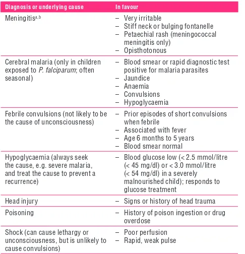
Table 3. Differential diagnosis in a child presenting with lethargy, unconsciousness or convulsions
Diagnosis or underlying cause In favour
Meningitisa,b – Very irritable
– Stiff neck or bulging fontanelle – Petaechial rash (meningococcal
meningitis only) – Opisthotonous
Cerebral malaria (only in children exposed to P. falciparum; often seasonal)
– Blood smear or rapid diagnostic test positive for malaria parasites – Jaundice
– Anaemia – Convulsions – Hypoglycaemia
Febrile convulsions (not likely to be the cause of unconsciousness)
– Prior episodes of short convulsions when febrile
– Associated with fever – Age 6 months to 5 years – Blood smear normal
Hypoglycaemia (always seek the cause, e.g. severe malaria, and treat the cause to prevent a recurrence)
– Blood glucose low (< 2.5 mmol/litre (< 45 mg/dl) or < 3.0 mmol/litre (< 54 mg/dl) in a severely malnourished child); responds to glucose treatment
Head injury – Signs or history of head trauma
Poisoning – History of poison ingestion or drug
overdose
Shock (can cause lethargy or unconsciousness, but is unlikely to cause convulsions)
– Poor perfusion – Rapid, weak pulse
• In a malarious area, perform a rapid malaria diagnostic test and prepare a blood smear.
• If the child is unconscious, check the blood glucose. If not possible, then treat as hypoglycaemia; if the level of consciousness improves, presume hypoglycaemia.
25
CHILD PRESENTING WITH LETHARGY, UNCONSCIOUSNESS OR CONVULSIONS
Table 3. Continued
Diagnosis or underlying cause In favour
Acute glomerulonephritis with encephalopathy
– Raised blood pressure – Peripheral or facial oedema – Blood and/or protein in urine – Decreased or no urine
Diabetic ketoacidosis – High blood sugar
– History of polydipsia and polyuria – Acidotic (deep, laboured) breathing
a The differential diagnosis of meningitis may include encephalitis, cerebral abscess or
tuber-culous meningitis. Consult a standard textbook of paediatrics for further guidance.
b A lumbar puncture should not be done if there are signs of raised intracranial pressure (see
section 6.3, p. 167 and A1.4, p. 346). A positive lumbar puncture may show cloudy cerebrospinal fl uid (CSF) on direct visual inspection, or CSF examination shows an abnormal number of white cells (usually > 100 polymorphonuclear cells per ml in bacterial meningitis). Confi rmation is given by a low CSF glucose (< 1.5 mmol/litre), high CSF protein (> 0.4 g/litre), organisms identifi ed by Gram staining or a positive culture.
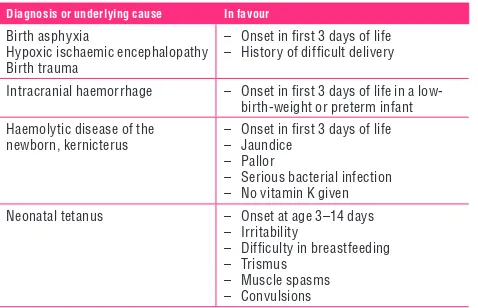
Table 4. Differential diagnosis in a young infant (< 2 months) presenting with lethargy, unconsciousness or convulsions
Diagnosis or underlying cause In favour
Birth asphyxia
Hypoxic ischaemic encephalopathy Birth trauma
– Onset in fi rst 3 days of life – History of diffi cult delivery
Intracranial haemorrhage – Onset in fi rst 3 days of life in a low-birth-weight or preterm infant
Haemolytic disease of the newborn, kernicterus
– Onset in fi rst 3 days of life – Jaundice
– Pallor
– Serious bacterial infection – No vitamin K given
Neonatal tetanus – Onset at age 3–14 days
– Irritability
– Diffi culty in breastfeeding – Trismus
26
1.
T
R
IA
G
E COMMON POISONING
Table 4. Continued
Diagnosis or underlying cause In favour
Meningitis – Lethargy
– Apnoeic episodes – Convulsions – High-pitched cry – Tense or bulging fontanelle
Sepsis – Fever or hypothermia
– Shock (lethargy, fast breathing, cold skin, prolonged capillary refi ll, fast weak pulse, and sometimes low blood pressure)
– Seriously ill with no apparent cause
For poisoning and envenomation see below and p. 34.
1.6 Common
poisoning
Suspect poisoning in any unexplained illness in a previously healthy child. Consult standard textbook of paediatrics for management of exposure to specifi c poisons and/or any local sources of expertise in the management of poisoning, for example a poison centre. Only the principles for managing inges-tion of few common poisons are given here. Note that tradiinges-tional medicines can be a source of poisoning.
Diagnosis
A diagnosis is based on a history from the child or carer, a clinical examination and the results of investigations, where appropriate.
■Obtain full details of the poisoning agent, the amount ingested and the time of ingestion. Attempt to identify the exact agent involved and ask to see the container, when relevant. Check that no other children were involved. The symptoms and signs depend on the agent ingested and therefore vary widely – see below.
■Check for signs of burns in or around the mouth or of stridor (upper airway or laryngeal damage), which suggest ingestion of corrosives.