http://dx.doi.org/10.12988/pacs.2015.41011
Supplementation of
Cantharanthus Roseus
Leaf
Extract as Anti-Inflammatory
Substance to Hypercholesterolemic Wistar
Derived-Rats
Sri Wahjuni1, Sri Rahayu Santi1 and A. A Ngurah Gunawan2
1Department of Chemistry
Faculty of Mathematics and Natural Science Udayana University, Bali, Indonesia
1Department of Chemistry
Faculty of Mathematics and Natural Science Udayana University, Bali, Indonesia
2Department of Physics
Faculty of Mathematics and Natural Science Udayana University, Bali, Indonesia
Copyright © 2014 Sri Wahjuni, Sri Rahayu Santi and A. A Ngurah Gunawan. This is an open access article distributed under the Creative Commons Attribution License, which permits unrestricted use, distribution, and reproduction in any medium, provided the original work is properly cited.
Abstract
supplemented with 0% CRLE; Group 2: 10% CRLE; Group 3: 15% CRLE; Group 4: 20% CRLE; and Group 5: 25% CRLE. CRLE supplementation was last for 1 month. TNF- αconcentration was determined using Sandwich Enzyme (ELISA) kit spectrophotometer (at π =500 nm) andconcentration of IL-6 was determined Cantharanthus roseus grown as ornament plant and contain tannin, tripernoids, and alkaloids. The component that acts an anti-inflammation is flavanoids.
Keywords: Leaf of Cantharanthus roseus, tumor necrosis factor (TNF), interleukin-6 (IL-6), anti-inflammation, ELISA
Introduction
In Indonesia, research work on traditional medicine is still preferable for combating animal diseases and human diseases. One of herbal plants that have been proved beneficial for curing various diseases is Cantharanthus roseus (C. roseus). It grown as ornament plants and its local name is “tapak dara”. As reported from many countries such as India and China, extract of C. roseus leaf can be used to cure many diseases (Singh and Singh, 2001). It has been discovered that extract leaf of C. roseus contains tannin, trypernoid, alkaloid, and flavonoids. Flavonoids occur in the form of glycoside and it has been shown in vivo that flavonoids may inhibit activity of NF-kB (nuclear factor kappa-B), leading to inhibition of inflammation reaction (Han et al., 2007).
Inflammation is a complex condition that starts from tissue damaging. Damage of the tissues are due to endogen factors (such as tissue necrosis) and to external factors such as contact with foreign particles or infections. Inflammation is generally a kind of response of vascular living tissues, that followed by important endothelium process. Process occurring in the endothelium of blood vessel lead in turn to development of atherosclerosis (Hosaya et al., 2005).Atherosclerosis may become the consequence of hypercholesterolemia, also called dyslipidemia – the increase in lipid content in the serum. Low density lipoprotein (LDL) particularly the oxidized form may significantly cause damage of the endothelium. On the other hand, high density lipoprotein (HDL) is the protective component (Robbin and Cataren, 2002). Furthermore, chronic hypercholesterolemia may result in cardiovascular diseases.
effect of supplementation of extract leaf of herbal plant C. roseus as anti-inflammatory substance, based on determination of pro-anti-inflammatory cytokines (TNF- α and IL-6) in the serum of experimental rats that underwent determination of TNF- α, Quintikine HS immunoassay kit for IL-6 determination, EDTA, and diethyl ether. Serum collected from the experimental animals were kept at -200C until assays were conducted. Prior to assays, serum samples were centrifuged at 2000 rpmfor 10 minutes.
Methods
Experimental design employed was a randomized pre-test and post-test. The experimental animals were divided into 5 groups according to the treatment applied, namely Group 1 (control): supplemented with 0% CRLE; Group 2: 10% CRLE; Group 3: 15% CRLE; Group 4: 20% CRLE; and Group 5: 25% CRLE.Prior to supplementation of CRLE, all experimental animals were initially raised under normal rat pellets for 1 month. Then, they are all subjected to high fat ration for 6 weekin order to make them suffering from hypercholesterolemia.Such feeding treatment has been shown to result in hypercholesterolemia and chronic inflamationin rats (Wahjuni,2011). At the end of the high fat feeding phase, concentrations of TNF-α and IL-6 were measured and the values were considered as the pre-test data. Supplementation of the various dosages of CRLE to the experimental animals lasted for 6 weeks. At the end of this phase, concentrations of TNF-α and IL-6 were again measured and the data was considered as the post-test values
The data was analyzed descriptively. Data selection including editing, coding and tabulation was performed using Windows file navigator statistical program (Triton, 2006; Pramesti, 2007). For normality analysis of TNF-α and IL -6 data, Shapiro Wilk test was employed at 5% level of significant (p = 0.05). H0 hypothesis: frequency of observation/ frequency of expectation. Based on this test, the data was considered to be normally distributed when P > 0.05. Moreover,
variant homogeneity was analyzed using the Levent’s test.Ho hypothesis:
Result and discussion
Decrease in TNF-α concentration
The average level of TNF-α measured from blood of 50 male rats suffering from hypercholesterolemia at pre-test and post-test was presented at Table 1. The pre-test TNF-α values for control group, Group 1, Group 2, Group 3 and Group 4
were (28.98 ± 6.00); (29.12 ± 5.79);(29.02 ± 5.34); (28.62 ± 4.72); dan (29,02 ±
5.06) pg/mLrespectively. Meanwhile, the post-test values for control group,
Group 1, Group 2, Group 3 and Group 4 were (28.11 ± 5.94); (27.32 ±5.01);(24.42 ± 5.74); (15,56 ±7,20); ( 26,02 ± 8,34 )pg/mL respectively. When
the pre-test and post-test values were compared, there was no significant reduction in concentration of TNF-α for the control group. On the other hand, supplementation of CRLE at 10%, 15%, 20% and 25% has resulted in decreasing level of TNF-α. Supplementation of CRLE at 10% and 15% led to reduction of TNF-α level up to 0.79 and 3.69 pg/ml, respectively. However, statistical analysis showed that such differences were not significant (P> 0.05). But a significant difference (P < 0.05) in TNF-α concentrations between the pre-test and post-test values was noted for rats subjected to supplementation of 20% CRLE (Group 3). For this group, TNF-α concentration has decreased at amount of 12.55 pg/ml. Furthermore, for rats under 25% CRLE supplementation, no significant different (P > 0.05) in TNF-α concentration was observed.
Tablel 1
Decrease level of TNF-α serum Wistar Rat Hiperkolesterolemia
Treatment Observation TNF-α (pg /mL) Pre-test Pos-test
Inflammation is generally a response to external and internal factors by vascular living tissues. The inflammation response is usually followed by an important process – endothelium process. Endothelium of blood vessel is the site where the deposition of fat occurs and known as atherosclerosis. Endothelium is the primary target for mechanical and chemical injuries caused by hypercholesterolemia. Chronic hypercholesterolemia has been known to result in the shift of acute pro-inflammation and prothrombosis responses into the chronic responses. These responses, in turn, are followed by infiltration of leucocytes, particularly of monocyte, to sub-endothelium, and subsequentlychanging into macrophage cells. The macrophage cells then phagocyte the remnant of LDL which is oxidized to form atheroma (Baraas, 2006).
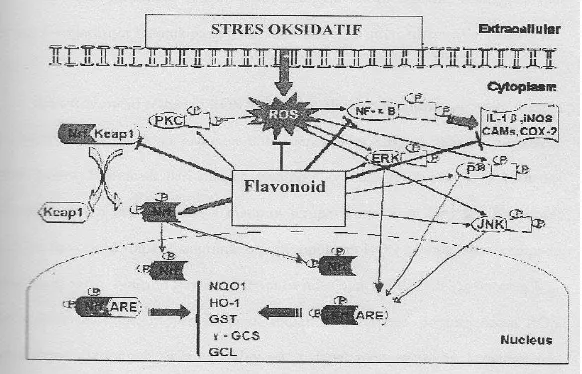
Simopolous (2002) reported that flavonoids as anti-inflammation play role as immunomodulator. Mechanismeis increas antioxtidan endogen and capcing free radikal.Inhibition stres oksidatif by some flavonoid incontain of cantharanthus roseus Leaf Extract (Figure 1). And than flavonoid cantharanthus roseus isolasitian metabolit sekunder compound, extraction, distilation, purification, dan identificationby GC-MS(Gas Chromatograph Mass Spectrometer) Shimadzu QP2010, identifay flavonoid what typeCantharanthus roseus Leaf Extract.
Figure 1: Habition Flavonoid 0f NF-kB (Nuclear Factor Kappa Beta)was decreas habition interleukin product pro inflamation, COX2 can habition reaction inflamation.
Decrease in IL-6 concentration
Concentrations of interleukin-6 (IL-6) in blood serum of rats suffering from hypercholesterolemia and then treated with CRLE at pre-test and post-test are presented in Table 2. The pre-test IL-6 concentrations for group supplemented with 0% CRLE (control), with 10%, 15%, 20%, and with 25% CRLE were
(134.58 ± 2.21); (134.24 ± 2.64); (134.75 ± 2.51); (134.64 ± 1.98); and (135.34 ±
4.57) pg/mL. Respectively. Moreover, their post-test concentration were(133,15 ±
4,01); (130,28 ± 3,59); (127,20 ± 5,56); (113,87 ± 4,30); and (120,87 ± 7,89)
pg/mL,respectively. Thus, the suplementation of CRLE to rats suffering from hypercholesterolemia, seperti juga terlihat pada Tabel 2 bahwa terjadi penurunan kadar IL-6 darah tikus Wistar Hypercholesterolemia.
Table 2
Decrease in IL-6 concentration in rats suffering from hypercholesterolemia following supplementation of CRLE
Treatment Observation IL-6(pg/mL)
Pre-test Pos-test
EDCR 0% (Kontrol) 134,58 ± 2,21 133,15 ± 4,01 EDCR 10 % 134,24 ± 2,64 130,28 ± 3,59 EDCR 15 % 134,75 ± 2,51 127,20 ± 5,56 EDCR 20 % 134,64 ± 1,98 113,87 ± 4,30 EDCR 25 % 135,34 ± 4,57 120,87 ± 7,89
Extract leaf of Cantharanthus roseus contains flavonoids in abundance that have anti-inflammatory property. When endothelium cells undergo inflammatory activation, there are increase in expression of selectin, VCAM-1 and ICAM-1 that promote adhesion of monocyte. Expression for molecule adhesion is induced by pro-inflammatory cytokines such as IL-1Band TNF-α ,by acute-phase protein CRP (produced in the liver as a response to IL-6), by protease activated receptor signaling, by uptake of oxLDL through oxLDL receptor-1 (LOX-1), and by interaction of CD40/CD40 ligands in tunica intima of the arteries (Bonetti et al., 2003).
Conclusion
Supplementation of Cantharanthus roseus leaf extract (CRLE) at 20% may significantly reduce the concentration of TNF-α in rats suffering from hypercholesterolemia that can be related with inflammation condition. As TNF-α is considered as a marker for the occurrence of inflammation, CRLE may be regarded as having anti-inflammatory property. Likewise, such dosage of CRLE (at 20%) may be used to reduce the concentration of cytokine interleukin-6 (IL-6) which is also considered as a marker for inflammation.
References
[1] Anonim, Tapak Dara. Wikipedia Bahasa Indonesia. Indonesia: Wikimedia Foundation Inc, 2009.
[2] Baraas - Faisa, kardiologi Molekuler Radikal Bebas, Disfungsi EndotelAterosklerosis, Antioksidan.Latihan Fisik dan Rehabilitasi Jantung. Bagian Kadiologi FKUI/ RS Jantung Harapan Kita, Jakarta, 2006.
[3] Bonetti, P. O.,et.al., Endothelial Dysfuction: A markerof Aterosclerosis Risk. Aterisoscler Thromb Vase Biol 23, (2003), 168-175.
http://dx.doi.org/10.1161/01.atv.0000051384.43104.fc
[4] Chen, G., and Goeddel, D. V., TNFR-1 Signaling:A Beautiful Pathway. Science. 296 (2002), 1634 – 1635. http://dx.doi.org/10.1126/science.1071924
[5] Coico, R., S.. J. and Benyamin, E., Element of Innate and Aquiered Immunity Immunology, In: Immunology Ashort Course 5th ed. New Jersey, John Wiley &Sons, (2003), 11 - 26.
[6] Goldberg A. C. Dyslipidemia (Hypercholesteolemia). The Mecrk Manual America, 2008.
[7] Han, S. N., Leka, L. S., Lichtenstein, A. H., Ausman, L. M., Schaefer, E. J., and Meydani, S. N., Effect of Hydrogenated and Saturated, Relative to Polyunsaturated, Fat on Immune and Inflammatory Responses of Adults with Moderate Hypercholesterolemia. Journal of Lipid Research. 43,( 2002),445 - 52.
[9] Hosoya, T.,Maruyama,A.,Kang,M.I.,Kawatani, .,Shibata,T.,Uchida,K.,Itoh,K., Yamamoto, M., Diffrential Response of the Nrf2 – Keap1 system to Laminar and Oscillatory Shear Stresses in Endothelial cells.The journal of Biochemistry chemistry. 29, ( 2005),27244 - 27250. http://dx.doi.org/10.1074/jbc.m502551200
[10] Kusningrum, R., Dasar Rancangan Percobaan dan Rancangan Acak Lengkap. Universitas Airlangga Surabaya, (1989), 20 - 21.
[11] Murray, K. Harper Biochemestry, twenty sixth edition. Mc Graw Hill Companie; New York, 2004.
[12] Natarajan V, Venugopal PV, Menon T., Effect of azadirachta (neem) on the growth pattern of dermatophytes. Indian J MedMicrobiol, 21, (2003), 98 - 101.
[13] Nayak B. S, Vinutha B, Geetha B, Sudha B., Experimental evaluation of Pentas lanceolata for Wound healing activity in rats. Fitotherapia, 76, (2006), 671 - 675. http://dx.doi.org/10.1016/j.fitote.2005.08.007
[14] Pramesti, G., AplikasiSPSS 15.0 dalamModel Linear Statistik.Jakarta Penerbit PT Alex Media Komputindo, 2007.
[15] Robbin and Cotran., Dasar Patologis Penyakit (terjemahan), Penerbit Buku Kedokteran, Sumber Agung podomoro Jakarta, 2002.
[16] Schultz, G. S., Ladwig, G., and Wysocki, A., Extracellular matrix: review of its roles in acute and chronic wounds. World Wide Wound, 2005.
[17] Shivananda Nayak B: Cecropia peltata L (Cecropiaceae) Has Wound Healing potential-A preclinical study in Sprague Dawley Rat model. International Journal of Lower Extremity Wounds, 5, (2006), 20 - 26.
http://dx.doi.org/10.1177/1534734606286472
[18] Singh SN, Vats P, Suri S, Shyam R, Kumria MML, Ranganathan S and Sridharan K., Effect of an antidiabetic extract of Catharanthus roseus on enzymic activities in streptozotocin induced diabetic rats. J Ethnopharmacol, 76, (2001), 269 - 77. http://dx.doi.org/10.1016/s0378-8741(01)00254-9
[19] Szmitko, P. E.,Wang, C. H.,Weisel,R. D., De-Almmeida, J. R.,Anderson, T. J., and Verma, S., New Marker of Inflammation and Endothelial Cell Activation.Circulation,108,( 2003), 1917 – 1923.
[21]Wahjuni, S, Asupan Minyak Ikan Lemuru (Sardinella longiceps) Melalui Penurunan TNF-α, IL-6,LDL, dan MDA, serta Peningkatan HDL Pada Tikus Wistar. Thesis Pasca Sarjana Universitas Udayana, 2011.