Newcastle Disease Virus Infection Study on Duck and Chicken in Subang
District
Panus A1,2, Setiyaningsih S3, Mayasari NLPI4
1Graduate of Microbiology Medic, Department of Animal Disease and Veterinary, Faculty of Veterinary Medicine, Bogor Agricultural University
2Laboratory of Virology, Institute of Veterinary Subang, Directorate General of Livestock and Veterinary Medicine, Ministry of Agriculture Jl. Terusan Garuda Blok Wepurebredari RT. 03 RW. 22 Subang, Jawa Barat
3Laboratory of Virology, Section of Microbiology Medic, Department of Animal Disease dan Veterinary, Faculty of Veterinary Medicine, Bogor Agricultural University, Gedung Fakultas Kedokteran Hewan Jl. Agatis Kampus IPB Dramaga, Bogor 16680 Jawa Barat
4Laboratory of Imunology, Section of Microbiology Medic, Department of Animal Disease dan Veterinary, Faculty of Veterinary Medicine, Bogor Agricultural University, Gedung Fakultas Kedokteran Hewan Jl. Agatis Kampus IPB Dramaga, Bogor 16680 Jawa Barat
E-mail: [email protected]
(received 12-01-2015; revised 13-03-2015; accepted 17-03-2015)
ABSTRAK
Panus A, Setiyaningsih S, Mayasari NLPI. 2015. Kajian infeksi virus Newcastle Disease pada bebek dan ayam di Kabupaten Subang. JITV 20(2): 134-147. DOI: http://dx.doi.org/10.14334/jitv.v20i2.1168
Tujuan dari penelitian ini adalah untuk mendeteksi dan mengetahui keragaman antigenik virus Newcastle disease (NDV) yang bersirkulasi di Kabupaten Subang. Sampel usapan kloaka, usapan orofaring dan serum diambil dari 393 ekor ayam dan 149 bebek dari penampungan, peternakan dan pasar unggas di 10 kecamatan di Kabupaten Subang. Screening NDV pada sampel pool (5-7 individu per pool) dengan real-time Reverse-Transcription Polymerase Chain Reaction (rRT-PCR) matrix (M) menunjukkan 19 dari 67 pool kloaka (28,3%) dan 8 dari 67 pool orofaring (11,9%) ayam terdeteksi NDV; 18 pool dari 67 pool ayam (26,9%) menunjukkan virus diekskresikan melalui kloaka dan orofaring. Sementara pada sampel itik, NDV terdeteksi hanya pada kloaka yaitu 8 dari 30 pool (26,7%). Total 18 isolat berhasil diisolasi dari sampel usapan kloaka dan usapan orofaring individu yang menunjukkan karakter antigenik yang homogen, namun beberapa isolat menunjukkan variasi dengan titre sampai 2 Log2 menggunakan antisera LaSota dan 2-4 Log2 dengan antisera Komarov. Mayoritas isolat menunjukkan afinitas lebih tinggi terhadap antisera Komarov, yang mengindikasikan semua isolat adalah NDV galur ganas. Karakterisasi patogenisitas dengan uji elusi hasilnya menunjukkan 3 isolat masuk ke kelompok galur mesogenik dan 15 isolat ke kelompok galur velogenik, sedangkan dengan rRT-PCR fusion (F) menunjukkan 100% isolat merupakan galur ganas (mesogenik/velogenik). Deteksi antibodi spesifik terhadap NDV pada 408 serum dengan uji HI menunjukkan 48 serum (12%) positif dengan kisaran titre 1 sampai 8 Log2; hanya sekitar 13% ayam yang divaksin menunjukkan titre protektif (≥3 Log2). Newcastle disease masih endemik di Kabupaten Subang dengan variasi antigenik galur virus yang bersirkulasi relatif tidak telalu bervariasi.
Kata Kunci: Newcastle Disease, rRT-PCR, Virulensi, Keragaman Antigenik, Antibodi
ABSTRACT
Panus A, Setiyaningsih S, Mayasari NLPI. 2015. Newcastle Disease Virus infection study on duck and chicken in Subang district. JITV 20(2): 56-69 DOI:http://dx.doi.org/10.14334/jitv.v20i2.1168
The objectives of this research were to study Newcastle Disease Virus (NDV) infection in Subang area and to examine the diversity of the circulating NDV. Swabs of cloacal and oropharynx, and serum were sampled from total of 393 chickens and 149 ducks in backyard farms and live bird markets located in 10 subdistricts. Screening of NDV in pool of 5-7 samples by real-time Reverse-Transcription Polymerase Chain Reaction (rRT-PCR) matrix (M) showed 19/67 (28.3%) cloacal and 8/67 (11.9%) pharyngeal pools of chicken samples; 18/67 (26.9%) of the pools excreted virus via cloaca and oropharynx, while the duck pools of 8/30 (26.7%) shed virus from cloaca. Virus isolation attempted on individual sample from positive pools yielded 18 isolates which the majority of the isolates showed homogeneous antigenic character, only some of these showed variations up to 2 Log2 with Lasota and 4 Log2 with Komarov antisera. Majority of isolates had a higher affinity to Komarov indicating their propencity to virulent strains. Pathogenicity examination using elution test showed 3 isolates virus were grouped to mesogenic strains and 15 isolates to velogenic strain, in agreement with rRT-PCR fusion results. HI test on 408 sera showed that NDV antibody was detected in 48 (12%) birds with titres ranging from 1 to 8 Log2; only about 13% of vaccinated chickens demonstrated protective
antibody titre (≥3 Log2). Newcastle disease is still endemic in Subang with relatively low antigenic variation among circulating strains.
INTRODUCTION
Newcastle Disease (ND) is one of serious diseases in poultry because it is very contagious, spread rapidly and attack some species of birds at all age. Mostly this outbreak attacks intensive poultry as follows: chicken, turkey, duck, quail, and pigeons. ND spread all over the world and potentially causes economy losses in poultry industry. In addition of poultry, this disease infect and causes death in wild birds as well. ND cases were firstly found and reported in the mid of 1920 in Indonesia (Java Island) and England (OIE 2012), then spreading in a few years later and becoming endemic in many countries (Ashraf & Shah 2014). Nowadays, almost all regions in Indonesia are affected and no one area or island is free from ND. In spite of mortality rate caused by ND was controlable, the effect in production is still a problem. Moreover, the impact of other losses is the costs for controlling the disease and also stopping export from ND endemic countries (Brown et al. 1999).
The outbreak caused by ND can be acute or chronic and infecting all species of birds especially chicken, both domestic and purebred. The outbreak occured in the field may caused by various strain of ND virus. According to the severity-level of the outbreak in chicken, Newcastle Disease Virus (NDV) was classified into three pathotypes namely lentogenic, mesogenic, and velogenic. Velogenic strain is distinguished into neurotropic and viscerotropic form (Aldous & Alexander 2001).
The loss caused by ND are morbidity and mortality which in infected poultry the rate may reach 100% caused by velogenic strain especially in sensitive chicken groups and under 10% in mesogenic strain (OIE 2008). In the developing countries where the livestock industry is growing very rapidly, the losses affected by NDV outbreak are not only mortality but also expenditure additionaly cost used for vaccination, biosecurity and depopulation. Even the free ND countries, have to spend on periodic testing in order to maintain free status from ND which needed for trading license. Moreover, in the developing countries as endemic ND, the impacts are not only economy losses but also affecting health and socioeconomic condition of lower-class society, whose quality and quantity of eggs and meat consumed decreased caused by ND (Alexander & Senne 2008). In 2002, ND outbreak in California, United States caused losses 200.000.000 US$ as an impact of depopulation (Kapczynski & King 2005). The losses affected by ND in layer are mortality and reduction of egg production, while causing growth disorder and reduction of body weight in boiler. Data of OIE (2009) showed in 2007, about 1500-8000 chickens were infected by ND every month in Indonesia. Moreover, according to Xiao (2012) in 2009 and 2010, ND outbreak occured in comercial chicken in Indonesia
causing 70-80% mortality. ND is still become a major problem in the poultry industry despite the vaccination carried out routinely (Samal 2011). Therefore, ND is a serious threat for poultry in Indonesia. Subang area in West Java is one of buffer zones of poultry production, particularly for broilers and layers. Totally 44.049.739 poultry population was reported in 2013 (DISNAK properly. Annually survey by Balai Penyidikan dan Pengujian Veteriner (BPPV) Subang in unvaccinated ND backyard birds in 2011, found 10 out of 131 serums tested were positive of ND with titre range 2-5 Log2 (BPPV 2011). In 2012, 12 out of 37 serums tested were positive of ND with titre range 1-4 Log2 (BPPV 2012), and in 2013, 184 out of 359 serums tested were positive of ND with titre range 2-8 Log2 (BPPV 2013). These results show that ND is still endemic in Subang area. As the basis of consideration for efective control measures and prevention, it is neccesary to conduct NDV isolation and detection of antibody against ND in ducks and chickens in Subang area.
For the time being, investigation of ND in Subang area is still limited. Commonly, the diagnosis was based on clinical symptoms, pathological alteration and serological test. Therefore, diagnostic technique with high sensitivity to detect and confirm NDV infection in ducks and chickens in Subang area is required.
MATERIALS AND METHODS
Samples
Samples were taken from 10 subdistricts in Subang, that were Binong, Ciasem, Cipendeuy, Cipunagara, Compreng, Pagaden, Pusaka Nagara, Subang, Sukasari and Tambak Dahan. These areas were selected because population of fowls were centralized in those locations (market, shelter, farm) and endemic area of ND as well.
Standard antigen and antisera and Kit
Collection of swabs and serum samples
Swabs of cloacal and oropharynx were taken from chickens and ducks from the bird’s shelter, livebirds market and poultry farms in 10 areas in Subang using sterile cotton swabs inserted in microtube 2 ml contains
Brain Heart Infusion Broth (BHIB). The temperature was kept cool (4-8°C) until arrival in the laboratorium.
Pooling swabs of cloacal and oropharynx samples consist of 5-7 individual samples in each pool was based on swab types, birds, location and time of sampling. The sample pool was subsequently used for rRT-PCR test using primer matrix (M). Blood was collected via branchial vein from each individu along with swab samples.
real time Reverse Transcription Polymerase Chain Reaction (rRT-PCR) Test
To detect the presence of genetic material of NDV extracted from swabs of cloacal and oropharynx, rRT-PCR test with NVSL protocol (2005) was done. RNA virus isolation was extracted based on QIAamp® Viral RNA Mini Kit (Qiagen) standard procedure. rRT-PCR amplification using Ag-Path IDTM One-Step RT-PCR kit from Life Technologies in Applied Biosystems 7500 Real Time PCR System was conducted. Cycles of rRT-PCR was performed in 45°C for 10 minutes, 95°C for 10 minutes, 95°C for 10 minutes, 56°C for 32 seconds, and 72°C for 10 seconds. The result was analyzed by
Applied Biosystems 7500 Real time PCR
SystemSoftware Version 1.4.0. Primer and probe were used are presented in Table 1.
Virus isolation in SPF embryonated chicken egg
Swabs of cloacal and oropharynx samples used as inoculum were from individual bird sample from positive rRT-PCR M pool. As much as 0.2 ml inoculum
containing penicillin-streptomycin (9:1) and incubated for 30 minutes at ambient temperature (25-27°C) was injected in allantoic cavity of Specific Pathogen Free (SPF) embryonated chicken egg. Eggs were incubated in incubator at 37°C for 4-7 days and observed 3 times a day to check the viability of embryo (OIE 2012). Isolates obtained from alantoic liquid were reconfirmed with rRT-PCR matrix (M).
Hemagglutination (HA) and Hemagglutination Inhibition (HI) Test
Procedures of HA and HI test was done by micro methods (OIE 2012), performed by adding 25 µl 0.85%
phosphate buffered saline (PBS) into micro plate in 1st-12th pit using micro pipet. In the 1st pit 25 µl serum standard was added and diluted, than moved into 2nd-11th pit. A total of 25 µl 4HAU ND virus suspension was added into each 1st-10th and 12th pit, and then incubated at ambient temperatute for 15 minutes and 25 µl suspension of 1% red blood cells (rbc) was added into 1st-12th pit, homogenized and incubated at ambient temperature (25-27°C) for 40 minutes. Positive result marked by occurance of resistance hemagglutination in form of precipitation of rbc on the bottom of micro plate pit. Titre of HI was determined based on the highest serum dilution that was still showed precipitation (agglutination inhibition). HA and HI test were performed 3 times.
Elution-time test
Test was performed based on Ezeibe & Ndip (2005) procedures. A total of 50 µl PBS solution was put into micro plate pit, then 50 µl virus suspension was added into 1st pit, and diluted into 1st-10th pit. As much as 50 µl PBS was added into each 1st-12nd pit, followed by 50 µl suspension of 0.6% rbc into 1st-12nd pit, homogenized and incubated at ambient temperature for
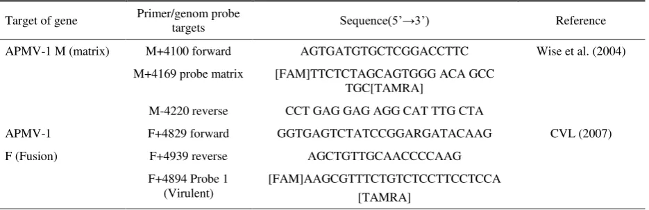
Table 1. Primers and Probes used for rRT-PCR matrix (M) and fusion (F)
Target of gene Primer/genom probe targets Sequence(5’→3’) Reference
APMV-1 M (matrix) M+4100 forward AGTGATGTGCTCGGACCTTC Wise et al. (2004)
M+4169 probe matrix [FAM]TTCTCTAGCAGTGGG ACA GCC
TGC[TAMRA]
M-4220 reverse CCT GAG GAG AGG CAT TTG CTA
APMV-1 F+4829 forward GGTGAGTCTATCCGGARGATACAAG CVL (2007)
F (Fusion) F+4939 reverse AGCTGTTGCAACCCCAAG
F+4894 Probe 1 (Virulent)
40 minutes, then hemagglutination was observed. Elution time was determined based on time of complete hemagglutination was observed on highest dilution until the precipitation of rbc showed (elution). Elution-time test was performed 3 times.
Data analysis
Data were analyzed descriptively and statistically to determine standard mean deviation (SD). The average of antibody titre was calculated by geometric mean titre (GMT) by the formula:
Log2 GMT = (Log2 t1)(S1) + (Log2 t1)(S1) + … + (Log2tn)(Sn) N
Information: N = Number of observed serum T = Antibody titre at the highest
dilution (which was still may inhibit agglutination of red blood cell)
S = Number of titrated serum n = Antibody titre of the nth
sample
Coefficient of variation/CV from immune response was expressed by following formula:
Detection of NDV in pool of swabs of cloacal and oropharynx with rRT-PCR matrix (M)
There were 542 samples succesfully collected consist of 149 ducks and 393 chickens sample comprised of 108 broilers, 148 broiler parent stocks, 15 layers and 122 lokal chickens from 10 areas in Subang district.
Testing for 97 pools swabs of cloacal resulted in 27 pools (29%) of positive which spreaded in 9 areas, 8 pools (7%) oropharynx swabs positive spreaded in 3 areas and 18 pools (18%) of cloacal and oropharynx swabs positive spreaded in 6 areas while none was positive from Cipendeuy area (Table 2). ND virus was only detected in cloacal swabs of ducks (8/30 pools), 19
pools cloacal swabs, 8 pools oropharynx swabs and 18 pools cloacal and oropharynx swabs (Table 1). Ducks tend to excrete the virus via cloaca according to the findings reported by Saepulloh & Darminto (2005) which were 14 (13%) isolates from cloacal and none from oropharynx swabs of 106 ducks in Kalimantan.
The highest number of positive M cloacal swabs pools (7) were obtained from Tambak Dahan area. The highest number of positive M oropharynx swabs pools (4) were obtained from Cipunagara area and the highest number of positive M cloacal and oropharynx swab pools (8) were obtained from Binong area, while in Cipendeuy area in pool of cloacal or oropharynx swabs, NDV were not detected.
Isolation of NDV in SPF embryonated chicken eggs
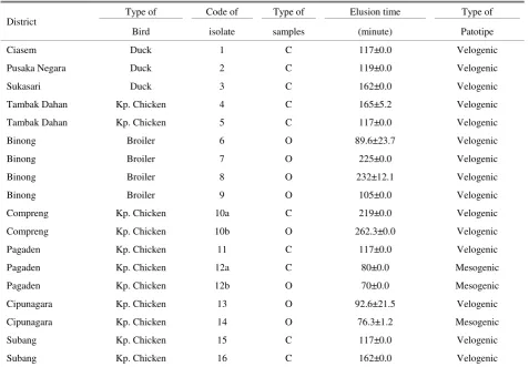
NDV virulence could be determined based on the infected embryo’s death time. According to Cattoli et al. (2011), NDV causing embryo's death in more than 90 hours after inoculation was grouped in to lentogenyc strain, and between 60-90 hours grouped in to mesogenyc strain, while less than 60 hours, was grouped into velogenyc strain.
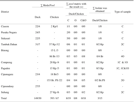
Table 2. The number of birds and pool sample from 10 subdistricts in Subang with rRT-PCR M test results and number of NDV isolates with antibody titre from individual of NDV detected
District
∑ Birds/Pool ∑ pool matrix with
the result (+) ∑ Isolate was
obtained Type of sample
Duck Chicken
Duck/Chicken
C O C&O Duck/Chicken
Ciasem 22/4 2 Kp/1 1/1 0/0 0/0 1/0 C
Pusaka Negara 24/5 - 2/0 0/0 0/0 1/0 C
Sukasari 22/5 - 5/0 0/0 0/0 1/0 C
Tambak Dahan 31/7 57 Kp /12 0/6 0/1 0/1 0/2 Kp 2C
Binong - 15 L /3 0/0 0/0 0/0 0/0 -
- 86 Br /13 0/3 0/3 0/8 0/4 Br 4O
Compreng - 20 Kp /4 0/1 0/0 0/1 0/2 Kp 1C & 1O
Pagaden - 15 Kp /3 0/1 0/0 0/1 0/3 Kp 1C, 1C&1O
Cipunagara 23/4 18 Br/3 0/0 0/0 0/0 0/0 -
- 153 Br. PS /22 0/4 0/4 0/5 0/2 Br.PS 2O
Cipeundeuy 27/5 - 0/0 0/0 0/0 0/0 -
Subang - 27 Kp /6 0/3 0/0 0/2 0/2 Kp 2C
Total 149/30 393 / 67 8/19 0/8 0/18 3/15
Kp= kampong; L= layer; Br= broiler; PS= parent stock; C= cloaca; O= oropharynx
According to Pertulla (2009)percentage of mortality due to infection of velogenyc NDV strain could reached 90% and usually the infected birds will die in 1-2 days after infection. Isolation NDV from swabs of bird cloacal and oropharynx samples from the field in Bangladesh have been done by Haque at al. (2010), and the result showed that 18 isolates were obtained from 20 cloacal swabs and 17 isolates from 20 oropharynx swabs.
ND virus found in unvaccinated native duck was originated from natural infection and usually mesogenyc or velogenyc NDV strain infection in duck showed no clinical symptoms (Saepulloh & Darminto 2005). The NDV isolated from unvaccinated native chickens was also from natural infection. Detection of NDV with RT-PCR has been performed by Kencana et al. (2012) from 10 native chickens in acute-field case with short incubation period (1-2 days) reported and the result showed ND positive. Additionally, Adi et al. (2010) also succed in isolating velogenic NDV strain from native chicken when ND outbreak occur in Bali, and stated that keeping of free-range chicken tends to
environment more than heterologous vaccine. Most of ND vaccine not prevent vaccinated birds from virulent NDV infection but vaccination significantly may decrease the amount of virus excreted through saliva and feces compared to unvaccinated birds (Kapczynski & King 2005; Miller et al. 2009).
NDV was successfully isolated from ducks and chickens showed with and without illness symptoms. According to Emilia (2013), virus was found in the sample from the birds that did not show clinical symptom, possibly due to effect of partial infection in birds, so clinical symptom did not appear, however the virus still excreted. Saepulloh & Darminto (2005) stated that if the NDV can be detected in sick bird feces (cloaca), then this indicated of systemic infection.
Only 18 isolates successfully isolated from 204 (9%) swabs of cloacal and oropharinx that positive of M and inoculated into embryonated chicken eggs (Table 1), it was due to a lot of NDV did not multiplicate in eggs because of the virus already inactive due to the handling and transport of samples were unfavorable. A similar incident also occured in Emilia (2013) study, from 20 samples of individual that positive of gen matrix (M) which were inoculated in eggs, only 11 isolates were successfully isolated. This showed that the tRT-PCR test may detect inactive virus, according to Indriani et al. (2014) one of the advantages of rRT-PCR is able to detect genetic materials of virus either active or inactive. Detection of Antibody with Hemagglutination InhibitionTest (HI).
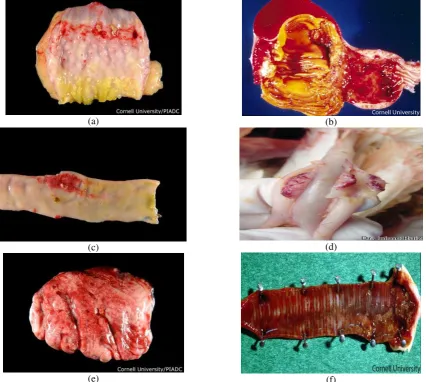
(a) (b)
(c) (d)
(e) (f)
HI test is often used in laboratories to examine specific antibody titres against NDV because its more specific and does not require special equipment so more economic (Syukron et al. 2013). The serums tested were selected based on representation of the flocks which detected positive and negative of NDV with rRT-PCR from each area. Distribution of antibody classified into three, namely: group 0, meaning that antibodies were
37 with ≥3 Log2 titre and 11 with <3 Log2 titre (Table
3). Antibodies against NDV detected in waterfowl (ducks), in the chickens were not vaccinated, and in vaccinated domestic birds. Antibodies against NDV detected in 9 of 18 duck serums from Pusaka Nagara area tested the spreading of 8 serums with ≥3 log2 titre and 1 serum with <3 Log2 titre, at 1 of 17 ducks serum and 1 of 32 chicken serums tested from Tambak dahan area with each ≥3 Log2 titre, at 10 of 62 broiler serums from Binong area tested with the spread of 5 serums with ≥3 log 2 titre and 5 serums with <3 log2 titre. in 1 of 13 chicken serum from Pagaden area tested with ≥3 log2 titre, in 26 of 135 broiler breeders (parents stock) ascertain whether the Cipeundeuy area is free of ND, it is necessary to do the detection and isolation of NDV and detection of specific antibodies against NDV in ducks and other birds such as native chicken and purebred chicken from other locations in Cipeundeuy area.
The percentage of total chicken serum that positively detected specific antibody against NDV (17%) was higher than the waterfowl (10%) and native chicken (2%). In vaccinated broilers, out of 212 serums tested only 36 serums containing antibodies against NDV, which 10 of them (28%) showed a low titre. This is because the serums were taken from culled broiler breeders which was not re-vaccinated of ND (live and killed vaccine) so antibody titre had been decreased, besides the serums were taken from broilers which had just only once ND vaccination and performed at day old with spray method using live ND vaccine and was not re-vaccinated (booster), so the possibility of uneveness of the titres is caused of antibody began to decline.
In waterfowl, only 3 isolates were successfully positive of M cloacal pools and 6 isolates obtained from 19% positive of M oropharinx pools (Table 3). There were not a lot of isolates of NDV were obtained, even there was some that not successfully obtained from positive of rRT-PCR M cloacal and oropharinx pools from flocks were also positively detected antibodies against NDV.
Value of coefficient of variation (CV) may used to describe the distribution of antibody titres in groups of animals. Mean and distribution of antibody titres in broilers using Geometric Mean Titre (GMT) and Coefficient of Varian (CV) calculations may be seen in Table 3.
Table 3. The result of detection antibody specific against NDV in bird serums from 10 areas in Subang area according to poultry commodities
Type/commodites
% Pool M + Isolate Programe
Vaccination
∑
Serum
∑
+ %
+
Antibody titre
Log2
C O C O Tested 0 <3 ≥ 3
Waterfowl 8 1 3 - No 102 10 10 92 1 9
Native chicken* 17 6 7 2 No 94 2 2 92 - 2
Purebred chicken 20 19 - 6 Yes 212 36 17 176 10 26
Total 45 26 10 408 48 12 360 11 37
C= cloacal; O= oropharynx; *the native chicken in Subang area were not vaccinated of ND since 2004
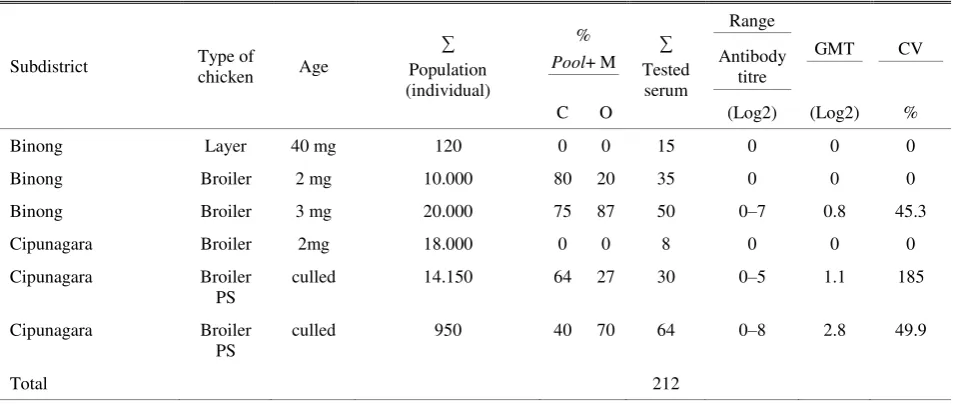
Table 4. The result of mean and distribution antibody titre of vaccinated broiler in Subang area using geometric mean tire (GMT) and coefficient of variation (CV)
Subdistrict Type of
chicken Age
∑
Population (individual)
% Pool+ M
∑
Tested serum
Range
GMT CV
Antibody titre
C O (Log2) (Log2) %
Binong Layer 40 mg 120 0 0 15 0 0 0
Binong Broiler 2 mg 10.000 80 20 35 0 0 0
Binong Broiler 3 mg 20.000 75 87 50 0–7 0.8 45.3
Cipunagara Broiler 2mg 18.000 0 0 8 0 0 0
Cipunagara Broiler
PS
culled 14.150 64 27 30 0–5 1.1 185
Cipunagara Broiler
PS
culled 950 40 70 64 0–8 2.8 49.9
Total 212
CV ≤35% = sebaran of homogenous titre; CV >35% = sebaran of not homogenous titre
it, where the flock with low antibody titre is protective against NDV, in field was still occur often (Yan et al. 2011). Besides, according to Reynolds & Maraga (2000) and Erf (2004) experimentally showed there was no directly correlation between antibody titre in serums against chicken immunity which was challenged with NDV.
Antibody titre response is strongly influenced by quality of vaccine, route and the implementation of application, the environment, individual’s factor and the species of bird (OIE 2012). Massal vaccination using live vaccine is often used than individually vaccine, because it is cheaper and easier to be applied (Senne et al. 2004). Vaccine from virulent strain (LaSota and B1) is commonly used around the world to overcome ND because it can provide protection to virulent NDV if the vaccination is done correctly (Kapczynski & King 2005; Cornax et al. 2012; Dortmans et al. 2012). In
the serology survey of unvaccinated domestic chicken in Sulaiman Province, Irak. The result showed that serum of 500 tested chickens, 172 serums (34%) was detected antibody. This shows that ND still endemic in Irak.
The existence of a positive antibody specific to NDV in unvaccinated duck and native chicken serums showed that in Subang was vulnerable of ND. This is because the duck and chicken were positive for antibody against NDV may be a carrier which became a source of Newcastle disease spreading (Saepulloh & Darminto 2005; Adi et al. 2010).
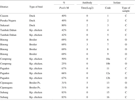
Observing the presence of antibodies in 16 birds that excreting NDV from 9 sub districts showed that only 4 individual from 3 sub districts were positive with chicken with illness symptom with titre 8 Log2 (isolate 8) and 12 weeks old vaccinated breeding broiler (parent stock) and showed illness symptom with titre 5 Log2 from Cipunagara (isolate 13) (Table 5). The existence of antibody in unvaccinated duck and was successfully isolated of NDV showed an immune response of ongoing NDV infection, while the detection of antibody in unvaccinated chickens and ducks where NDV was and turtle doves) showed antibody titre 0 Log2 was due to infection process has not been occurred, so the antibody has not been produced. In 3 vaccinated domestic chickens and detected of antibody against NDV, showed high titre antibody in the range 3-8 Log2 may still excrete virus through the cloaca and oropharynx. This may be affected by ineffective vaccination due to vaccine virus with field NDV was not homologous, so virus shedding occurred. Antibody response was not formed due to suboptimal vaccination, so that the chickens may be infected by NDV according to no detected antibody of vaccinated domestic chickens and isolated of NDV. The result of testing sample serum by HI test which successfully isolated from NDV may be seen in Table 4.
Antigenic diversity
Homologous antiserum will has an higher affinity with the viral surface epitopes so its more optimal inhibiting hemagglutination activity. Characterization
of 18 antigen isolates with the HI test using antisera Lasota (lentogenic) obtained the average (mean) of HI titres between 6-8 log2 with antisera and Komarov (mesogenic) between 9-13 log2. Isolates affinity with LaSota antisera showed relatively homogeneous antigenic character, only a few isolates that showed variation reaches 2 log2 (1st, 2nd, and 3rd isolate), as well as antisera Komarov, only a few isolates that showed variation reaches 4 log2 (3rd, 6th, and 16th isolate). All NDV isolates showed a higher affinity against antisera Komarov compared with Lasota antisera that indicates all the NDV tend to get into a virulent NDV strain (Figure 2). According to Alexander & Senne (2008) differences of antigenic between NDV strains which may be recognized by specific antibodies determined by a hemagglutinin (HN) protein. Beside it, according Adu (1985) and Ibu et al. (2008) antigenic variation in NDV from the same strainoccurs because of various functions of the external proteins due to mutation.
Study of the antigenic diversity of NDV using polyclonal antisera had been conducted by Emilia (2013) using antisera strain Lasota, Komarov, the G7 and the ITA against four isolates Serpong area, West Java. The result showed that varies affinity with HI titres ranging between 3-5 log2 (Lasota), 5-10 log2 (Komarov), 6-8 log2 (G7) and 3-7 log2 (ITA). Hsiang-Jung et al. (2004) examined the variation of antigenic against 36 isolates from Taiwan NDV obtained between 1969-1996 using 22 monoclonal antibodies (MAB) and was able to separate the 36 isolates into 18 groups antigenic and based on the nucleotide sequences of gene F were grouped into 15 genotypes. Characterization of NDV antigen may also be done with a monoclonal antibody (MBA). Hu et al. (2010) showed using four types of MBA, that NDV antigenic variation may occur due to mutations of residues K (Lysine) at position 347 in the HN protein.
Characterization of physical properties of ND Virus with Elution Time Test
Table 5. Result of detection of specific antibody against NDV with HI test in 16 bird’s serums which successfully isolated of NDV in Subang area
District Type of bird
% Antibody Isolate
Pool+ M Titre(Log2) Code Type of
sample
Ciasem Duck 40% 0 1 C
Pusaka Negara Duck 40% 0 2 C
Sukasari Duck 80% 2 3 C
Tambak Dahan Kp. chicken 42% 0 4 C
Tambak Dahan Kp. chicken 42% 0 5 C
Binong Broiler 69% 0 6 O
Binong Broiler 69% 3 7 O
Binong Broiler 69% 8 8 O
Binong Broiler 69% 0 9 O
Compreng Kp. chicken 50% 0 10a C
Compreng Kp. chicken. 25% 0 10b O
Pagaden Kp. chicken 67% 0 11 C
Pagaden Kp. chicken 66% 0 12a C
Pagaden Kp. chicken 67% 0 12b O
Cipunagara Broiler Ps. 31% 5 13 O
Cipunagara Broiler Ps. 31% 0 14 O
Subang Kp. chicken 83% 0 15 C
Subang Kp. chicken 83% 0 16 C
Kp= kampong; Ps= parent stock; C= cloaca; O= oropharynx
Code of Isolates
Figure 2. Comparison of antigenic of 18 isolates with HI test. The majority of HI titres looks homogeneous, only a few isolates that showed a variation with the antisera Lasota 2 log2 and with antisera Komarov 4 log2. HI titre comparison of both antisera showed isolates tend to lead to a virulent strain
Lasota Komarov
Ti
ter HI
(Lo
g
2
Table 6. The result of elusion time test of NDV isolates that obtained from chicken and duck in 10 areas in Subang area
District
Type of Code of Type of Elusion time Type of
Bird isolate samples (minute) Patotipe
Ciasem Duck 1 C 117±0.0 Velogenic
Pusaka Negara Duck 2 C 119±0.0 Velogenic
Sukasari Duck 3 C 162±0.0 Velogenic
Tambak Dahan Kp. Chicken 4 C 165±5.2 Velogenic
Tambak Dahan Kp. Chicken 5 C 117±0.0 Velogenic
Binong Broiler 6 O 89.6±23.7 Velogenic
Binong Broiler 7 O 225±0.0 Velogenic
Binong Broiler 8 O 232±12.1 Velogenic
Binong Broiler 9 O 105±0.0 Velogenic
Compreng Kp. Chicken 10a C 219±0.0 Velogenic
Compreng Kp. Chicken 10b O 262.3±0.0 Velogenic
Pagaden Kp. Chicken 11 C 117±0.0 Velogenic
Pagaden Kp. Chicken 12a C 80±0.0 Mesogenic
Pagaden Kp. Chicken 12b O 70±0.0 Mesogenic
Cipunagara Kp. Chicken 13 O 92.6±21.5 Velogenic
Cipunagara Kp. Chicken 14 O 76.3±1.2 Mesogenic
Subang Kp. Chicken 15 C 117±0.0 Velogenic
Subang Kp. Chicken 16 C 162±0.0 Velogenic
Kp= kampong, 10a and 10b; 12a and 12b= from the same individual
10a and 10b) and isolates obtained from cloacal and oropharyng swabs from 1 native chicken in Pagaden area (isolate 12a and 12b) were included to the same strain that was mesogenic/mesogenic and velogenic/velogenic, and HI titre from each isolate also had the same value. It indicated that both of excreted isolates from cloacal and oropharinx from 1 individual were the same NDV, but to confirm the virus identity, it needs to be sequenced. Elution time may be affected by temperature. According to Hussain et al. (2008) the elution time will be longer at 4°C and will be faster when the temperature is raised, therefore in order to obtain an accurate results, the elution test should be performed at a steady temperature. Besides, the elution test was also influenced by the concentration of red blood cells, if there are too many red blood cells during the testing, the red blood cells are not able to be bound by the virus, so that looks like a reaction to release of red blood cells by the virus and this will reduce the efficiency of elution test (Ezeibe & Ndip 2004).
Characterization of ND Virus by rRT-PCR fusion (F)
sequence. The incompatibility of primer oligobucleotide cause hybridization between primer/probe with RNA of virus not occurs, so it was not detected by real-time PCR system software.
CONCLUSION
Newcastle disease was still endemic in Subang area and the infection may be subclinical. Eighteen ND viruses which were found, majority had relatively homogeneous character, just a few isolates that showed diversity of pathogenicity and antigenicity (antisera LaSota: isolate 1, 2, and 3; antisera Komarov: isolate 3, 6 and 16). The ducks from Cipeundeuy area had not been exposed of NDV. The distribution of antibody titres in vaccinated chicken was unequal. The result of this study contributed information about ND in Subang area and may be used as feedback for Subang government to determine the prevention and control programs of ND infection in poultry in Subang.
RECOMMENDATION
Further research needs to be done to see the acid-base sequences of cleavage site of fusion (f) protein by sequencing or pathotyping test (MDT, ICPI, and IVPI) in order to obtain more information about pathogenicity and NDV strains which spread in Subang area. Sequencing needs to be done to see mutation in hemaglutinin. Strict sanitary and vaccinate use a combination of homologous live and inactive vaccine with field NDV can be applied to prevent ND infection in birds in Subang. Besides, more extensive surveys with more samples needs to be done to get information about infection of NDV that cover all areas in Subang area.
REFERENCES
Adi AAAM, Astawa NM, Putra KSA, Hayashi Y, Matsumoto Y. 2010. Isolation and characterization of a pathogenic Newcastle disease virus from a natural case in Indonesia. J Vet Med Sci. 72:313-319.
Adu FD. 1985. Characteristics of Nigerian strains of Newcastle disease virus. Avian Dis. 29:49-851.
Al-Garib SO, Gielkens ALJ, Gruys E, Hartog L, Koch G. 2003. Immunoglobulin class distribution of systemic and mucosal antibody responses to Newcastle disease in chickens. Avian Dis. 47:32-40.
Aldous EW, Alexander DJ. 2001. Detection and differentiation of Newcastle disease virus (avian paramyxovirus type 1). Avian Pathol. 30:117-128. Alexander DJ, Bell JG, Alders RG. 2004. A Technology
Review: Newcastle Disease, with Special Emphasis on
Its Effect on Village Chickens. Rome: FAO Animal Production and Health.
Alexander DJ, Senne DA. 2008. Newcastle disease and other paramyxovirus infections. In: A Laboratory Manual for the Isolation, Identification and Characterization of Avian Pathogens. In: Zavala LD, editor. Athens, GA American Association of Avian Pathologists. p. 135-141.
Ashraf A, Shah MS. 2014. Newcastle Disease: Present status and future challenges for developing countries. Afr J Microbiol Res. 8:411-416.
Aziz TAG, Ahmed TA. 2010. Serological survey of Newcastle disease in domestic chickens in Sulaimani province. JZS. 13:31-38.
[BPPV] Balai Penyidikan dan Pengujian Veteriner Subang. 2011. Laporan Tahunan 2011. Subang (Indones): Balai Penyidikan dan Pengujian Veteriner.
[BPPV] Balai Penyidikan dan Pengujian Veteriner Subang. 2012. Laporan Tahunan 2012. Subang (Indones): Balai Penyidikan dan Pengujian Veteriner.
[BPPV] Balai Penyidikan dan Pengujian Veteriner Subang. 2013. Laporan Tahunan 2013. Subang (Indones): Balai Penyidikan dan Pengujian Veteriner.
Boven Mv, Bouma A, Fabri THF, Katsma E, Hartog L, Koch G. 2008. Herd immunity to Newcastle disease virus in poultry by vaccination. Avian Pathol. 37:1-5.
Brown C, King DJ, Seal BS. 1999. Pathogenesis of Newcastle disease In chickens experimentally Infected with viruses of different virulence. Vet Pathol. 36:125-132. live LaSota vaccine strain-induced protection in chickens upon early challenge with a virulent Newcastle disease virus of heterologous genotype. Avian Dis. 56:464-470.
[CVL] Central Veterinary Laboratory. 2007. Standard operating procedure for real time polymerase chain reaction detection of virulent Newcastle disease virus in clinical specimens.
Daulay S. 2005. Peluang dan potensi burung liar dalam penyebaran Newcastle disease di Sulawesi Selatan (Disertasi). [Bogor (Indones)]: Institut Pertanian Bogor.
[DISNAK] Dinas Peternakan Kabupaten Subang. 2010. Kasus ND di Kabupaten Subang. Laporan Tahunan Dinas Peternakan Subang Tahun 2010. Subang (Indones): Dinas Peternakan Kabupaten Subang.
[DISNAK] Dinas Peternakan Kabupaten Subang. 2013. Laporan Tahunan Dinas Peternakan Subang Tahun 2013. Subang (Indones): Dinas Peternakan Kabupaten Subang.
Dortmans JCFM, Peeters BPH, Koch G. 2012. Newcastle disease virus outbreaks: Vaccine mismatch or inadequate application?. J Vet Microbiol. 160:17-22.
Emilia. 2013. Isolasi dan karakterisasi biologik Virus Newcastle Disease (NDV). (Tesis). [Bogor (Indones)]: Institut Pertanian Bogor.
Erf GF. 2004. Cell-mediated immunity in poultry. Poult Sci. 83:580-590.
Ezeibe MCO, Ndip ET. 2005. Differences in the red blood cell elution times of strains of Newcastle disease virus. AJOL. 22:99-101.
Ezeibe MCO, Ndip ET. 2005. Red blood cell elution time of strains of Newcastle disease virus. J Vet Sci. 6:287-288.
Haque MH, Hossain MT, Islam MT, Zinnah MA, Khan MSR, Islam MA. 2010. Isolation and detection of Newcastle disease virus from field outbreaks in broiler and layer chickens by reverse transcription-polymerase chain reaction. Bangladesh J Vet Med. 8:87-92.
Hsiang-Jung T, Kuo-Hui C, Chun-Hsien T, M FK, Manvell RJ, Alexander DJ. 2004. Antigenic and genotypical characterization of Newcastle disease viruses isolated in Taiwan between 1969 and 1996. Vet Microbiol. vaccine candidate with high yield in embryonated chicken eggs. Avian Dis. 55:391-397.
Hussain M, Mehmood MD, Ahmad A, Shabbir MZ, Yaqub T. 2008. Factors affecting hemagglutination activity of Avian Influenza virus subtype H5N1. J Vet Anim Sci. 1:31-36.
Ibu JO, Okoye JOA, Fasina FO, Aba-Adulugba EP, Abechi AS, Woma YT. 2008. Antigenic relatedness among Newcastle disease virus isolates from Nigerian feral birds and the La sota strain. Vet Sci. 13:79-84.
Indriani R, Dharmayanti NLPI, Adjid RMA. 2014. Efikasi penerapan vaksin AI H5N1 clade 2.1.3 pada itik Mojosari terhadap tantangan virus AI H5N1 clade 2.3.2 pada kondisi laboratorium. JITV. 19:59-66.
Kapczynski DR, King DJ. 2005. Protection of chicken against overt clinical disease and determination of viral shedding following vaccination with commercially
available Newcastle disease virus vaccines upon challenge with highly virulent virus from the California 2002 exotic Newcastle disease outbreak. Vaccine. 23:3424-3433.
Kencana GAY, Kardena IM, Mahardika IGNK. 2012. Peneguhan diagnosis penyakit Newcastle disease lapang pada ayam bupurebred di Bali menggunakan teknik RT-PCR. J Kedokteran Hewan. 6:28-31.
Kim LM, Afonso CL, Suarez DL. 2006. Effect of Probe-Site Mismatches on detection of virulent Newcastle disease viruses using a fusion-gene real-time reverse transcription polymerase chain reaction test. J Vet Diag Invest. 18: 519-528.
Miller PJ, Afonso CL, Attrache JE, Dorsey KM, Courtney SC, Zijing G, Kapczynski DR. 2013. Effects of Newcastle disease virus vaccine antibodies on the shedding and transmission of challenge viruses. Dev Comp Immun. 41:505-513.
Miller PJ, Estevez C, Yu Q, Suarez DL, J KD. 2009. Comparison of viral shedding following vaccination with inativated and live Newcastle disease vaccines formulated with wild-type and recombinant viruses. Avian Dis. 53:39-49.
[NVSL] National Veterinary Services Laboratories. 2005. Real-Time RT-PCR for detection of virulent Newcastle disease virus in clinical samples.
Numan M, Zahoor MA, Khan HA, Siddique M. 2005. Serologi status of Newcastle disease in broilers and layers in Faisalabad and surrounding districts. Pak Vet J. 25:55-58.
[OIE] Office International des Epizooties. 2008. Manual of diagnostic tests and vaccines for terrestrial animals.
[OIE] Office International des Epizooties. 2009. NDV in Indonesia.
[OIE] Office International des Epizooties. 2012. Newcastle Disease.
Perozo F, Marcano R, Afonso CL. 2012. Biological and phylogenetic characterization of a genotype VII Newcastle disease virus from Venezuela: efficacy of field vaccination. J Clin Microbiol. 50:1204-1208.
Pertulla L. 2009. Epidemiology and Characterization of Newcastle disease in Smallholder Poultry in Mozambique. 1-21.
Reynolds DL, Maraqa AD. 2000. Protective immunity against Newcastle disease: the role of antibodies specific to Newcastle disease virus polypeptides. Avian Dis. 44:138-144.
Rezaeianzadeh G, Dadpurebred H, Safar A, Ali M, Hossein M, Nazemshirazi. 2011. Serological and molecular study of Newcastle disease virus circulating in village chicken of Fars Province, Iran. J Vet Med Anim Health. 3:105-111.
Samal SK. 2011. Newcastle disease and related avian paramyxovirus. In: The Biology of Paramyxovirus. Norfolk (UK): Caister Academic Press. p. 69-114.
Senne DA, King DJ, Kapczynski DR. 2004. Control of Newcastle disease by vaccination. Dev Biol. 119:165-170.
Sheffield FW, Smith W, Belyavin G. 1954. Purification of Influenza virus by red-cell adsorption and elution. 35:214-222.
Syukron MU, Suartha IN, Dharmawan NS. 2013. Serodeteksi penyakit tetelo pada ayam di Timor Leste. Indones Med Vet. 2:360-368.
Wise MG, Suarez DL, Seal BS, Pedersen JC, Senne DA, King DJ, Kapczynski DR, Spackman E. 2004. Development of a Real-Time Reverse-Transcription PCR for detection of Newcastle disease virus RNA in clinical samples. J Clin Microbiol. 42:329-338.
Xiao S. 2012. Complete Genom Sequences of Newcastle Disease Virus Strain Circulating in Chickens Populations of Indonesia. J Virol. 86:5969-5970.