p-
ISSN 0854-4263Vol. 23, No. 1, November 2016
e-
ISSN 4277-4685INDONESIAN JOURNAL OF
CLINICAL PATHOLOGY AND
MEDICAL LABORATORY
Majalah Patologi Klinik Indonesia dan Laboratorium Medik
EDITORIAL TEAM
Editor-in-chief: Puspa Wardhani
Editor-in-chief Emeritus: Prihatini
Krisnowati
Editorial Boards:
Maimun Zulhaidah Arthamin, AAG Sudewa, Rahayuningsih Dharma, Mansyur Arif, July Kumalawati, Nurhayana Sennang Andi Nanggung, Aryati, Purwanto AP, Jusak Nugraha, Sidarti Soehita,
Endang Retnowati Kusumowidagdo, Edi Widjajanto, Budi Mulyono, Adi Koesoema Aman, Uleng Bahrun, Ninik Sukartini, Kusworini Handono, Rismawati Yaswir, Osman Sianipar
Editorial Assistant: Dian Wahyu Utami
Language Editors:
Yolanda Probohoesodo, Nurul Fitri Hapsari
Layout Editor: Akbar Fahmi
Editorial Adress:
d/a Laboratorium Patologi Klinik RSUD Dr. Soetomo Jl. Mayjend. Prof. Dr Moestopo 6–8 Surabaya, Indonesia Telp/Fax. (031) 5042113, 085-733220600 E-mail: [email protected], [email protected]
Website: http://www.indonesianjournalofclinicalpathology.or.id
Printed by Airlangga University Press. (OC 316/11.16/AUP-75E). Kampus C Unair, Mulyorejo Surabaya 60115, Indonesia. Telp. (031) 5992246, 5992247, Fax. (031) 5992248. E-mail: [email protected]
Kesalahan penulisan (isi) di luar tanggung jawab AUP
p-
ISSN 0854-4263Vol. 23, No. 1, November 2016
e-
ISSN 4277-4685INDONESIAN JOURNAL OF
CLINICAL PATHOLOGY AND
MEDICAL LABORATORY
Majalah Patologi Klinik Indonesia dan Laboratorium Medik
CONTENTS
RESEARCHS
Molecular Aspect Correlation between Glycated Hemoglobin (HbA1c), Prothrombin Time (PT) and Activated Partial Thromboplastin Time (APTT) on Type 2 Diabetes Mellitus (T2DM)
(Aspek molekuler Hubungan Kadar Hemoglobin Terglikasi (HbA1c), Prothrombin Time (PT) dan Activated Partial Thromboplastin Time (APTT) di Diabetes Melitus Tipe 2)
Indranila KS ... 1–6
Platelet-Lymphocyte Ratio (PLR) Markers in Acute Coroner Syndrome
(Platelet Lymphocyte Ratio (PLR) Sebagai Petanda Sindrom Koroner Akut)
Haerani Harun, Uleng Bahrun, Darmawaty ER ... 7–11
The Mutation Status of Kras Gene Codon 12 and 13 in Colorectal Adenocarcinoma
(Status Mutasi Gen Kras Kodon 12 dan 13 di Adenocarcinoma Colorectal)
Gondo Mastutik, Alphania Rahniayu, Anny Setijo Rahaju, Nila Kurniasari, Reny I’tishom ... 12–17
Creatine Kinase Related to the Mortality in Myocardial Infarction
(Creatine Kinase terhadap Angka Kematian di Infark Miokard)
Liong Boy Kurniawan, Uleng Bahrun, Darmawaty Rauf, Mansyur Arif ... 18–21
Application of DNA Methylation on Urine Sample for Age Estimation
(Penggunaan Metilasi DNA Dalam Perkiraan Umur Individu di Sampel Air Kemih)
Rosalinda Avia Eryatma, Puspa Wardhani, Ahmad Yudianto ... 22–26
Lipid Profile Analysis on Regular and Non-Regular Blood Donors
(Analisis Profil Lipid di Pendonor Darah Reguler dan Non-Reguler)
Waode Rusdiah, Rachmawati Muhiddin, Mansyur Arif ... 27–30
Percentage of CD3+ T Lymphocytes Expressing IFN-γ After CFP-10 Stimulation
(Persentase Limfosit T-CD3+ yang Mengekspresikan Interferon Gamma Setelah Stimulasi Antigen
CFP-10)
Yulia Nadar Indrasari, Betty Agustina Tambunan, Jusak Nugraha, Fransiska Sri Oetami ... 31–35
Characteristics of Crossmatch Types in Compatibility Testing on Diagnosis and Blood Types Using Gel Method
(Ciri Inkompatibilitas Uji Cocok Serasi Metode Gel terhadap Diagnosis dan Golongan Darah)
Irawaty, Rachmawati AM, Mansyur Arif ... 36–41
Diagnostic Values of Mycobacterium Tuberculosis 38 kDa Antigen in Urine and Serum of Childhood Tuberculosis
(Nilai Diagnostik Antigen 38 kDa Mycobacterium tuberculosis Air Kemih dan Serum di Tuberkulosis Anak)
Agustin Iskandar, Leliawaty, Maimun Z. Arthamin, Ery Olivianto ... 42–49
Erythrocyte Indices to Differentiate Iron Deficiency Anemia From β Trait Thalassemia
(Indeks Eritrosit Untuk Membedakan Anemia Defisiensi Besi Dengan Thalassemia β Trait)
Thanks to editors in duty of IJCP & ML Vol 23 No. 1 November 2016
Kusworini Handono, Prihatini, Purwanto AP, July Kumalawati, Jusak Nugraha, Ida Parwati, Adi Koesoema Aman, Edi Widjajanto, AAG. Sudewa, Nurhayana Sennang AN HbA1c Levels in Type 2 Diabetes Mellitus Patients with and without Incidence of Thrombotic Stroke
(Kadar HbA1cPasien Diabetes Melitus Tipe 2 Dengan dan Tanpa Kejadian Strok Infark Trombotik)
Dafina Balqis, Yudhi Adrianto, Jongky Hendro Prayitno ... 56–60
Comparative Ratio of BCR-ABL Genes with PCR Method Using the Codification of G6PD and ABL Genes in Chronic Myeloid Leukemia Patients
(Perbandingan Angka Banding Gen BCR-ABL Metode PCR Menggunakan Baku Gen Glucosa-6-Phosphate Dehidrogenase dan Gen Abelson Kinase di Pasien Chronic Myeloid Leukemia)
Tonggo Gerdina Panjaitan, Delita Prihatni, Agnes Rengga Indrati, Amaylia Oehadian ... 61–66
Virological and Immunological Response to Anti-Retroviral Treatment in HIV-InfectedPatients
(Respons Virologis dan Imunologis Terhadap Pengobatan Anti-Retroviral di Pasien Terinfeksi HIV)
Umi S. Intansari, Yunika Puspa Dewi, Mohammad Juffrie, Marsetyawan HNE Soesatyo,
Yanri W Subronto, Budi Mulyono ... 67–73
Comparison of sdLDL-C Analysis Using Srisawasdi Method and Homogeneous Enzymatic Assay
Method onHypertriglyceridemia Condition
(Perbandingan Analisa sdLDL-C metode Srisawasdi dan Homogeneous Enzymatic Assay di Kondisi Hipertrigliseridemia)
Gilang Nugraha, Soebagijo Poegoeh Edijanto, Edhi Rianto ... 74–79
Pattern of Bacteria and Their Antibiotic Sensitivity in Sepsis Patients
(Pola Kuman dan Kepekaan terhadap Antibiotik di Pasien Sepsis)
Wahyuni, Nurahmi, Benny Rusli ... 80–83
The Correlation of Naive CD4+T Lymphocyte Cell Percentage, Interleukin-4 Levels and Total
Immunoglobulin E in Patients with Allergic Asthma
(Kenasaban antara Persentase Sel Limfosit T-CD4+ Naive dengan Kadar Interleukin-4 dan Jumlah
Imunoglobulin E Total di Pasien Asma Alergi)
Si Ngr. Oka Putrawan, Endang Retnowati, Daniel Maranatha ... 84–89
LITERATURE REVIEW
Antibiogram
(Antibiogram)
Jeine Stela Akualing, IGAA Putri Sri Rejeki ... 90–95
CASE REPORT
Pancreatic Cancer in 31 Years Old Patient with Normal Serum Amylase Level
(Kanker Pankreas di Pasien Usia 31 Tahun Dengan Kadar Amilase Serum Normal)
96
CASE REPORT
PANCREATIC CANCER IN 31 YEARS OLD PATIENT WITH NORMAL
SERUM AMYLASE LEVEL
(Kanker Pankreas di Pasien Usia 31 Tahun dengan Kadar Amilase Serum Normal)
Melda F. Flora, Budiono Raharjo, Maimun Z. Arthamin
ABSTRAK
Kanker pankreas adalah keganasan sel di jaringan pankreas. kejadiannya meningkat pada usia di atas 60 tahun. Namun, sekitar 20% dapat terjadi di usia muda. Patogenesis terjadinya masih belum jelas, dikemukakan bahwa mutasi genetik dan faktor eksogen seperti merokok berhubungan dengan terjadinya keganasan sel pankreas. Kasus adalah seorang laki-laki perokok berusia 31 tahun dengan keluhan utama nyeri ulu hati menjalar ke punggung, disertai mual, muntah, nafsu makan turun. Pada pemeriksaan fisik didapatkan sklera ikterik, perkusi redup dan ronkhi di paru, distensi abdomen dan asites. Pada pemeriksaan laboratorik didapatkan leukositosis, trombositopenia, peningkatan aspartate aminotransaminase (AST) lebih dari 10 kali Upper Range Limit (URL), hiperbilirubinemia direk, peningkatan alkaline phosphatase (ALP), Gamma Glutamyl Transferase (GGT) dan lipase serum, sedangkan amilase serum normal. Terdapat juga peningkatan kadar CA19-9. Pada computed tomography scan (CT scan) dan Magnetic Resonance Imaging (MRI) didapatkan gambaran kanker pankreas primer yang telah bermetastasis ke pleura dan hati. Kadar amilase normal di pasien dapat disebabkan karena awal peningkatan dan penurunan kadar amilase terjadi lebih cepat dan pada saat diperiksa telah turun mencapai kadar normal. Simpulan, kanker pankreas dapat terjadi di usia muda. Amilase yang normal dapat terjadi di kanker pankreas.
Kata kunci: Kanker pankreas, usia muda, perokok, kadar amilase serum normal
ABSTRACT
Pancreatic cancer is a malignancy of cells in pancreatic tissue. Its incidence increase in age over 60 years. However, about 20% can occur in young adult. The pathogenesis of pancreatic cancer is still unclear. It has been known that genetic mutation and exogenous factor such as smoking are associated with pancreatic cell malignancies. This was a case of a 31 years old male smoker, with the chief complaint of epigastric pain, radiating to the back, accompanied by nausea, vomiting and decreased appetite. There were icteric, dim percussion and ronchi, abdominal distension and ascites. The laboratory findings: there were leukocytosis, thrombocytopenia, increased AST more than 10 times URL, conjugated hyperbilirubinemia, increased serum alkaline phosphatase (ALP), gamma glutamyl transferase (GGT) and lipase, whereas serum amylase was within normal range. There was also elevated CA19-9 level. On CT Scan and MRI found images of primary pancreatic cancer which had metastatic to the pleura and liver. Normal serum amylase level in patient because of onset of increasing and decreasing amylase level occur rapidly and when the examination was performed the level was decreased to normal level. Conclusion, pancreatic cancer can occur in young adult and amylase level may be within normal range in pancreatic cancer.
Key words: Pancreatic cancer, young adult, smokers, normal serum amylase level
2016 November; 23(1): 96–00 p-ISSN 0854-4263 | e-ISSN 4277-4685 Available at www.indonesianjournalofclinicalpathology.or.id
Department of Clinical Pathology, Faculty of Medicine, Brawijaya University/Dr. Saiful Anwar Hospital, Malang, Indonesia. E-mail:[email protected]
INTRODUCTION
Pancreatic cancer is a malignancy of cells in pancreatic tissue. Pancreatic cancer has become the fifth highest cause of death in the United States with a number of 27,000 cases per year. The incidence of
pancreatic cancer is about 12 cases per 100,000 people with a ratio of 1:3 between male and female patients.1
97
Pancreatic Cancer in 31 Years Old Patient - Flora, et al.
In Indonesia, Hadi4 reported that there were 18
cases of pancreatic cancer, similar to 0.19% of total patients, from January 1976 to June 1979 at UPF of the Internal Clinic of Dr. Hasan Sadikin Hospital, Bandung. The ratio of male and female patients suffering from pancreatic cancer was 1.6.1 Those patients were mostly
at the age of 23 to 69 years old (with the mean of 53.8 years old). Some other researches on Dr. Kariadi Hospital in Semarang (1985-1989) found that there were 24 patients with pancreatic cancer with a ratio of 1:1 between male and female patients aged between 45–65 years old (with a mean of 54.3 years).1–4
Unfortunately, the cause of pancreatic cancer is still unclear. Genetic mutation and various factors can trigger malignant cell changes. For instance, mutation in Ki-Ras oncogenes is associated with 90% of pancreatic adenocarcinoma incidence, and approximately 60-80% of this case was triggered by defects in the p53 tumor suppressor gene. Furthermore, exogenous risk factors are also allegedly closely associated with pancreatic cancer, such as smoking, while drinking coffee and exposure to carcinogens still has not been proven. On the other hand, endogenous factors include diabetes mellitus, chronic pancreatitis, pancreatic calcification, and pancreatolithiasis.2–4
Therefore, this research aimed to report a male patient aged 31 years old with pancreatic cancer metastasizing to his liver and pleura with normal serum amylase levels. This report can be considered as a rare case, commonly found in patients with pancreatic cancer at the age of >50 years.
CASE
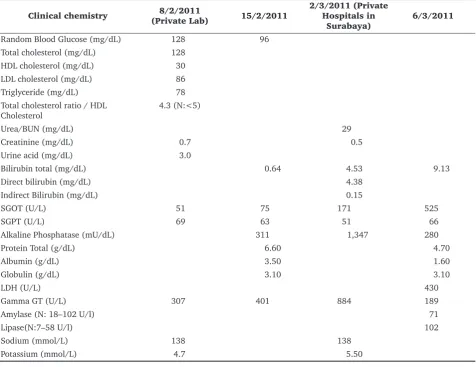
A male patient aged 31 years came with a chief complaint of heartburn radiating to the back accompanied by nausea, vomiting, and decreased appetite since one month before entering the hospital. A week earlier, his belly was getting bigger, and he had yellow eyes without any complaints of defecate. He had smoking habit about ten cigarettes a day since the age of 9 years. Based on physical examination conducted, there was jaundice. Meanwhile, based on lung examination, there were also dim and rhonchi on his right lung, as well as abdominal distention and ascites. The results of laboratory tests can be seen in Table 1, 2, and 3 below.
On February 17, 2011 the results of Computed Tomography (CT scan) test showed that multiple nodule lesions in both lobes of the liver, which could be a metastatic deposit. Gallbladder wall was also thick, contracted and had some materials that could be a
precipitate or a small calculus. Besides, there was a mass in the pancreatic cauda, most likely as a primary cancer. Ascites fluid also emerged in the pelvis, but without enlarged lymph nodes of pancreas, aorta, or pelvis.
The roentgen thorax photograph, furthermore, showed lung and heart within normal limits. On March 6, 2011, the results of magnetic resonance imaging
(MRI) then showed pleural effusion and consolidation in both lower lobes of the lung, free fluid in the cavity
of the abdomen, and multiple nodules in both lobes of the liver. The results also showed intrahepatic bile duct that was not stretched, the portal vein that seemed not involved, thick gall bladder, contraction, the possibility of a calculus material, and larger lien, but no focal lesions. Lesions of the pancreatic cauda indicated cancer and both kidneys appeared normal.
In addition, the results of pleural fluid cytology examination on March 7, 2011 found atypical cells indicating a malignancy. On March 12, 2011, the results of Fine Needle Aspiration Biopsy (FNAB) then showed hepatic malignant cells indicating a metastasis.
DISCUSSION
Indonesian Journal of Clinical Pathology and Medical Laboratory, 2016 November; 23(1): 96–101 98
Table 1. Results of complete blood and hemostasis tests
Complete blood tests 8/2/2011
(Private Lab.)
15/2/2011 (Singapore)
6/3/2011 (Singapore)
Haemoglobine (g/dL) 12.8 13.3 13.9
Leukocyte (103/μl) 13,640 14,540 31,770
Thrombocyte (103/μl) 461,000 346,000 136,000
HCT (%) 39.6 41.6 41.0
MCV (N:80–100 pg) 78.8 81.5 77.2
MCH (N:26–34g/dL) 25.5 26.2 26.2
MCHC (N: 32–36 g/dL) 32.3 32.1 33.9
RDW (N:11.5–14.5%) 13.1 13.3 14.9
ESR (N:0–15 mm/hour) 73
Differential count (eosinophil/ basophil/ stab/segment/ lymphocyte/monocytes)
2/1/–/83/8/6 –/1/85/10/4 –/–/–/80/11/7 mielocyte 2%
Hemostasis 2/3/2011 9/3/2011 (Singapore)
PPT 15.10” (K:13.50”) 19.7” (N: 10.0–14.0)
INR 1,21
APTT 43.40 (K:33.60”) 56.7 (N: 25.0–41.0)
BT (N:1–3 minutes) 2 minutes
CT (N: 5–15 minutes) 10 minutes
Table 2. Results of blood chemistry test
Clinical chemistry 8/2/2011
(Private Lab) 15/2/2011
2/3/2011 (Private Hospitals in
Surabaya)
6/3/2011
Random Blood Glucose (mg/dL) 128 96
Total cholesterol (mg/dL) 128 HDL cholesterol (mg/dL) 30 LDL cholesterol (mg/dL) 86
Triglyceride (mg/dL) 78
Total cholesterol ratio / HDL Cholesterol
4.3 (N:<5)
Urea/BUN (mg/dL) 29
Creatinine (mg/dL) 0.7 0.5
Urine acid (mg/dL) 3.0
Bilirubin total (mg/dL) 0.64 4.53 9.13
Direct bilirubin (mg/dL) 4.38
Indirect Bilirubin (mg/dL) 0.15
SGOT (U/L) 51 75 171 525
SGPT (U/L) 69 63 51 66
Alkaline Phosphatase (mU/dL) 311 1,347 280
Protein Total (g/dL) 6.60 4.70
Albumin (g/dL) 3.50 1.60
Globulin (g/dL) 3.10 3.10
LDH (U/L) 430
Gamma GT (U/L) 307 401 884 189
Amylase (N: 18–102 U/l) 71
Lipase(N:7–58 U/l) 102
Sodium (mmol/L) 138 138
99
Pancreatic Cancer in 31 Years Old Patient - Flora, et al.
Generally, patients with pancreatic cancer have common complaints, especially epigastric abdominal pain spreading to the back and stabbing pain, which will be reduced if the patients are sitting and bowing. These complaints are similar to pains in acute pancreatitis or cholangitis. These attack pains can be intermittent or continuous. Tumors in the head or around of the ampulla of Vater often cause severe jaundice due to pressure on choledochal duct and symptoms of diabetes mellitus are found in about 60% of pancreatic cancer patients. In this patient, the main complaint obtained was in the form of heartburn radiating to the back accompanied by nausea, vomiting and decreased appetite as found in most patients with pancreatic cancer. The patient also had yellow eye symptom that may be caused by a disturbance in the choledochal duct.1,5–7
In physical examination, moreover, mass in the epigastric region, jaundice, and palpable enlargement of the gallbladder are often found. The presence of ascites shows tumor invasion into the peritoneum. The physical examination of patients with pancreatic cancer investigated in Dr. Hasan Sadikin Hospital shows several symptoms. Those symptoms are thin or cachectic body (100%), palpable liver more than one finger under arcus costae (100%), jaundiced skin (87.5%), a palpable tumor in the epigastric that is difficult to move (75%), gallbladder swell (62.5%), mass in the left hypochondrium (12.5%) and thrombophlebitis more often found in the corpus and cauda carcinoma. In this patient, the physical examination detected some symptoms, namely jaundice, stomach getting bigger, abdominal distension and ascites, as well as dim and ronchi sounds of the right lung. The involvement of the liver and right lung was alleged to have metastasis invasion to the liver and lung.2,4,5,8
Laboratory results of pancreatic cancer patients, furthermore, may indicate anemia caused by nutritional deficiencies or per anal bleeding or chronic bleeding. Thrombocytopenia may also occur and is associated with splenic vein thrombosis. In general, serum amylase elevates and serum lipase more often elevates. In patients with extrahepatic cholestasis symptoms, levels of serum direct bilirubin and alkaline phosphatase also elevate. Blood glucose levels can also be elevated up to 20% and urine gives a positive reduction in some patients, especially pancreatic cancer accompanied by diabetes mellitus. In this patient’s blood test results, leukocytosis was found. It means that thrombocytopenia in this case was caused by the occurrence of paraneoplastic syndrome. Paraneoplastic syndrome is a clinical disorder with symptoms and signs that arise far from the primary tumor. The causes of paraneoplastic syndrome are not known for certain, but in general, there are four mechanisms, namely: Abnormal secretion of hormones; Metabolic conversion of steroid hormones; Production and secretion of cytokines; Stimulation of autoimmune antibody production. Hematology results of paraneoplastic syndrome can be either anemia or erythrocytosis, leukemoid reactions, t hromboc y topenia, or thrombocytosis. Paraneoplastic syndrome found in this patient was thrombocytopenia. Thrombocytopenia is suspected because of the presence of antibodies to thrombocyte.9–11
Besides that, in this patient, jaundice symptoms accompanied by increased levels of total bilirubin and direct bilirubin, levels of alkaline phosphatase and levels of gamma glutamyl transferase were found. It indicates cholestasis, a disruption of the flow of bile into the duodenum. In cholestasis, troubles with hepatic transport can occur, and disruptions in
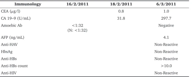
Table 3. Results of immunology test
Indonesian Journal of Clinical Pathology and Medical Laboratory, 2016 November; 23(1): 96–101 100
secretion processes of the canaliculi (inversion of the polarity function of hepatocytes can occur so that the movement of materials, such as conjugated bilirubin, bile acids and fats can be eliminated) into the bile through the plasma membrane surface of sinusoid can be disrupted.
In cholestasis, prolonged effects of detergents derived from bile acids will cause interference with cytochromes P-450, oxidation function, glucuronidase, sulfation and conjugation. Protein synthesis, such as alkaline phosphatase and GGT, will increase, while the production of albumin-globulin serum protein will decrease. In this patient, cholestasis occurred was caused because of calculus or pancreatic cancer triggering extrahepatic cholestasis. A blockage in the bile duct is more common in carcinoma in the pancreatic head, but does not cover the possibility of carcinoma in the pancreatic cauda. Aspartate aminotransaminase (AST) enzyme in this patient increased more than 10 times the upper range limit
could also be caused by extrahepatic obstruction as often found in patients with extrahepatic bile duct blockage, in which AST enzymes increase about 3–10 times URL values.2,5.8,9
In addition, in this patient, lipase levels also increased, whereas amylase levels were normal. Lipase is an enzyme secreted by pancreas into duodenum to destroy triglycerides into fatty acids. Together with amylase, lipase appears in the blood vessel damage or disease associated with pancreatic acinar cells. Previously, lipase is predicted to be simply excreted by pancreas, resulting in increased levels of lipase considered a pathological condition of the pancreas. But, now lipase levels are also associated with several conditions, such as infarction or bowel obstruction. Increased lipase in non-pancreatic disease cases is typically less than 3 times URL values. But, the increasing levels of lipase in the non-pancreatic disease cases are higher than those in pancreatic diseases (such as acute pancreatitis, chronic pancreatitis reoccurrence, pancreatic cancer, pancreatic pseudocyst), biliary diseases (such as acute cholecystitis, cholangitis, bile duct obstruction ), chronic kidney diseases, intestinal diseases (such as infarction and intestinal obstruction), or inflammation of salivary gland tumors, and peptic ulcers.1,9
Amylase, on the other hand, is an enzyme secreted by pancreatic acinar cells into the pancreatic duct and duodenum. Amylase enzyme helps catabolism of carbohydrates into simple sugar components in the gut. Pancreatic acinar cell damage or pancreatic obstruction (pancreatic cancer or bile duct stones) can trigger the release of enzymes into the lymphatic
system of intrapancreas and free peritoneum. Serum amylase examination is a sensitive examination for pancreatic disorders, but not specific ones. Amylase is also increased in other cases, such as peptic ulcer involving the pancreas, gastrointestinal disease, ruptured ectopic pregnancy, renal failure, and diabetic ketoacidosis.2,10,11
The fact that in this patient, lipase levels increased, but amylase levels were normal, moreover, is different from the theory explaining that increased levels of serum amylase can elevate levels of serum lipase due to the increase of amylase onset faster than the increase of lipase. In patients with acute pancreatitis, amylase levels usually begin to increase at 2–12 hours after onset, reach its peak 12–72 hours later, and return to normal within one week with a sensitivity of 75–92% and a specificity of 20–60%. Serum amylase levels then decrease faster because renal clearance is so rapid that the sensitivity becomes less than 30% after 2–4 days of the onset, whereas lipase serum levels increase in 4–8 hours after the onset of illness, reach its peak in 24 hours, and decrease in 8–14 day. Examination of lipase specificity and sensitivity is better than examination of amylase, which is in the amount of 55-99% and 86-100%, so the serum lipase levels decrease longer than amylase levels.2,5,9,12
The results of the immunological examination, furthermore, showed elevated CA 19-9, which was a glycoprotein of 36 kD associated with mucin.
Carbohydrate antigen (CA 19-9) is synthesized by normal cells of the pancreas and bile duct, gastric mucosa, colon, bronchial, salivary glands, endometrium, and prostate. But, in healthy individuals there is few CA 19-9. Consequently, the increasing of this antigen is associated with pancreatic cancer and
biliary tract cancer. The increasing of this antigen also occurs in other malignancies and benign conditions. Nevertheless, carbohydrate antigen (CA 19-9) is not used for pancreatic and hepatobiliary cancer screening, but can be used to support diagnosis, monitor response to therapy, and detect recurrence.2,8
In other words, pancreatic cancer diagnosis can be made based on clinical, physical, and laboratory examinations. It also often requires some means of support, such as radiology. Radiological examination, therefore, is needed to confirm the mass and location of the pancreas.
101
Pancreatic Cancer in 31 Years Old Patient - Flora, et al.
on both the duodenum, and changes in the anterior wall of the stomach. Third, Endoscopic Retrograde Cholangio Pancreatography (ERCP) examination was conducted with duodenoscopy using contrast media in the pancreatic head cancer. The results showed signs of obstruction or stenosis of the pancreatic duct. Thus, pancreatic fluid cytology examination could also be conducted. Fourth, percutaneous transhepatic cholangiography (PTC), especially for pancreatic cancer patients with extrahepatic cholestasis mark, was performed. Another important examination was ultrasonography (USG). However, the presence of intraluminal air and thick abdominal fat tissue can reduce the accuracy of the examination. The results of the examination showed irregular partial enlargement in pancreas depended on the location. Bile duct blockage mainly due to pancreatic head cancer showed signs of obstruction, such as the widening of the main bile duct, intrahepatic bile duct, and gall bladder enlargement. CT scan, on the other hand, illustrates the cross-section of the body and organs in our body. With the addition of contrast media, then the image generated will be better and accurate. Thus, it can show an irregular enlargement of the head of the pancreas and gallbladder enlargement accompanied by widening the intra and extrahepatic of the bile duct (positive Courvoisier signs).1,5,8
Finally, based on the results of the CT scan examination, magnetic resonance imaging (MRI) showed that there were pancreatic cauda, multiple nodule lesions in both lobes of the liver, deposition or infinitesimal calculus in the gallbladder, fluid ascites, as well as pleural effusion and consolidation on both lower lobes of the lung. Based on the results of the cytologic examination, there were also atypical cells in pleural fluid, indicating a malignancy. Based on the results of the fine-needle aspiration biopsy (FNAB) there were also malignant cells in the liver. Therefore, it can be concluded that the patient had a mass on his pancreatic cauda suspected as primary cancer of the pancreas with cholestasis metastasizing to the lung and liver.
CONCLUSION
This research reported a 31 years old male smoker with a chief complaint of pains in the pit of his stomach radiating to his back with a diagnosis of pancreatic cancer metastasizing to his liver and pleura. Thrombocytopenia can be considered as one of the
paraneoplastic syndrome symptoms, and normal serum amylase level can decrease due to a rapid decrease in amylase level. Thus, by the time of the examination, amylase levels had decreased within normal limits.
REFERENCES
1. Rasyad HS. Tumor Pankreas. In: Sulaiman H.A, Akbar H.N, Lesmana LA, Noer HMS. Buku Ajar Ilmu Penyakit Hati. 1st Ed., Jakarta, Jayabadi, 2007; 599–607.
2. Cubilla AL, Fitzgerald P.J. Morphological Patterns of Primary Nonendocrine Human. Aacrjournals. 1975; 35: 2234–2246. 3. Riess H, Goerke A, Oettle H. Pancreatic cancer. Germany,
Springer, 2008; 1–120.
4. Hadi S. Gastroenterologi. Ed ke-2., Bandung, P.T Alumni, 2002; 862–897.
5. Padmomatono FS. Tumor pankreas. In: Sudoyo A.W, Setiyohadi B, Alwi I, Simadibrata M, Setiati S. Buku Ajar Ilmu penyakit dalam. Jakarta, Interna Publishing, 2009; 739–746.
6. Sjamsuhidajat R, Jong WD. Buku Ajar Ilmu Bedah. 2nd Ed., Jakarta, EGC. 2005; 594–606.
7. Toskes PP, Greenberg NJ. Approach to the patient with pancreatic disease. In: Fauci AS, Braunwald E, Kasper DL, Longo DL, hauser SL, et al. Harrison’s principle of internal medicine. 17th Ed., Philadelphia, McGraw-Hill companies, 2008; 2001–2017. 8. Wilson DD. Manual of Laboratory and diagnostic tests. United
State, McGraw-Hill companies; 2008; 122–124.
9. Pagana KD, Pagana TJ. Manual of Diagnostics and laboratory tests. 3rd Ed., United State, Mosby Elsevier, 2006; 63–65, 349–351.
10. Blut h M H, Ha rdi n R E , Ten ner S, Zen i l ma n M E , Threatte GA. Laboratory Diagnosis of Gastrointestinal and Pancreatic Disorders. In: McPerson, Pincus. Henry Clinical Diagnosis and management of laboratory methods. 21st Ed., Brooklyn, W.B Saunders Company, 2006; 280–290.