Antimicrobial and Antioxidant Activity of Endophyte Bacteria Associated
with Curcuma longa Rhizome
Sulistiyani
1*, Tri Ardyati
2, Sri Winarsih
31
Master Program of Biology, Faculty of Mathematic and Natural Sciences, University of Brawijaya, Malang, Indonesia 2
Department of Biology, Faculty of Mathematic and Natural Sciences, University of Brawijaya, Malang, Indonesia 3
Department of Microbiology, Faculty of Medicine, University of Brawijaya, Malang, Indonesia
Abstract
Most cases of bacterial resistance towards antibiotics, encourage various efforts to gain new sources of antibiotics. Endophyte bacteria is a micoorganism has important role as the producer of bioactive compounds. Endophyte bacteria from Curcuma longa with antimicrobial and antioxidant activities have not been studied yet. Curcuma longa has been utilized as the main ingridients of traditional herbal medicines (jamu). The objective of this research was to investigate the antimicrobial and antioxidant activity of endophyte bacteria associated with Curcuma longa rhizome. Based on morphological characteristics of bacterial colonies, eight endophyte bacteria was isolates from Curcuma longa rhizome. Screening of endophyte isolate has antimicrobial activity was done using agar well diffusion method. The culture supernatant of each endophyte isolate was dropped on agar well against pathogenic bacteria Salmonella enterica ser. Typhi, Staphylococcus aureus and yeast Candida albicans. Three endophyte isolates K3, K2 and M1b showed antimicrobial activity against pathogenic bacteria and yeast. Isolate K3 showed strong antimicrobial activity againts C. albicans and S.
aureus, however isolate K2 and isolate M1b showed antimicrobial activity againts Salmonella enterica ser. Typhi and S. aureus, respectively. Those endophyte bacteria also had antioxidant activity shown by scavenging ability toward DPPH radical with consecutive percentage of isolate K3 (72.3 %), K2 (51.3 %) and M1b (64.6 %). Isolate K3 showed the highest antimicrobial and antioxidant activity. Based on biochemical characteristics using Microbact 24E kit, isolate K3 was identified as Paenibacilus alvei and isolate K2 as Enterobacter agglomerans.
Keywords: antimicrobial, antioxidant, Curcuma longa’s rhizome, endophyte bacteria.
INTRODUCTION
Recent main health care issues include the rise of antibiotic resistances and the rise of chronic and degenerative disease in countries throughout the world regardless of income level. The rise of antimicrobial resistance need the discovery and/or production of novel anti-microbial. Antioxidants, that have capability scavenging free radicals, are known to play important roles in preventing the degenerative, ROS-linked diseases. As the human population growth and the increase awareness on healthy life, people prefer natural compounds. Thus, the exploration of novel source of natural bioactive compound is unavoidable. One of the most promising source of natural bioactive compound is endophyte [1].
Endophytes are microorganisms, often bacte-ria, actinomycetes or fungi that live in healthy plant tissue intercellularly and/or intracellularly without causing any apparent symptoms of disease. Endophyte bacteria are found in roots,
Correspondence author: Sulistiyani
Email : [email protected]
Address : Master Program of Biology, University of
tubers, rhizome, nodule, stems, leaves, flowers, ovules, seeds and fruits of various plant species. In general roots have greater numbers of endophytes than above ground tissues [2]. Many evaluations of bacterial endophytes have shown that they are widespread in numerous plant kingdom. A single plant may have several different endophyte bacteria. The structure of bacterial endophyte communitas are varied, dynamic overtime, and attributed to plant source, plant age, tissue type, time of sampling, season and environment [3].
metabolites are found to apply as agrochemicals like insecticidal, growth-promoting, and their potential in the pharmaceutical like antibiotics, antioxidants, antitumor, antidiabetics, antipara-sitics, antithrombotic, anticancer and immuno-suppressants agents [4].
Medicinal plant is well known as source of precious bioactive compound. Endophytes that have long time associate with medicinal plant may participate in metaboloc pathway or gain some genetic information to produce specific bioactive compound similar to the host plant. Plant that have ethnobotanical history should be sourced of endophyte microbe. Therefore, it is to analyze the antimicrobial and antioxidant ac-tivity of endophyte bacteria associated with tur-meric rhizome.
MATERIALS AND METHODS Study Area
Sample of turmeric rhizomes were collected from Mondo Village, Mojo District, Kediri Regen-cy. The soil type in the research site is aluvial, along the area of Brantas Watershed. Soil acidity in the plant site is 6.34. Optimum pH of soil for most plants range 5.5 – 7.0 and nutrient will ad-sorbed well in the range pH 5.5 – 6.5 [5]. Refer to the soil pH at the sample site, it has qualified for the plant to grow well. Healthy ten months old plants were selected as source of rhizome for endophyte bacteria isolation.
Surface Sterilization of Turmeric rhizome
Rhizomes of turmeric were washed with run-ning tap water. The procedure includes sequen-tial immersion of rhizomes parts in 70% ethanol for 3 minutes, sodium hypochlorite 2% for 5 mi-nutes and 70% ethanol for 30 seconds, then rhi-zomes was washed using sterilized distilled water for five times [6]. The last twice washing solu-tions were plated on Nutrient Agar (NA) to con-firm the effectiveness of sterilization treatments. The surface of turmeric rhizomes were pilled out using aseptic technique and the inner tissues of rhizomes were macerated using a sterile mortar and pestle [7].
Isolation of Endophyte Bacteria from Curcuma longa Rhizome [8,9]
Total of 10 g turmeric rhizomes were ex-tracted then performed a serial dilution in saline solution (0.85% NaCl) and plated out in Nutrient Agar (NA) to recover endophyte bacteria present in the rhizome. All the plates were incubated at 28-30°C (room temperature) for 48 hours. The isolated bacteria were preliminary characterized according to their morphological characteristics. The distinct colony types were picked up from Gram-positive bacteria Staphylococcus aureus and Gram-negative Salmonella enterica ser. Typhi clinical isolates were used as test microorganism in this study. All pathogenic strains were ob-tained from Department of Microbiology, Medi-cal Faculty, University of Brawijaya. After 18-24 hours of incubation at 37⁰C (for bacterial strains) in NA and 30⁰C (for yeast strain) in PDA, a loop-ful of each test strains was suspended in sterile distilled water water until obtained 1 x 10⁶ cfu.mL-1 for bacteria and 105 cfu.mL-1 for yeast.
Assays of Antimicrobial Activity
Assay of Antioxidant Activity by Scavenging DPPH Free Radical
Endophyte bacteria culture (in NB medium) were centrifuged at 4000 rpm for 15 minutes, 4⁰C and then the supernatants were assayed their antioxidant activity by scavenging DPPH free radical methode [14] described with any modification. The supernatant (0.5 mL) was add-ed to 3 mL of 0.1 mM DPPH in methanol solu-tion. Methanol 1.5 mL was then added thus the final volume of solution was 5 mL. For control, supernatant of each sample was replaced by steril Nutrient Broth (NB). Methanol was used as blank. Discoloration of DPPH radical solution was measured at 517 nm in triplicate after incubation in the dark for 5 hours. Ascorbic acid was used as the positive control. Percentage of scavenged DPPH radical was calculated using following for-mula
% Scavenging = 𝐴₀−𝐴₁
𝐴₀ ∗ 100
A0 is the absorbance of control and A1 is the absorbance of sample (supernatant of endophyte bacteria culture) or standard. Ascorbic acid was taken at various concentrations as a known antioxidant for comparative analysis. Then the percentage of scavenging were plotted against respective concentrations used, and from the graph, EC50 was calculated.
Statistical analysis
The experimental results of biological activity tests were expressed as mean ± standard deviation (SD) of three replicates. The results were processed using Microsoft Excel 2007 and SPSS software. The data of antimicrobial activity assay results was analyzed using Kruskal-Wallis test followed by t-test and Tukey test whereas antioxidant activity using Anova following Tukey test.
RESULTS AND DISCUSSION
Based on the morphology characteristics of colony, we obtained eight isolates K1, K2, K3, K4, M1a, M1b, M5 and M6 of endhopytes bacteria. Each pured isolate was tested further for the antimicrobial and antioxidants activities.
Antimicrobial Activity
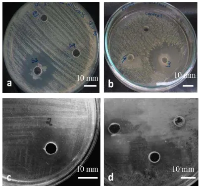
Three of eight isolates of turmeric endophyte bacteria has inhibition activity towards pathogen-ic test mpathogen-icroorganism. Isolate K3 inhibit the pa-thogenic yeast C. albicans (Fig. 1a) and
pathogen-K2 inhibit the pathogenic bacteria Salmonella
enterica ser. Typhi(Fig. 1c) and isolate M1b inhibit S. aureus (Fig. 1d).
Figure 1. Inhibition zone of endophyte bacteria isolates Isolate K3 towards Candida albicans (a) and Staphylococcus aureus (b); Isolate K2 towards
Salmonella enterica ser.Typhi (c); Isolate M1b
towards Staphylococcus aureus (d).
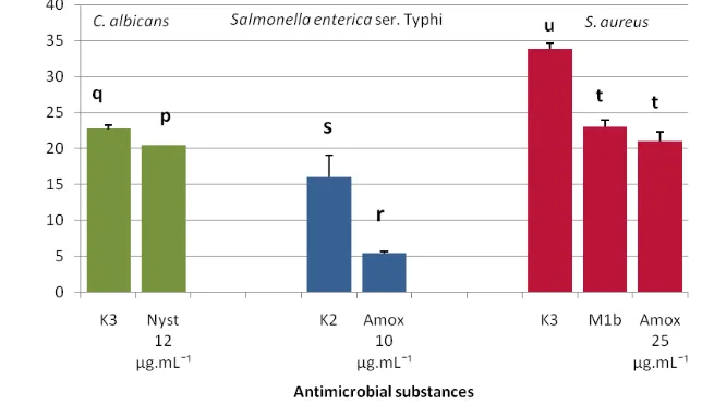
The results showed that the inhibition zone of isolate K3 towards C. albicans was greater than antifungal nystatin (12 µg.mL-1). It also showed similar results for inhibition zone of isolate K3 to S. aureus, which is greater than amoxicilin (25 µg.mL-1) and isolate K2 to Salmonella enterica ser. Typhi than amoxicilin (10 µg.mL-1). Other-wise, the inhibition zone of isolate M1b to S.
au-reus was relatively similar to the inhibition zone of amoxicillin (25 µg.mL-1) (Fig 2).
Microbes produce any substance for defense systems or survival mechanism. These include antibiotics, bacteriocins, metabolic by-products, lytic agents, numerous types of protein exotoxins, and short chain fatty acid [15]. This study found that Isolate K3 and M1b (Gram positive bacteria ) showed inhibition of growth towards the pathogenic bacteria S. aureus as Gram-positive bacteria and showed no inhibition towards Gram-negative bacteria S. enterica ser. Typhi. In contrast, isolate K2 showed inhibition to Gram- negative bacteria S. enterica ser. Typhi and showed no inhibition to the Gram-positive bacteria S. aureus and yeast C. albicans. These properties are similar or corresponding to the nature of bacteriocins that they have a relatively narrow killing spectrum and are toxic only to bacteria closely related to the producing strain.
1 cm
10 mm
10 mm
10 mm 10 mm
substance is bacteriocin. In addition more than 99% of bacteria can produce at least one bacteriocin and within a species tens or even
hundreds of different kinds of bacteriocins are present [15].
Figure 2. Antimicrobial Activity of Endophyte Bacteria Turmeric Rhizomes towards the Pathogenic Test Microorganism
Antioxidant Activity
The test of antioxidant activity on the eight isolates of endophyte bacteria showed that all isolates has the ability of scavenging to DPPH radical (Fig. 3). Three isolates with the highest antioxidant activity are isolate K3, K2, and M1b. Bacterial growth curve were made for the three isolates to obtain optimum time for sampling to test the antioxidant activity.
Antioxidant compound produced by the en-dophyte bacteria, as reported by scholar, is con-sisted of various substances. Antioxidant sub-stances produced by endophyte bacteria are EPS [16], surfactin [17], L-asparaginase [18], carote-noid pigment [19], and several enzymes [20]. Most of the compounds were produced maximal-ly at the end of exponential phase. Thus the sample for antioxidant activity was collected at the 14th hour (the end of exponential phase).
Before the test of antioxidant activity, OD of liquid culture of endophyte bacteria was equated. The test of antioxidant activity showed that isolate K3 has the highest ability of antioxi-dant activity compared to the other two isolates (Table 1) and then the EC50 of K3 isolate was determined.
Efficient Concentration or EC50 value is defined as the concentration of substrate that causes 50% loss of the DPPH activity (colour)[21]. Isolate K3 supernatant had EC50 value 70.26 µL.mL-1 and vitamin C (as standard) had EC50 value 3.71 µg.mL-1. In this study isolate K3 supernatant was still in original liquid and had not evaporated yet or extracted in to concentrate, so it was intelligible that the EC50 value was too lower than vitamin C. For next study may be required further processing of the supernatant.
10 mm
Figure 3. Antioxidant Activity of Curcuma longa Endophyte Bacteria Isolates Tabel 1. Antioxidant activity of 3 isolates
No Isolates Scavenging DPPH radical (%)
1 K2 51.3 ± 3.1
2 K3 72.0 ± 1.7
3 M1b 64.7 ± 2.5
Note: Each value is represented as mean ± SD (n=3)
Species Identification
Isolates K3 and K2 had both antimicrobial and antioxidant activity significantly , therefore need to characterize these isolates furthermore. Iso-late K3 and K2 was characterized based on the biochemical characteristic using Kit Microbact System 24E. Biochemical characteristics of isolate K2 were analyzed by the software Microbact System 24E and isolate K3 were ana-lyzed refer to Identification flow chart from Microbiology Laboratorium, The University of Ottawa Canada base on Bergey’s Manual of Determinative Bacteriology [22]. The results of characterization showed that isolate K2 was as-sumed as Enterobacter agglomerans with 99.9% similarity, whereas isolate K3 is Bacillus alvei or
Paenibacillus alvei with91.2% similarity.
Paenibacillus alvei are rod-shaped, Gram-positive, motile, spore-forming , catalase-positive bacteria and grow on simple media (NA/NB). Paenibacillus alvei are common found in honeybee colonies , soil , milk, mosquito larvae, the wax mot, humans and very rarely pathogenic for vertebrates. It has reported that Paenibacillus alvei produce antimicrobial substance: paenibacillin P and paenibacillin N [23], peptide AN5-1 [24], cyclic lipopeptides [25], depsipeptide [26]. Some of the antimicrobial substance show active againts pathogen S. aures and C. albicans [23,24,26,27] and consistent with this findings, this study showed endophyte bacteria from Curcuma longa’s rhizome, isolate K3 that assumed as Paenibacillus alvei show antimicrobial activity to S. aureus and C. albicans. Isolate K3 also show antioxidant activity, it promote previous research that Paenibacillus alvei’s metabolite have antioxidant activity Exopolysaccarides (EPS) [16,28,29]. Enterobacter agglomerans are rod shape, Gram negative, motile, non-sporforming bacteria. These bacteria first were isolated from plants, vegetable, fruits, seeds, and they are also commonly found in the ecological niches such as water, soil, sewage, feculent material, foodstuffs, clinical specimens [30]. It has reported that Enterobacter agglomerans produce antimicrobial substance that have inhibition growth to any pathogen
bacteria and fungi. Some of them have inhibition growth to Salmonella sp. like herbicolin O [31], phenazine [32], and consistent with these finding, isolate K2 that assumed as Enterobacter
agglomerans has antimicrobial activity to Salmonella enterica ser.Typhi. Isolate K2 also has antioxidant activity, it promote previous research that Enterobacter agglomerans has free radicals-scavenging ability [33].
CONCLUSION
The study obtained eigth isolates of endophyte bacteria from the Curcuma longa rhizomes. Three isolates of endophyte bacteria have antimicrobial activity, i.e. isolate K3 to C. antimicrobial to Gram positive pathogenic bacteria activity was showed by isolate K3 which identified as Paenibacillus alvei. The strong antimicrobial activity to Gram negative pathogenic bacteria and had high relative antioxidant activity was showed by Isolate K2 which identified as Enterobacter aglomerans by biochemical characterization.
REFERENCES
[1] Bintang, M., U.M.S. Purwanto, D.E. Kusumawati, J.J. Yang. 2015. Study of endophytic bacteria as novel source of antioxidant agent based on GC-MS Analysis. Int. J. Chem. Environ. Biol. Sci. 3(5). 368-369 [2] Anjum, N., R. Chandra. 2015. Endophyte bacteria: optimizaton of isolation procedure from various medicinal plants and their preliminary characterization. Asian J. Pharm. Clin. Res. 8(4). 233-238.
[3] Zinniel, D.K., P. Lambrectht, N.B. Harris, Z. Feng, D. Kuczmarski, P. Higley, C.A. Ishimaru, A. Arunakumari, R. G. Barletta, A.K. Vidaver. 2002. Isolation and charac-terization of endophyte colonizing bacteria from agronomic crops and prairie plants. Appl. Environ. Microbiol. 68(5). 2198-2208. [4] Huawei, Z., C. Ying, X. Bai. 2014.
Advancement in endophyte microbes from medicinal plants. Int. J. Pharm. Sci. Res. 5(5). 1589-1600.
and Soil Science. Available at: https:// pss.uvm.edu/ppp/pubs/oh34.htm.
[6] Ratti, R.P., N.F.G. Serrano, C.O. Hokka, C.P. Sousa. 2008. Antagonistic propertis of some microorganisms isolated from Brazilian tropical savannah plants against Staphylococcus coagulase-positive strain. J. Venom. Anim. Toxins incl. Trop. Dis. 14(2). 294-302.
[7] Sulistiyani, T.R., P. lisdiyanti, Y. Lestari. 2014. Population and diversity of endophytic bacteria associated with medicinal plant Curcuma zedoaria. Microbiol. Indonesia. 8. 65-72.
[8] Pranoto, E., G. Fauzi, Hingdri. 2014. Isolation and characterization of Endophyt Bacteria on highland productive and Young Tea Plant (Camellia Sinensis (L.) O. Kuntze) GMB 7 Clone. Biospecies. 7(1). 1-7.
[9] Wulandari, H., Zakiatulyaqin, Supriyanto. 2012. Isolation and antagonistic test of endophytic bacteria from pepper (Piper nigrum l.) againt velvet blight pathogen (Septobasidium sp.). J. Perkebunan dan Lahan Tropika. 2(2). 23-31.
[10] Tawiah, A., S.Y. Gbedema, F. Adu, V.E. Boamah, K. Annan. 2012. Antibiotic- producing microorganism from River Wiwi, Lake Bosomtwe and the Gulf of Guinea at Doakor Sea Beach, Ghana. Bio Med. Central Microbiol. 12(234). 1-8.
[11] Agricultural Research Service, United States Department of Agriculture (USDA). 2016. Table Breakpoints Used for Susceptibility Testing of Salmonella and E. coli. Available at: https://www.ars.usda.gov/ARSUserFiles /60400520/NARMS/ABXSalm.pdf.
[12] Sahputera, A. 2014. Uji efektifitas ekstrak madu karet dalam menghambat pertumbuh an S. aureus. Research Report. State Islamic University of Jakarta.
[13] Ellabib, M.S., I.A. El Jariny. 2001. In vitro activity of 6 antifungal agents on Candida species isolated as causative agents from vaginal and other clinical specimens. Saudi Med. J. 22(10). 860 – 863.
[14] Afify, A.M.M.R., R.M. Romeilah, S.I.M. Sultan, M.M. Hussein. 2012. Antioxidant activity and biological evaluations of probiotic bacteria strains. Int. J. Academic Res. Part A. 4(6). 131-139.
[15] Riley, M.A., M.A. Chavan. 2007. Bacteriocins: ecology and evolution. Springer. Verlag Berlin Heidelberg.
[16] Liu, J., J.G. Luo, H. Ye, Y. Sun, Z.X. Lu, X.X. Zeng. 2009. Production, characterization and antioxidant activities in vitro of exo-polysaccharides from endophyte bacterium P. polymyxa EJS - 3. Carbohydr. Polym. 78. 275–281.
[17] Yalçın, E., K. Çavuşoğlu. 2010. Structural analysis and antioxidant activity of a biosurfactant obtained from Bacillus subtilis RW-I. Turkish J. Biochem. 35(3). 243–247. [18] Nongkhlaw, F.M.W., S.R. Joshi. 2015.
L-asparaginase and antioxidant activity of endophytic bacteria associated with ethnomedicinal plants. Indian J. Biotech. 14. 59-64.
[19] Mohana, D.C, S. Thippeswamy, R.U. Abhishek. 2013. Antioxidant, antibacterial, an ultraviolet protective properties of carotenoids isolated from Micrococcus spp. Radiat. Prot. Environ. 36(4). 168-174. [20] Li, S., Y. Zhao, L. Zhang, X. Zhang, L. Huan.,
D. Li, C. Niu, Z. Yang, Q. Wang. 2012. Antioxidant activity of Lactobacillus plantarum strains isolated from traditional. Chinese foods. Food Chem. 135. 1914– 1919.
[21] Molyneux, P. 2004. The use of the stable free radical diphenylpicrylhydrazyl for estimating antioxidant activity. Songklana-karin J. Sci. Technol. 26(2). 211-219. [22] Microbiology Laboratorium, The University
of Ottawa Canada. 2016. Identification Flow Chart - Bergey’s Manual of Determinative Bacteriology. Available at: https://mysite. science.uottawa.ca/jbass/mirolab/IDFlowch arts.
[23] Anandaraj, B. 2008. Co-Production of two new peptide antibiotics and specific protease by a bacterial isolate Paenibacillus alvei NP 75, through ribosomal and non-ribosomal mediated protein synthesis machinery. Master Thesis. Faculty of Technology. Anna University, Chennai. [24] Alkotaini, B., N. Anuar, A.A.H. Kadhum,
A.A.A. Sani. 2013. Detection of secreted antimicrobial peptides isolated from cell-free culture supernatant of Paenibacillus alvei AN5. J. Indian Microbiol. Biotechnol. 40. 571–579.
from Paenibacillus. J. Am. Soc. Mass Spectrometry. 26(10). 1768-1779.
[26] Chevrot, R., S. Didelot, L. Van den Bossche, F. Tambadou, T. Caradec, P. Marchand, E. Izquierdo, V. Sopéna, E. Rosenfeld. 2013. Probiotics Antimicrob. Proteins. 5(1). 18-25. [27] Pancevska, N.A, I. Popovska, K. Davalieva, J.
Kunguloski. 2016. Screening for antimicrobial activity of Bacillus subtilis and Paenibacillus alvei isolated from rotten apples compost. Acta Microbiologica Bulgarica. January. 56-64.
[28] Aziz, S.M.A., H.A. Hamed, M. Fadel, M.E. Moharam. 2015. Properties and role of exopolysaccharides produced by Paenibacillus alvei NRC14 for cell protection. J. Appl. Sci. 1(3. 35-47.
[29] Selim M. S., S.S. Mohamed, R. H. Shimaa, M.E. El Awady, O.H. El Sayed. 2015. Screening of bacterial antioxidant exopolysaccharides isolated from Egyptian habitats. J. Chem. Pharm. Res. 7(4). 980-986.
[30] Quecine, M.C., W.L. Araujo, P.B. Rossetto, A. Ferreira, S. Tsui, P.T. Lacava, M. Mondin, J.L. Azevedo, A.A. Pizzirani-Kleinera. 2012. Sugarcane growth promotion by the
endophytic bacterium Pantoea
agglomerans 33.1. Appl. Environ. Microbiol. 78. 7511–7518.
[31] Ishimaru, C.A., E.J. Klos, R.C. Brubaker. 1988. Multiple antibiotic production by Erwinia herbicola. Dis. Control Pest Man-age. 78(6). 746-750.
[32] Lim, J.A., D.H. Lee, B.Y. Kim, S. Heu. 2014. Draft genome sequence of Pantoea agglomerans R190, a producer of antibiotics against phytopathogens and foodborne pathogens. J. Biotechnol. 188. 7-8.