A
Flavonoid Glycoside Compound Isolated from
Macaranga gigantifolia
Merr Leaves
Gian Primahana1, Akhmad Darmawan1,*
1
Research Center for Chemistry, Indonesian Institute of Sciences, Kawasan PUSPIPTEK, Serpong, Banten, Indonesia.
*Corresponding author : [email protected]
Received 3 August 2016 ; Revised 6 January 2017 ; Accepted 9 January 2017
ABSTRACT
Apigenin-8-C-glycoside (1), a flavonoid glycoside compound has been isolated from the leaves of Macaranga gigantifolia Merr. Isolation and purification was conducted by chromatography methods and chemical structure characterization was carried out by spectroscopic methods. Cytotoxicity of apigenin-8-C-glycoside has been tested against Murine leukemia P-388 cell lines and has moderate activity with IC50 values 55.4 μg/mL.
Key words: Apigenin-8-C-glycoside, Macaranga gigantifolia Merr., Murine leukemia P-388 cell lines.
INTRODUCTION
Macaranga gigantifolia Merr. (Euphorbiaceae), known locally as mahang-mahangan (Indonesia) is one of about 300 species member of genus Macaranga [1]. Some of species,
including Macaranga tanarius, Macaranga denticulate, Macaranga adenantha, and
Macaranga alnifolia has been extensively studied, indicated that flavonoid and terpenoid are the major compounds of the Macaranga genus [1-10]. Many part of this species has been used as traditional medicines. Root of M. denticulate has been used in Traditional Chinese Medicine for treatment icteric hepatitis [7], M. indica used for treatment anemia and tumor in India and Bangladesh [11], M. peltala used to relieve from venereal sores and also reported that active as antioxidant, antimicrobial and cytotoxic activity [12].
As part of our study about natural products drug discovery, including exploration and the potentiality bioactive plants origin from Indonesia, ethyl acetate fraction of methanol extract of M. gigantifolia Merr. leaves were separated using column chromatography method. Apigenin (5,7,4’-trihydroxyflavone) and its glycosides which has been reported active as anticancer, anti-inflammatory and antioxidant [13-16], is naturally abundant component from common flavonoids plants found in flowers, fruits, vegetables, beans and tea. To the best of author knowledge,there is lack of paper published about chemical content of M. gigantifolia
EXPERIMENT
Chemicals and instrumentation
All chemical used were obtained from Merck with pro analytical grade. Solvent used for maceration and column chromatography is technical grade and redistilled before use. 1Dand 2D-NMR spectra were recorded on JEOL JNM-ECA 500 spectrometer with TMS as internal standard. LC-MS were measured with Mariner Biospectrometry-Finnigan instrument. Column chromatography method was carried out with silica gel (200-300 mesh, Kieselgel 60, Merck) using n-hexane:ethyl acetate as solvent system for isolation and silica gel 60 F254
(Merck) for TLC with 5% H2SO4 in ethanol as compound detection reagent.
Plant material
The leaves of Macaranga gigantifolia Merr was collected from Mekongga Forest, District of Kolaka, Southeast Sulawesi, Indonesia in March 2012. The plant was determined at Herbarium Bogoriense, Research Center for Biology, Indonesian Institute of Sciences, Bogor, Indonesia.
Isolation of compound I
About 80 grams MeOH extract was partitioned with n-hexane and EtOAc successively.
The EtOAc soluble fraction (17.4 g) was chromatographed over silica gel column, and eluted with gradient solvent system of n-hexane:EtOAc started from 20% ethyl acetate to 40% ethyl acetate with 5% increasement to obtain 5 fractions (F1-F5). Compound 1 (28 mg) was crystallized in the F1 bottle (eluted with 20% ethyl acetate) after evaporation and re-diluted with EtOAc and further successively purified with CHCl3, acetone and MeOH.
Compound 1: yellow powder, ESI-MS m/z 431.0963 [M-H], NMR (JEOL JNM-ECA
RESULT AND DISCUSSION
Dried M. gigantifolia leaves extracted with methanol and further were partitioned successively with n-hexane and EtOAc. EtOAc fraction was chromatographed over silica gel column chromatography, eluted with a gradient solvent system of n-hexane:EtOAc (8:2 to 6:4) and was obtained 5 fractions (F1-F5). Compound 1 was crystallized in the F1 bottle (eluted with 20% ethyl acetate) after evaporation and re-diluted with EtOAc and further successively purified with CHCl3, acetone and MeOH.
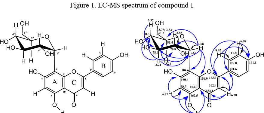
The 1H-NMR and 13C-NMR spectra of compound 1, showed 4 aromatic protons at δH 6.88 (2H, d, J=8.9 Hz))/δC 115.8, and 8.02 (2H, d, J=8.9 Hz)/δC 129.0, indicated the presence of A2B2 ring system. In addition, singlet signals at δH 6.27/δC 98.1, and 6.78/δC 102.4, were
belong to two proton atom which are attached to ring A and C, respectively. The position of two singlet proton which mentioned above was supported by the correlation data of HMBC (Fig. 2). Based on this chemical shift data, compound 1 was predicted as apigenin derivative. The anomeric proton (H-1”) at δH 4.68 (d, 1H, J=9.7 Hz) correlated to carbon peak at δC 73.4 indicated that the linkage between the sugar residue and aglycone (flavone) is C-glycosidic system at C-8 of the aglycon carbon atom. The C-glycosidic linkage was confirmed by
HMBC correlations between anomeric proton at δH4.68 with carbon atom at δC 104.6 (C-8)
and 156.0 (C-8a) (Fig. 2). Mass spectroscopic analysis using LC-MS (Fig. 1) showed that compound 1 has molecular peak at m/z 431.0963 [M-H] correspond to molecular weight 432.0963 with molecular formula C21H20O10. From the 1D- and 2D-NMR data, confirmed by
mass spectroscopic data and compared with the reference (Table 1) [21], compound 1 was identified as Apigenin-8-C-glycoside (Fig. 2). This is the first report apigenin-8-C-glycoside isolated from M. gigantifolia Merr. plant.
Figure 1. LC-MS spectrum of compound 1
Figure 2. Chemical structure of apigenin-8-C-glycoside (1) and their HMQC-HMBC Correlation
Cytotoxicity activity of compound 1 against Murine Leukimia P-388 cancer cell lines was performed using MTT assay. Calculation of IC50 showed that Apigenin-8-C-glycoside
has moderate activity with IC5055.4 μg/mL.
CONCLUSION
After isolation, purification and characterization of the phenolic compound isolated from ethyl acetate fraction of the methanol extract M. gigantifolia leaves, it can be concluded that compound 1 is apigenin-8-C-glycoside, which have moderate cytotoxicity activity against
Murine leukemia P-388 cell lines with IC5055.4 μg/mL.
ACKNOWLEDGMENT
This work was supported by Ministry of Research and Technology, Republic of Indonesia and LIPI Competitive Project. We also thank to Mr. Ismail Rahman for collected and determinate the Macaranga gigantifolia plant.
REFERENCES
[1] Blattner, F. R., Weising, K., Banfer, G., Maschwitz, U., Fiala, B., Mol. Phylogen. Evol.,
2001, 19, 331-334.
[3] Tanjung, M., Mujahidin, D., Juliawaty, L. D., Makmur, L., Achmad, S. S., Hakim, E. H., Syah, Y. M., Proceeding of The International Seminar on Chemistry, Padjajaran University, Jatinangor, October 30-31, 2008, 252-255.
[4] Dinh, V., Zhang, H. P., Duc, N. M., Tuu, N. V., Qin G. W., J. Asian Nat. Prod. Res.,
2006, 8(2), 155-158.
[5] Zakaria, I., Ahmat, N., Ahmad, R., Jaafar, F.M., Ghani, N.A., Khamis, S., World Appl. Sci. J., 2010, 9(9), 1003-1007.
[6] Tseng, M. H., Chou, C. H., Chen, Y. M., Kuo, Y. H., J. Nat. Prod., 2001, 64, 827-828. [7] Yang, D. S., Li, Z. L., Wang, X., Yan, H., Yang, Y. P., Luo, H. R., Liu, K. C., Xiao, W.
L., and Li, X. L., Fitoterapia, 2014, 99, 261–266.
[8] Yoder, B. J., Cao, S., Norris, A., Miller, J. S., Ratovoson, F., Razafitsalama. J., Andriantsiferana, R., Rasamison, V. E. and Kingston, D. G. I., J. Nat. Prod., 2007, 70(3), 342–346.
[9] Phommart, S., Sutthivaiyakit, P., Chimnoi, N., Ruchirawat, S., Sutthivaiyakit, S., J. Nat. Prod., 2005, 68, 927-930.
[10] Yang, D. S., Wang, S. M., Peng, W. B., Yang, Y. P., Liu, K. C., Li, X. L., Xiao, W. L.,
Nat. Prod. Bioprospect., 2015, 5, 105–109.
[11]Amina, K., Mahmudur, R., Afroza, A., Sumiya, I., Mahfuja, A., Sumaiya, K., World J. Pharm. Res., 2014, 3(10), 172-182.
[12] Meenakshi, V. M., Raj, P. V., Chandrasekhar, H. R., Rao, J. V. and Udupa, N.,
International Conference on Biomedical and Pharmaceutical Engineering, Dec 2-4,
2009, 1-3.
[13] Redaelli, C., Formentini, L., Santaniello, E., Phytochemistry. 1980, 21(7), 1828-1830.
[14] Bao, Y. Y., Zhou, S. H., Fan, J. and Wang, Q. Y., Future Oncol., 2013, 9(9), 1353-1364.
[15] Cai, J., Zhao, X. L., Liu, A. W., Nian, H., Zhang, S. H., Phytomedicine, 2011, 18, 366-373.
[16] Ma, J., Li, Q., Zhao, J., Ying, G., Su, Q., Ji, Z., J. Nanjing Med. U., 2007, 21(2), 94-98. [17] Darmawan, A., Kosela, S., Kardono, L. B. S., Syah, Y. M., J. Appl. Pharm. Sci., 2012,
2(12), 175-177.
[18] Darmawan, A., Wahyudi, P. S., Kosela, S., Kardono, L. B. S., Fajriah, S., Indones. J. Pharm., 2015, 26(1), 52-56.
[19] Fajriah, S., Megawati, M., Darmawan, A., J. Trop. Life. Sci., 2016, 6(1), 7-9.
[20] Sahidin, S., Hakim, E. H., Juliawaty, L. D., Syah, M. Y., Din, L. B., Ghisalberti, E. L., Latif, J., Said, I. M., Achmad, S. A., Z. Naturforsch., 2005, 60(9-10), 723-727.
![Table 1. 1H- and 13C-NMR Data of Compound 1 and apigenin-8-C-glycoside Apigenin-8-C-glucoside [21]](https://thumb-ap.123doks.com/thumbv2/123dok/2862021.1694491/3.595.99.500.436.710/table-nmr-data-compound-apigenin-glycoside-apigenin-glucoside.webp)