ABSTRACT
Aim: to investigate which recent infection could have caused the present dengue-like symptoms, in adult patients clinically fulfilling the WHO criteria for dengue, in which serologically were not confirmed for dengue virus infections.
Methods: prospective study. During an outbreak of dengue (between May 1995 and May 1996) 118 consecutive adults (>13 years) suspected by the WHO 1997 case definition of DF or DHF were investigated. Patients were examined for history of illness, physical and laboratory findings consisting of full blood counts, prothrombin time (PT), activated partial thromboplastin time (aPTT), liver function (bilirubin, ASAT, ALAT), renal function (creatinine), and serological assays included dengue, hantavirus, chikungunya, R. typhi, R. tsutsugamuchi, rubella virus, influenza A virus, and leptospira.
Results: in 58 of the total 118 patients, recent dengue virus infection was serologically confirmed. In 20 of the remaining 60 patients, we found serological evidence of another recent infection: hantavirus (5), chikungunya virus (2), R. typhi (5), R. tsutsugamuchi (2), rubella virus (3), influenza A virus (1), and leptospira (2). No evidence for recent infection with any of the mentioned agents was detected in the remaining 40 specimens.
Conclusion: we conclude that based on clinical characteristics alone, it is not easy to diagnose dengue. Specific laboratory tests to differentiate dengue from other febrile illnesses are needed. Among these, in Indonesia hantavirus infection should be considered as well.
Key words: hantavirus, dengue virus infection, rickettsia, chikungunya.
Hanta Virus Infection During Dengue Virus Infection
Outbreak in Indonesia
Catharina Suharti*, Eric C.M. van Gorp**, Wil M.V. Dolmans***, Jan Groen****,
Suharyo Hadisaputro*, Robert J. Djokomoeljanto*, Osterhaus Ab D.M.E.****,
Jos W.M. van der Meer***
* Department of Internal Medicine, Faculty of Medicine, Diponegoro University-Dr. Kariadi Hospital. Jl. Dr. Soetomo no. 18, Semarang, Indonesia, ** Department of Internal Medicine, Slotervaart Hospital, Amsterdam,
*** Department of Medicine, University Medical Centre St Radboud, Nijmegen, The Netherlands, **** Institute of Virology, Erasmus University Hospital, Rotterdam, The Netherlands.
Correspondence mail to: [email protected].
INTRODUCTION
Indonesia has a tropical climate and humidity which is conducive to perpetuation of Aedes aegypti, the main vector of dengue. Dengue virus belongs to the genus
Flavivirus, family Flaviviridae and may cause a wide spectrum of illness: asymptomatic infection, dengue
fever (DF) and dengue hemorrhagic fever (DHF).1
Clinical signs and symptoms of dengue fever are not specific and include sudden onset of fever, weakness, headache, joint pain, muscle pain, retro-orbital pain, nausea, vomiting, and rash. The less severe grades of DHF (grade I and II) manifest similar to DF with signs of spontaneous bleeding, and are difficult to distinguish from other viral illnesses found in tropical areas such as chikungunya virus infection (which is also transmitted by Ae. Aegypti), influenza, mild hantavirus infection and rubella, as well as bacterial infections such as mild leptospirosis and rickettsiosis.1-7
Epidemiological studies have demonstrated that during outbreaks of dengue 10.5% to 42.7% of cases can be detected by virus isolation,8,9 46.3% by IgM/IgG
ELISA,9 41.8% by hemagglutination inhibition (HI)
tests,10 41.4 to 66% by IgM ELISA and/or virus
isolation, 11,12 and 73% by HI and/or virus isolation.13 Thus,
the rest of these patients may actually suffer from another febrile illnesses. Alternatively, the sensitivity of tests used may have been low, or a wrong sampling time-point for the test was used.
be the cause of their present illness in the remaining patients. Since infections with chikungunya virus,
hantavirus, Rickettsia typhi, R. tsutsugamushi,
leptospira, rubella virus and influenza virus may present with the same clinical characteristics as dengue and may be encountered in Indonesia, the specimens were examined for serological evidence of these infections. We also investigated whether clinical symptoms and signs may help to differentiate between these infections.
METHODS
Patients and Data Collection
The study was performed in Dr. Kariadi Hospital, the university hospital of Diponegoro University, Semarang, Indonesia. Between May 1995 and May 1996, during an outbreak of dengue, 118 consecutive adults (>13 years), clinically suspected by the WHO 1997 case definition(1) of DF or DHF, were examined prospectively.
Of each patient, data were collected on a clinical record form, listing for each patient during hospital stay: name, age, sex, history of illness (date of onset of disease and date of admission), symptoms, physical findings and laboratory findings, including blood tests for cell counts, prothrombin time (PT), activated partial thromboplastin time (aPTT), liver function (bilirubin, ASAT, ALAT), renal function (creatinine). Chest x-ray, ECG and ultrasonography were not done routinely.
For serological assays, 4 ml of blood was collected in vacutainer tubes (Becton Dickinson, Rutherford, NJ 07417) on the day of admission, on the day of discharge, and a week after discharge. The third specimen was omitted when the second specimen was two weeks or more after the first. Specimens were kept at minus 800C
until assayed.
Serological Assays
All assays were performed at the virology laboratory of Erasmus University Hospital, Rotterdam, The Netherlands. Air transport of the samples from Semarang to The Netherlands was done on dry ice.
Samples were tested for the presence of specific antibodies against dengue virus by IgM capture/IgG
enzyme linked immunosorbent assay (ELISA).14
A recent dengue infection was diagnosed if IgM antibodies were demonstrated or if a fourfold rise of IgG antibodies between admission and at least two weeks later was demonstrated. All samples in which no recent dengue virus infection was found, were tested for antibodies against chikungunya virus, hantavirus, influenza A virus, leptospira, and Richettsia typhi. For
(IgG), Hantavirus (IgM and IgG) and influenza A (IgA), different ELISA-based systems were used, as described previously.3,15-17 A greater than two times rise of IgG
antibody titer against chikungunya virus between admission and at least two weeks later, was considered to represent a recent infection. If IgM antibodies against hantavirus were detected in ELISA, those were confirmed by immunofluorescence (IFA); the presence of IgM antibodies plus confirmed by IFA, was considered to represent a recent infection. A rising IgA titer against influenza A between admission and at least two weeks later was considered to represent a recent infection with influenza A virus. For the diagnosis of leptospirosis a haemagglutination inhibition (HI) test was
done.(18) Only seroconversion between admission and a
sample of at least two weeks later was considered to
represent recent infection. IgM antibodies against R.
typhi were measured by immunofluorescence (INDX, Baltimore, USA); an antibody titer > 1:32 was considered to represent recent infection.
The sera in which no evidence for a recent infection with dengue virus, nor with any of the above mentioned other agents were demonstrated, were tested for the presence of IgM antibodies against rubella virus, using a IgM capture assay (Organon Technika, Boxtel, The Netherlands) and for the presence of IgM antibodies
against R. tsutsugamushi by immunofluorescence
(INDX, Baltimore, USA; positive titer >1:32).
Statistical Analysis
Continuous data were described as median (range) and nominal data were described in number (%). All statistical analyses were performed with SPSS for Windows version 9.0.
RESULTS
Patients with Confirmed Recent Dengue Virus Infection (n=58)
In 58 patients, recent dengue virus infection was serologically confirmed. According to the 1997 WHO criteria, the classification of the 58 patients were: DF (42), DHF I (4), DHF II (7), DHF III (4), DHF IV (1). Their median age (range) in years was 19 (14-53) and sex ratio (M/F) 28/30.
Patients without Serological Evidence of Recent Dengue Virus Infection (n=60)
60 patients, we found serological evidence of another recent infection: hantavirus (5), chikungunya virus (2),
R. typhi (5), R. tsutsugamushi (2), rubella virus (3), influenza A virus in (1), and leptospira (2). No evidence for recent infection with any of the mentioned agents was detected in the remaining 40 specimens. Evidence for infection with chikungunya virus in the past was found in 12 patients (IgG antibodies, but no rising titer). Three of these 12 patients had recent infections with dengue virus (1), hantavirus (1), and R. typhi (1). Also, evidence for hantavirus infection in the past was found in two patients.
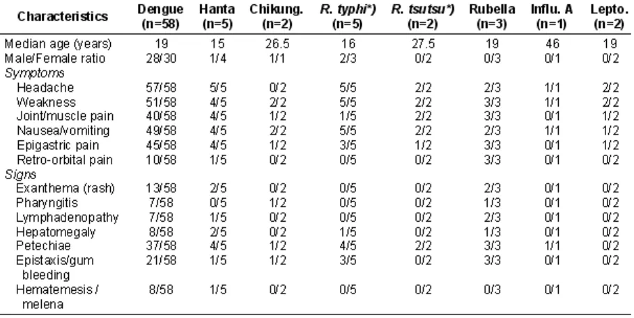
Clinical Characteristics
Table 1 shows the distribution of clinical variables of 58 patients with confirmed recent dengue and 20 with evidence of recent infection with chikungunya virus, hantavirus, R. typhi, R. tsutsugamushi, rubella virus, influenza A virus or leptospira. Headache, weakness, joint and/or muscle pain, epigastric pain, nausea, vomiting, were the most commonly presented symptoms/ signs in all patients regardless of their final diagnosis. Bleeding manifestations were usually mild consisting of petechiae, gum bleeding and epistaxis. Hematemesis and/ or melena were found in some patients with dengue and one with hantavirus infection classified on admission as DHF III. One patient with confirmed recent dengue virus infection (DHF IV) died, the remaining 117 patients recovered fully after a hospital stay of 2 to 18 days (median 6 days).
Laboratory Results
As shown in table 2 the median levels of hemoglobin, haematocrites and leukocytes were normal in all cases. Marked thrombocytopenia was found in patients with dengue and in some patients with hantavirus and R. tsutsugamushi infection. A prolongation of coagulation parameters was seen in some patients with dengue (aPTT, PT), chikungunya (PT), hantavirus infection (aPTT) and R. typhi infection (PT). A mild to moderate elevation of ASAT and ALAT were found in dengue virus infection and in some patients with rubella
virus, hantavirus, chikungunya virus and R. typhi
infection. A moderate increase of creatinine (up to 4.3 mg/dl) was found in some patients with dengue virus infection. Three of five patients with hantavirus infection had proteinuria and microscopic hematuria , but in all of them serum creatinin was normal.
DISCUSSION
virus, influenza A virus, or leptospira was demonstrated. The clinical symptoms and signs of these 20 patients (table 1) were very similar to those of the 58 patients whose present illness was due to dengue. Most of these patients had mild dengue virus infection (DF or DHF I or II). Our findings in adult patients with suspected dengue contrasted with those in 50 children with dengue shock syndrome (DHF III or DHF IV), in whom the diagnosis dengue virus infection was confirmed in all cases.19 The explanation probably is that the clinical
picture of severe dengue virus infection (DSS) is more specific.
IgM antibodies against hantavirus were detected in 5 patients indicating recent infection, and in two patients with IgG with no rising titer indicating infection in the past. On admission, four of these five patients with recent hantavirus infection were clinically diagnosed as DF, whereas one patient had a more severe clinical presentation, and was originally diagnosed as DHF III. None of these patients had traveled outside Indonesia. These are the first reported hantavirus infections in humans from Indonesia. Hantavirus infection may manifest as mild or severe disease. The different
and disease severity.(20) Severe disease may manifest
itself as Hantavirus Pulmonary Syndrome (HPS) or Hemorrhagic Fever with Renal Syndrome (HFRS). HPS may present as a non-specific illness with fever, myalgia, headache, and gastrointestinal symptoms, followed by a non-productive cough, culminating rapidly in respiratory insufficiency. Severe HFRS is characterized by extreme albuminuria and impaired renal function.21-24 Three of five patients with hantavirus
infection had proteinuria and microscopic hematuria for several days, but no impaired renal function.
We demonstrated antibodies against chikungunya in 14 patients but only in two patients the antibody profile indicated recent infection. On admission, these two patients were clinically diagnosed as DF and DHF I. Chikungunya is difficult to differentiate from DF or mild DHF due to the overlap in clinical symptoms and signs (table 2) as also was described previously.1,3,25
We detected IgM antibodies against R. typhi in 5
patients. This disease is known to occur in Indonesia.26-28
Our five patients had mild rickettsiosis and had clinically
been suspected of DF. Antibodies against R.
We detected IgM antibodies against rubella virus in 3 patients. These patients were originally diagnosed as DF in one and DHF II in two patients. A similar finding has been reported previously.29
Antibodies against influenza A virus were found in one patient, clinically diagnosed as DF on admission. Also previous studies demonstrated that influenza can mimic mild dengue infection.4,30
We found antibodies against leptospira in two patients. Manifestations of leptospirosis may vary from a mild flu-like illness to an acute life-threatening condition with renal failure (Weil’s disease). Our two patients had mild leptospirosis and were mistaken for DF on admission. Both dengue and leptospirosis are endemic in Indonesia. During a large outbreak of leptospirosis in Salvador, Brazil, 42 percent of cases were misdiagnosed as dengue fever in the outpatient clinic.6
Also, substantial misdiagnosis of dengue and leptospirosis occurred in Barbados. During 1995, 48 of 108 sera and during 1996, 21 of 64 sera from leptospirosis-negative patients were found to have IgM antibodies to dengue
virus.5 On the other hand, after hurricane-generated
floods in Puerto Rico, leptospirosis was diagnosed in patients suspected clinically of dengue, but testing dengue-seronegative.31
In the remaining 40 patients, we failed to demonstrate the presence of another recent infection. This may be due to the presence of yet another disease, not tested for. For instance, in patients clinically suspected of dengue, typhoid fever with a positive
culture for Salmonella typhi (Hadinegoro SR,
unpublished), and measles32 have been diagnosed.
We did not test for Japanese encephalitis, also caused by a flavivirus, but none of the patients had any manifestations of this disease, which in most cases runs a severe course and is often fatal. The other flavivirus causing Yellow Fever does not occur in Indonesia.
CONCLUSION
It is difficult to clinically distinguish dengue from other febrile illnesses. This applies particularly to dengue fever and the milder grades of dengue hemorrhagic fever (DHF I and II) in adults. Which specific laboratory tests are to be used depends on the geographi-cal distribution of diseases. Differentiating other infectious agents not only is of epidemiological but also of clinical interest, because adequate antibiotic treatment can be instituted in some of these infections, reducing morbidity and mortality. Hantavirus infection should be added to the differential diagnosis of dengue virus infection in Indonesia.
ACKNOWLEDGEMENTS
Participants in this project, besides the authors, were Dr. Budi Riyanto, Dr. M. H. Gasem and Dr L.Widiono from the Department of Internal Medicine, Dr. Kariadi Hospital, Diponegoro University, Semarang, Indonesia. We thank Y.T. van der Heide from the Clinical Chemistry and Hematology Laboratory, Slotervaart Hospital, Amsterdam, The Netherlands, for managing the blood samples. Ms P. Koraka and C. Copra of the Virological laboratory, Erasmus University Hospital Rotterdam, The Netherlands for excelent technical assistance. We thank also Royal Netherlands Academy of Arts and Science for financial support.
REFERENCES
1. World Health Organization. Dengue hemorrhagic fever: diagnosis, treatment, prevention and control. 2nd ed. Geneva: WHO; 1997. p. 12-47.
2. Gubler DJ. Dengue and dengue hemorrhagic fever. Clin Microbiol Rev. 1998; 11: 480-96.
3. Nur YA, Groen J, Heuvelmans H, Tuynman W, Copra C, Osterhaus AD. An outbreak of West Nile Fever among migrants in Kisangani, Democratic Republic of Congo. Am J Trop Med Hyg. 1999;61:885-8.
4. Silarug N, Foy HM, Kupradinon S, Nisalak A, Pongsuwant Y. Epidemic of fever of unknown origin in rural Thailand, caused by influenza A (HINI) and dengue fever. Southeast Asian J Trop Med Pub Hlth. 1990;21:61-7.
5. Levett PN, Branch SL, Edwards CN. Detection of dengue in patients investigated for leptospirosis in Barbados. Am J Trop Med Hyg. 2000;62:112-4.
6. KO AI, Galvao Reis M, RibeiroDourado CM, Johnson WD Jr, Riley LW. Urban epidemic of severe leptospirosis in Brazil, Salvador Leptospirosis Study Group. Lancet. 1999;354:820-5.
7. Clement J, Neild G, Hinrichsen SL, Crescente JA, Van Ranst M. Urban leptospirosis versus urban hantavirus infection in Brazil. Lancet. 1999;354:2003-4.
8. Sharp TW, Wallace MR, Hayes CG, Sanchez JL, DeFraites RF, Arthur RR, Thornton SA, Batchelor RA, Rozmajzl PJ, Hanson RK, Wu SJ, Iriye C, Burans JP. Dengue fever in U.S. troops during operation restore hope, Somalia, 1992-1993. Am J Trop Med Hyg. 1995;53:89-94.
9. Innis BL, Nisalak A, Nimmannitya S, Kusalerdcharya S, Chongswasdi V, Suntayakorn S, Puttisri P, Hoke CH. An enzyme -linked immunosorbent assay to characterize dengue infections where dengue and Japanese Encephalitis co-circulate. Am J Trop Med Hyg. 1989;40:418-27.
10. Chairulfatah A, Setiabudi D, Ridad A, Colebunders R. Clinical manifestations of dengue hemorrhagic fever in children in Bandung, Indonesia. Ann Soc Belg Med Trop. 1995;75:291-5. 11. Deparis X, Murgue B, Roche C, Cassar O, Chungue E. Changing clinical and biological manifestations of dengue during the dengue-2 epidemic in French Polynesia in 1996/ 1997-description and analysis in a prospective study. Trop Med Int Health. 1998;3:859-65.
outbreak of dengue hemorrhagic fever in Irian Jaya, Indonesia. Am J Trop Med Hyg. 1997;57:49-55.
13. Kuberski T, Rosen L, Reed D. Mataika J. Clinical and laboratory observations on patients with primary and secondary dengue type 1 infections with hemorrhagic manifestations in Fiji. Am J Trop Med Hyg. 1977;26:775-83. 14. Groen J, Velzing J, Copra C, Balentien E, Deubel V, Vorndam V, Osterhaus AD. Diagnostic value of dengue virus-specific IgA and IgM antibody detection. Microbes Infect. 1999;13:1085-90.
15. Groen J, Gerding M, Jordans JG, Clement JP, Osterhaus AD. Class and subclass distribution of Hantavirus-specific serum antibodies at different times after the onset of nephropathia epidemica. J Med Virol. 1994;43:39-43.
16. Rothbarth PH, Groen J, Bohnen AM, de Groot R, Osterhaus AD. Influenza virus serology: a comparative study. J Virol Methods. 1999;78:163-9.
17. Moll van Charante AW, Groen J, Mulder PGH, Rijpkema SG, Osterhaus ADME. Occupational risks of zoonotic infections in Dutch forestry workers and muskrat catchers. Eur J Epidemiol. 1998;14:109-16.
18. Levett PN, Whittington CU. Evaluation of the indirect hemagglutination assay for diagnosis of acute leptospirosis. J Clin Microbiol. 1998;36:11-4.
19. Suharti C, Setiati TE, van Gorp ECM, Djokomoeljanto RJ, Trastotenoyo MS, van der Meer JWM, Dolmans WMV. Clinical picture and risk factors for mortality in dengue shock syndrome, submitted for publication.
20. Mc Caughey C, Hart CA. Hanta viruses. J Med Microbiol. 2000;49:587-99.
21. Khan AS, Ksiazek TG, Peters CJ. Hantavirus pulmonary syndrome. Lancet. 1996;347:739-41.
22. Bonn D. Hantaviruses an emerging threat to human health? Lancet. 1998; 352: 86.
23. Aker S, Ivens K, Pilaski J, Grabensee B, Heering P. Acute renal failure in hantavirus infections. Med Klin. 2000;95:213-7.
24. Ferreira MS, Nishioka SD, Santos TL, Santos RP, Santos PS, Rocha A. Hantavirus pulmonary syndrome in Brazil: clinical aspects of three new cases. Rev Inst Med Trop Sao Paulo. 2000;42:41-6.
25. Thaikruea L, Charearnsook O, Reanphumkarnkit S, Dissomboon P, Phonjan R, Ratchbud S, Kounsang Y, Buranapiyawong D. Chikungunya in Thailand: a re-emerging disease? Southeast Asian J Trop Med Public Health. 1997;28:359-64.
26. Richards AL, Soeatmadji DW, Widodo MA, Sardjono TW, Yanuwiadi B, Hernowati TE, Baskoro AD, Roebiyoso, Hakim AL, Soendoro M, Rahardjo E, Putri MP, Saragih JM, Strickman D, Kelly DJ, Dasch GA, Olson JG, Church CJ, Corwin AL. Seroepidemiologic evidence for murine and scrub typhus in Malang, Indonesia. Am J Trop Med Hyg. 1997;57:91-5. 27. Corwin AL, Soeprapto W, Widodo PS, Rahardjo E, Kelly DJ,
Dash GA, Olson JG, Sie A, Larasati RP, Richards AL. Short report: surveillance of rickettsial infections in Indonesian military personnel during peace keeping operations in Cambodia. Am J Trop Med Hyg. 1997;57:569-70.
28. Parola P, Vogelaers D, Roure C, Janbon F, Raoult D. Murine typhus in travelers returning from Indonesia. Emerg Infect Dis. 1998;4:677-80.
29. Tan DSK, Chew V, Nuruddin NM. Rubella cases mistaken for dengue fever. Singapore Med J. 1980;21:769-70.
30. Thaung U, Ming CK, Thein M. Dengue hemorrhagic fever in Burma. Southeast Asian J Trop Med Public Health. 1975;6: 580-91.