www.elsevier.nlrlocateraqua-online
ž
Triploid catarina scallop Argopecten
Õ
entricosus
/
Sowerby II, 1842 : growth, gametogenesis, and
suppression of functional hermaphroditism
Cesar A. Ruiz-Verdugo
´
a, Jose L. Ramırez
´
´
a,
Standish K. Allen Jr.
b, Ana M. Ibarra
a,)a
Centro de InÕestigaciones Biologicas del Noroeste, S.C., Programa de Acuacultura y Biotecnologıa Marina,´ ´
Laboratorio de Genetica Acuıcola, Apdo Postal 128, La Paz, Baja California Sur, 23000, Mexico´ ´
b
Aquaculture Genetics and Breeding Technology Center, Virginia Institute of Marine Sciences, P.O. Box 1346, Gloucester Point, VA 23062, USA
Accepted 20 November 1999
Abstract
Triploidy was induced in catarina scallop, ArgopectenÕentricosus, using two concentrations of
Ž .
cytochalasin-B CB . Growth of triploid scallops exceeded that of diploids in all evaluated traits. The largest percent difference between the diploid control group and the treated groups was seen after diploid scallops reached the peak of sexual maturation and began spawning. The gonad of triploid scallops was easily recognizable by visual inspection because of a brownish discoloration and lack or few egg masses present. That characteristic allowed for the separation of ‘putative
Ž .
triploids’ PTs , which when compared with diploid scallops from within the same treatment Žtreated diploids , had a significantly larger muscle weight than the differences seen between. treated and control groups. Gametogenesis and the condition of hermaphroditism in this normally functional hermaphrodite were greatly affected by the triploid condition. In the female portion of the gonad, few oocytes developed. The male portion of the gonad was arrested early during development, and the male acini were replaced by female acini, producing all female gonads in increasing percentages during the culture period, suppressing the normal condition of hermaphroditism. Oocytes of diploids were larger than those of triploids early in the culture, probably because of delayed maturation in triploids. Later during the culture, oocytes of triploids
)Corresponding author. Tel.:q52-1-125-3633; fax:q52-1-125-3625.
Ž .
E-mail address: [email protected] A.M. Ibarra .
0044-8486r00r$ - see front matterq2000 Elsevier Science B.V. All rights reserved. Ž .
were larger than those of diploids only when compared with treated diploids.q2000 Elsevier
Science B.V. All rights reserved.
Keywords: Triploid; Hermaphrodite; Gametogenesis; Growth; Mollusk; Scallop; ArgopectenÕentricosus
1. Introduction
Ž .
The catarina scallop Argopecten Õentricosusscircularis, Sowerby II 1842 is
commercially one of the most important bivalve resources in Baja California Sur, MX. This functional hermaphrodite has been studied for 15 years, and there have been
Ž .
considerable efforts to grow it commercially Felix-Pico, 1991 . Recently, studies to
Ž
establish the best genetic improvement method for this species were undertaken Cruz et
.
al., 1998; Ibarra, 1999; Ibarra et al., 1999 . These demonstrated that potential improve-ment by selective breeding was high. Additional research on age–size at first sexual maturity in catarina scallop has also shown that the maturation process can have a significant effect on productivity, because commercial harvest occurs at age 1 year
Ž6-cm length , and the age of first sexual maturation can be as early as 4 months 2-cm. Ž
. Ž .
length Cruz, Rodriguez-Jaramillo and Ibarra, unpublished results . For commercial-scale culture, it is necessary to look for ways to achieve maximum growth of market products, and the use of triploids is one alternative. The principal value of triploid organisms is their larger size, thought to be caused by their total or partial sterility such
Ž .
that more energy is available for growth Allen and Downing, 1986 , or caused by an
Ž .
increased heterozygosity in triploids Beaumont and Fairbrother, 1991 , especially in
Ž .
triploids produced by blocking meiosis I Hawkins et al., 1994 , or because of cell
Ž
gigantism caused by polyploidy, with lack of cell-number compensation Guo and Allen,
.
1994a .
Research to improve the culture potential of fish and shellfish with commercial value has led to the viable production of many polyploid organisms. For bivalves, triploids have been successfully produced and evaluated for growth or gametogenesis for many
Ž
commercially important species, e.g., oysters Stanley et al., 1981, 1984; Allen and Downing, 1986, 1990; Allen et al., 1989; Yamamoto et al., 1988; Nell et al., 1994; Hand
. Ž
et al., 1998 , pearl oysters Wada et al., 1989; Komaru and Wada, 1990; He et al.,
. Ž .
1996 , mussels Yamamoto and Sugawara, 1988; Beaumont and Kelly, 1989 , clams
ŽAllen et al., 1982, 1986; Eversole et al., 1996 , and scallops Tabarini, 1984; Komaru. Ž .
and Wada, 1989; Zeng et al., 1995; Heasman et al., 1998 . Gonad maturation and
Ž
sterility have been studied for several triploid species. In Mya arenaria Allen et al.,
. Ž .
1986 and Chlamys nobilis Komaru and Wada, 1989 , sterility was confirmed. How-ever, total sterility has not been the rule among triploids; spermatozoa have been detected in triploid Crassostrea gigas, Pinctada fucata, Mulinia lateralis, and
Sac-Ž
costrea commercialis Komaru and Wada, 1990; Guo and Allen, 1994a; Cox et al.,
. Ž
1996 . Furthermore, for C. gigas, fertilization of triploid gametes is possible Guo and
.
Allen, 1994b . Although gametogenesis has been reasonably well-described for diploid hermaphroditic scallops, in triploid scallops it has been previously studied only for the
Ž .
histologi-cal descriptions of gonad morphology for triploids of any functional hermaphroditic bivalve species.
In this study, we report on the results of triploid research in catarina scallop. Two
Ž .
concentrations of cytochalasin-B CB were evaluated for triploid induction, and the effects of triploidy on growth, gametogenesis, and gonadal maturity were studied for the functional hermaphroditic catarina scallop, A.Õentricosus.
2. Materials and methods
2.1. Maturation and spawning
Spawners for this study were taken from the lagoon of Rancho Bueno at Bahıa
´
Magdalena, B.C.S., MX. Scallops larger than 40 mm and with similar gonadal develop-ment were visually selected and transported to the CIBNOR Genetics Laboratory at La Paz, B.C.S. Conditioning for spawning consisted of adding a mixture of Isochrysis
galbana, Chaetoceros muelleri, and Thalassiosira pseudonana at a total concentration of
6 Ž
4=10 cellrscalloprday, using a continuous drip feeding system Racotta et al., 1998;
.
Ramirez et al., 1999 . Water temperature was kept constant at 198C and salinity at 38 ppt. After 15 days, when the breeding organisms reached sexual maturity, they were
Ž .
induced to spawn by injecting 0.2 ml of serotonin 0.5 mM intramuscularly.
2.2. Egg collection
The catarina scallop is a functional hermaphrodite that sheds sperm and eggs intermittently during the same spawning. However, when serotonin is used, it induces the release of sperm allowing for the partial separation of gametes. After discharge of sperm, the scallops were kept in a 1000-l tank until they began releasing eggs. They were then washed and each was placed in a 2-l container to release the remaining eggs. Eggs were washed using a 15-mm screen and allowed to stand until verification of lack
Ž .
of or reduced self-fertilization was microscopically made. All water used in the Genetics Laboratory is filtered to 1mm and UV-sterilized before use.
2.3. Triploid induction
Mass production of triploids was done by mixing the eggs of 10 females fertilized with a mixture of stripped sperm from five males. To synchronize development after
Ž .
fertilization, sperm was added at a high concentration approximately 20 sperm per egg .
Ž
CB was evaluated for its effects in inducing triploidy at two concentrations 0.1 and 0.5
.
mgrl , in both cases applied for 15 min after fertilization, beginning when 50% of the
Ž
eggs had released the first polar body to inhibit extrusion of the second polar body PB
.
II . Water temperature was kept at 218C during the treatments. The CB was dissolved in
Ž .
the same amount of DMSO for stock solutions Allen et al., 1989 . Three replicates for
Ž .
Eyed-larvae. Ten spat from each replicate were shipped to the Haskin Shellfish Research Laboratory to determine percent triploidy by flow cytometry.
2.4. LarÕal culture
After fertilization, zygotes were held in 20-l containers until they reached the D-larvae stage, after which, they were transferred to a 50-l tank for larvae culture. The larvae were grown at densities between 10 and 12 per ml, and fed I. galbana,
Monochrysis lutheri, and C. muelleri at varying concentrations depending on age;
30,000–50,000, 60,000–80,000, and 90,000–120,000 cells mly1, for larvae at ages 3–6, 8–11, and 12–15 days, respectively. Water was exchanged every other day, keeping it between 198C and 218C.
2.5. Growout study
Ž .
Spat 2–4 mm were transported to the Lagoon of Rancho Bueno, where they were kept for 382 days. Thirty individuals per replicate were sampled at 81, 118, 146, 205, 280, and 382 days of growout. Growth was evaluated by measuring shell length, shell
Ž .
width, shell height, total weight, biomass wet tissue weight, gonad weight, and adductor muscle weight. Gonad and muscle indices were estimated from days 118 to 382. For both, the index was obtained for each individual by dividing either the gonad weight or the muscle weight by its wet tissue weight and expressing it as a percentage. Until day 205, sampling was made from triploid and control groups without
verifica-Ž .
tion of the ploidy of samples. That is, ‘triploid’ data T5 comprises both, diploids and
Ž .
triploids. On the final three sampling dates days 205, 280, and 382 , scallops within the
Ž .
0.5 mgrl treated group were classified into two groups: ‘putative triploids’ PTs and
Ž . Ž .
‘treated diploids’ 2N-T5 . The same was not done for the 0.1 mgrl group T1 because of the low percentage of triploids in this group. Putative triploids were those individuals for which the gonad presented a characteristic morphology by visual inspection, where rather than having the common orange-reddish and white-cream colors in the gonads of diploids, it was of a uniform brownish discoloration and the gonadal sac was large. Treated diploids were organisms from within the 0.5 mgrl CB-treated group with the characteristic gonadal morphology of diploids. Whereas all analysis were done compar-ing the T5, T1, and control groups, further unplanned analysis were done later on PTs, 2N-T5, and control groups. Verification of the classification method based on gonad morphology as PT or as 2N-T5 was done in a group of individuals from a second
Ž .
triploid induction unpublished results . Scallops were sampled, individually numbered, and classified as either PT or diploid by their gonad morphology. Then they were analyzed by flow cytometry to determine their ploidy status. The certainty in visually classifying scallops as PTs was 91%.
2.6. Gonadal deÕelopment and histology
Gonadal development was evaluated macroscopically during sampling and data
Ž .
Table 1
Effects of CB on survival and percent triploidy in catarina scallop
Treatment Number Number of Survival to Spat Survival to Percent
U U
of eggs"S.E. larvae"S.E. D-larvae number"S.E. spat triploidy"S.E.
3 3 3
Percent survival of triploid was calculated relative to the control at that larvae stage"standard error.
Ž .
Different letters are used when significantly different ANOVA, P-0.05 .
processing. Sections 5- to 7-mm thick were cut, stained with hematoxylin-eosin, and counterstained with eosin Y. The microscopic scale for classification of gametogenic
Ž .
stages was that of Villalejo-Fuerte and Ochoa 1993 for this same species, but the term
Ž .
‘follicle’ was substituted by ‘acinus’ following the Beninger and LePennec 1991
Ž .
statement that sex cells in scallops are produced in acini female and male and not follicles: Stage Isinitial gametogenesis — characterized by abundant interfollicular
connective tissue, evident female acini development with 54 acini per mm2, and abundant spermatocytes; Stage IIsadÕanced gametogenesis — interfollicular
connec-tive tissue is scarce, with 32 female acini per mm2, oocytes are polygonal and pyriform, and spermatocytes are abundant in the periphery of the male acinus; Stage IIIsmaturity
— connective tissue is absent, 12–16 female acinirmm2, each full of oocytes, and abundant spermatocytes and spermatides; Stage IVsspawning — connective tissue is
scarce and scattered, 18 female acinirmm2 partially empty, and groups of spermato-zoids in central position of the male acinus; Stage Vsspent — abundant interfollicular
connective tissue, with 34 female acinirmm2, and isolated oocytes and spermatozoids. Oocytes of diploid and triploid scallops were measured by using an image analyzer to determine differences between ploidy groups. Two measurements, ‘pole to pole’, and the largest distance perpendicular to the ‘poles’ were taken to obtain an average diameter even in those oocytes that were not circular.
Table 2
Means from ANOVA results for each studied growth trait are the average of means at age during growout. Means for derived indices are averages from days 118 to 382 of growout
Traits Control CB-treated groups
Ž . Ž .
T1 0.1 mgrl T5 0.5 mgrl
a a b
Ž .
Shell length mm 43.3 43.2 46.2
a a b
Ž .
Shell width mm 21.3 20.8 23.2
a a b
Ž .
Shell height mm 42.5 42.4 45.3
a a b
Ž .
Total weight g 27.9 26.3 34.5
a a b
Ž .
Tissue weight g 10.0 9.4 12.8
a a b
Ž .
Muscle weight g 3.6 3.3 5.3
a a a
Ž .
Gonad weight g 0.7 0.7 0.8
a a b
Ž .
Muscle Index % 25.8 25.7 28.1
a a b
Ž .
2.7. Statistical analyses
Ž
Each of the response variables evaluated for larval stages number of eggs, number of
. Ž .
D-larvae, number of eyed-larvae were analyzed using a single factor ploidy condition , model I analysis of variance. For growout data on direct traits and indices, two-factor
Žploidy and age with interaction analyses of variance were used for each trait. Indices.
were transformed to arcsin for the analysis. Means were compared using Tukey tests
ŽSokal and Rohlf, 1981 . Significance for all tests was preset at P. -0.05.
Ž . Ž .
3. Results
3.1. Triploid induction
Treatment with CB produced triploids of catarina scallop with both CB
concentra-Ž .
tions, although 0.5 mgrl T5 was most effective, resulting in an average of 58%
Ž . Ž .
triploids among replicates. For 0.1 mgrl T1 , triploid induction was very low 8%
ŽTable 1 . No triploid spat were detected in the control group. Eggs treated with 0.5.
mgrl CB had lower survival generally, but this was not statistically different from 0.1
Ž .
mgrl; both showed a significantly lower survival than controls Ps0.007 , for both larvae and spat.
3.2. Growth
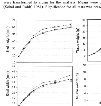
For the total growout period, all metric traits evaluated indicated significant effects of
Ž .
ploidy condition but only in the T5 group Table 2 . When comparisons were done for each growout age, no differences between groups for any of the traits evaluated were seen up to 81 days of growout, but from 118 days to the end of the experimental period,
Table 3
Ž .
Means and percent differences at age between the T5 0.5 mgrl treated group and the control groups, and between PTs and their internal controls or 2N-T5
Trait Growout T5 Control Percent T5 group Percent
days group group differences PTs 2N-T5 differences
scallops in the T5 group were significantly larger and heavier than both the control
Ž .
group and the T1 group Fig. 1 . Among the different traits evaluated, the largest differences were seen for weight traits, especially for wet tissue weight and muscle weight. At 205 days, total weight of the T5 group was 28% greater than the control, tissue weight was 37% heavier, and adductor-muscle weight showed the largest gain,
Ž .
63% heavier than the controls Table 3: T5 vs. control . Shell height and shell width increased by 9% and 10% at that same age, respectively. From 280 to 382 days, a reduction of the percent differences between scallops in the T5 group and the control
Ž
group was seen for tissue weight and muscle weight, but not for gonad weight Table 3:
Ž . Ž .
.
T5 vs. control; Fig. 1 . Scallops in the T5 group had a gonadal sac of similar size to the control and T1 groups during all the culture time, but gonads of triploid scallops were clearly distinguished from gonads of diploid scallops because of a brownish discol-oration, where no eggs or sperm could be seen. This allowed for the separation into PTs and 2N-T5 within the T5 group. By separating diploids from triploids on this basis, PTs were larger than those estimated from the whole T5 group containing a mixture of both triploid and diploid scallops. At 280 days, when the largest differences between the T5 group and the control group were seen, PTs had a total weight 86% greater than 2N-T5, a tissue weight 111% heavier, a muscle weight 182% heavier, and a gonad weight 101%
Ž .
heavier Table 3: PTs vs. 2N-T5 .
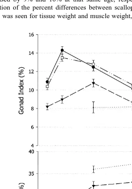
3.3. Gonad and muscle indices
Gonad and muscle indices of scallops in the T5 group were significantly different from both the control and the T1 groups. The gonad index of the T5 group was lower
ŽTable 2 , whereas the muscle index was larger than the control and T1 groups. An.
inverse relationship between muscle index and gonad index was characteristic of the
Ž .
control and T1 groups up to day 280 Fig. 2 , with the highest value of gonad index on day 146 corresponding with the lowest value of muscle index. The same was not true for the T5 group, for which the gonad index increased from days 118 to 205 in parallel with
Ž . Ž .
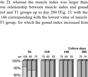
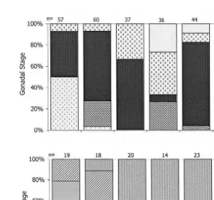
Fig. 3. Visual macroscopic maturation index of gonad development for the control, 0.1 mgrl T1 , and 0.5
Ž . Ž .
Ž .
the increase in muscle index Fig. 2 . When the indices are calculated again with the separated PTs, their gonad index was the lowest when compared with the control, and with the T5 group from which they were separated, and their muscle index was the
Ž .
highest Fig. 2 .
3.4. Visual maturation scale
After almost 4 months of growout, beginning days 118 up to 382, visual differences in maturation index were observed between the groups. Gonads of scallops in the control
Ž .
and T1 groups were partially mature or mature Fig. 3 , and egg and sperm portions of the gonadal sac were easily identified. For the T5 group, a larger percentage of the
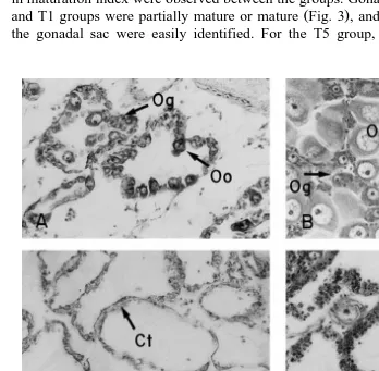
Fig. 4. Female gametogenesis in diploid and triploid catarina scallops. Only the female portion of the gonad
Ž . Ž .
for the functional hermaphrodite catarina scallop is shown. A initial stage of diploid, 20=; B maturity
Ž . Ž . Ž .
stage of diploid, 20=; C inactive stage of triploid, 20=; D early active ‘a’ stage, 20=; E early active ‘b’
Ž .
Ž
gonads was classified as immature, although a few scallops were mature possibly being
.
diploids, because of the large percent of diploids within the T5 group . For scallops within the T1 group, only a small percentage of gonads were classified as immature or undifferentiated up to day 280. The spent stage was not seen. Some scallops with partial spawns were classified as mature.
3.5. Gametogenesis
Ž .
Histological analysis showed that maturation in the control diploid scallops was normal. At 81 days of growout, about 50% of the scallops were either mature or
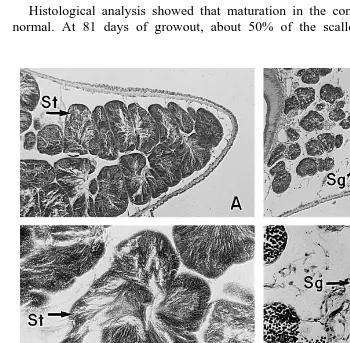
Fig. 5. Male gametogenesis in diploid and triploid catarina scallop. The male portion of the gonad for the functional hermaphroditic catarina scallop is shown in a, b, c, and d. Both, female and male portion of the
Ž . Ž .
gonad are shown in e and f. A Advanced stage of diploid, 10=; B early active stage of triploid male gonad
Ž . Ž . Ž .
part, 10=; C maturity stage of diploid, 20=; D early active stage of triploid, 20=; E maturity stage of
Ž .
spawned. Maturing individuals were seen during the whole study period thereafter. The first spent scallops were seen on day 205, indicating that the first massive spawn took place between days 146 and 205. By day 280, a large proportion of the individuals were
Ž .
seen again in an intermediate gametogenesis Figs. 4A,B and 5A,C,E . All diploid individuals were hermaphrodites. At the microscopic level, triploid gonads were easily distinguished from diploids because gametogenesis in the triploid scallops was abnormal and different from diploids. Most scallops in the T5 group had an abnormal gonad development, but 30–45% had normal development, presumably because they were diploids. This percentage agreed well with what was seen with the macroscopic visual
Ž .
maturation index Fig. 3 , and with the triploid percentage determined by flow cytome-try for spat.
Because triploids within the T5 group could be easily detected as such at the microscopic level, their gametogenesis was followed separately from that in the diploids within the group. The abnormal development of gametogenesis in triploids was sepa-rated into four stages, described in Table 4. Using those stages to define gonad development for triploids, we can see that the female portion of the gonad in triploid
Ž .
scallops was maturing but it was delayed when compared with diploids Fig. 6 . On day
Ž . Ž .
81, most of the triploid PTs gonads were in the inactive stage Fig. 4C . By day 118, a
Ž .
large proportion were in early active ‘a’ stage Fig. 4D but some still remained in the inactive stage. By day 146, less than 20% of female gonad part were inactive, and the
Ž
remaining were divided about equally between early active ‘a’ and early active ‘b’ Fig.
.
4E stages of development. The male gonad part did not mature further than the early
Ž .
active ‘a’ stage Fig. 5B,D . By days 205–280, most triploid female gonads were in early active ‘b’ stage. By day 382, all triploid scallops were in an indeterminate stage of
Ž .
development Fig. 4F .
Table 4
Gametogenic stages in triploid scallops, defined from histological data
Stage Characteristics Figure
A, inactive In the female part of the gonad, there were abundant acini, with Fig. 4C extensive layers of oogonies in their walls, but no oocytes were present.
acini were retarded showing few spermatogonies with a reduced size.
B, early active ‘a’ The difference with Stage A was the presence of few oocytes in the Fig. 4D female acini, often abnormal, although some oocytes were apparently
normal and mature.
In the male portion of the gonad, development of the acini were retarded Fig. 5B,D showing few spermatogonies with a reduced size.
C, early active ‘b’ This stage has the same morphology of Stage B for the female gonad, Fig. 4E with the presence of a few maturing and mature oocytes, but the acini
were reduced.
The male portion of the gonad was often occupied with female acini, Fig. 5F where some oocytes were developing. Scallops in this stage was
classified as ‘female’ sex.
D, indeterminate The gonad was empty, with abundant connective tissue dispersed and Fig. 4F the acini filled with abundant haemocytes. There was no evidence of
Ž . Ž .
Fig. 6. Frequency of each gametogenic stage for the female part of the gonad in a diploids and b triploids for the growout period. Ploidy condition was detected on the basis of histological inspection of gonadal
Ž .
development. Numbers sampled n are included on each column.
Ž .
When gonads of triploid 3N-T5 were histologically compared with those of diploids
Ž .
from the untreated control control for oocytes, there were differences in mean oocyte
Ž . Ž . Ž
size early in the culture 118 days Table 5 . At that age, the oocytes of triploids 27.8
. Ž . Ž .
mm were smaller than diploid oocytes 40.3mm Table 5 . When oocytes of 3N-T5
Table 5
Ž . Ž . Ž .
Oocyte size mm comparisons at age for triploids 3N-T5 and diploids from the control group Control , and
Ž . Ž .
for triploids 3N-T5 and diploids from the treated 0.5 mgrl group 2N-T5
Different letters between columns indicate significant differences between diploid and triploids.
Ž . Ž .
Days of growout Oocyte size mm Oocyte size mm
Diploid Triploid Diploid Triploid
Control 3N-T5 2N-T5 3N-T5
c a b a
118 40.3 27.8 32.1 27.8
b b,c c,d c,d
146 36.5 37.4 39.0 37.4
b,c b,c c,d d
205 37.6 40.5 38.2 40.5
c c b,c d
Table 6
Observed sex ratios during growout for triploid scallops within the T5 group, estimated from histological data
2
Ž . Ž .
Days of culture Percent hermaphrodites Percent females only x test significance P
81 100 0 1.00
were compared with 2N-T5, the differences at 118 and 280 days were significant, but
Ž .
only at 280 days were the oocytes of 3N-T5 scallops larger 41.6 mm than those of
Ž .
2N-T5 35.3 mm . At 146 and 205 days, oocytes of 3N-T5 were not significantly different from oocytes of 2N-T5. By day 382, there were a large number of oocytes in the diploid scallops, but no oocytes were seen in triploid scallops.
3.6. Functional hermaphroditism
Within each sampling period and for the triploid scallops within the T5 group, histological data showed an abnormal decrease in the number of gonads that were hermaphroditic. The frequency of hermaphroditic triploid scallops went from 100% at
Ž . Ž
day 81 to only 4% at day 280 Table 6 . For diploid scallops within the T5 group as
.
well as within the control group , all gonads were hermaphroditic for all sampling times. There was only one exception within the T5 group, where one ‘only male’ scallop was seen.
4. Discussion
4.1. Growth
Whereas some differences as early as larval stages have been shown to exist between
Ž .
diploid and triploid Yamamoto et al., 1988; Beaumont and Kelly, 1989 , it is generally accepted that triploid shellfish are not much different from diploids before beginning
Ž .
maturation Guo and Allen, 1994a . This was true for the catarina scallop, for which the
Ž .
growth advantage was first seen after about 3 months of growout )118 days , when diploid scallops were largely mature. The largest difference between the control and the T5 groups was seen at 205 days, after the maturity peak at 146 days. At 205 days, tissue weight was 37% larger and muscle weight was 63% larger in the T5 group. Triploid
Ž
mollusks were significantly larger than diploids in almost all species studied Guo and
.
Allen, 1994a . Triploid advantage was observed in Argopecten irradians: tissue weight
Ž .
36% larger and muscle weight 73% larger Tabarini, 1984 . Also, in C. nobilis triploids,
Ž .
wet and dry tissue weights were 32–52% heavier Komaru and Wada, 1989 ; in
ŽJiang et al., 1993 ; in C. gigas, tissue weight was 80% heavier Akashige, 1990 , and in. Ž .
Ž .
M. lateralis, tissue weight was 72% heavier Guo and Allen, 1994a .
Another reported characteristic in mollusks associated with triploid sterility is the
Ž .
reduction in gonad size. An exception was noted by Tabarini 1984 who found that triploid scallops actually had a heavier wet gonad weight than diploid scallops. In our results, lack of statistical differences in gonad weight between the treated and the control groups contrasted with visual observations, which indicated that triploid scallops had a gonadal sac larger than diploids, although it had few or no eggs, and no macroscopically visible sperm as diploids did. The significance of the larger gonadal sac in triploids was confirmed only when PTs were compared against diploids from the treated groups
Žtreated diploidss2N-T5 ; gonads of PTs were 100% heavier than those of diploids. In.
spite of the larger gonadal sac of triploids, gonad index of the T5 scallops was reduced when compared with the control group from 118 to 280 days. Because that was paralleled with a high muscle index, the low gonad index can be attributed to a large muscle weight rendering the wet tissue weight of triploids larger than for the diploid
Ž .
control. Whereas, Tabarini 1984 found no decreased gonad index for triploid bay
Ž .
scallop, Komaru and Wada 1989 found diploids to have a 62% greater gonad index than triploids of C. nobilis, which is very close to our results of a gonad index 61%
Ž .
greater in diploid than in triploid catarina scallops at the peak of maturity 146 days . Up
Ž
to 280 days of growout, PTs, followed by the T5 group mixture of triploids and
.
diploids , had the lowest gonad indices and highest muscle indices observed. However,
Ž .
for all groups, both indices decreased later during the culture 382 days , probably because of the abnormal environmental conditions present that year of El Nino 1997–
˜
1998, when unusually high temperatures were present. High temperatures are known to
Ž .
affect gonad maturation in scallops Sastry, 1963; Barber and Blake, 1991 , which would result in a decreased gonad index, and this is probably associated with the
Ž
expected decrease in phytoplankton biomass during years of El Nino Lluch-Cota et al.,
˜
. Ž . Ž .
1999 . Tran et al. 1993 cited by Lluch-Cota et al., 1999 reported a 60% decrease in phytoplankton biomass during El Nino 1982–1983. A similar, or even larger decrease
˜
might have occurred during El Nino 1997–1998, which would explain the decrease in
˜
muscle index, even in triploids.
4.2. Growth of PTs
When PTs were compared against 2N-T5, the differences in muscle weight and tissue
Ž .
weight between triploid and diploid scallops were striking Table 3 . The catarina scallop is known to have an early sexual maturity when environmental conditions are
Ž .
appropriate Cruz, Rodriguez-Jaramillo, and Ibarra, unpublished data , with partial
Ž .
apparently just stored but seldom used for maturation of gametes because few gametes develop, which could result in a continuous increase in size of the adductor muscle.
A hypothesis to explain the large difference found between triploid and diploid
Ž .
mollusks was proposed by Guo and Allen 1994a : the differences are caused by the occurrence of polyploidy gigantism in triploids resulting from an increased cell volume paired with lack of cell-number compensation. For scallops, particularly for adductor-muscle weight, an additional hypothesis can be proposed to explain the large increase in that tissue over the others. In scallops, one of the main sites for storage of energy
Žglycogen is the adductor muscle Martinez and Mettifogo, 1998; Barber and Blake,. Ž .
1991 . If glycogen is not used in triploids for maturation, its concentration will increase over time. Glycogen accumulation in cells could result in an increased size of the muscle because of cell gigantism, which will not necessarily be caused only by an additional set of chromosomes, but also by the unused and accumulated glycogen as it is known to
Ž .
occur in the liver of vertebrates, where glycogen is stored Stryer, 1981 . Support for
Ž .
this hypothesis comes from Tabarini’s 1984 results, finding glycogen concentrations in adductor muscle of triploid A. irradians as high as 135% of that from adductor muscle of diploid scallops. Furthermore, in the present study, muscle weight of triploid scallops also decreased as it did in diploid scallops when both were kept up to 382 days. Environmental conditions during our study were unusual, because El Nino 1997–1998
˜
Ž
was in progress, with increased temperatures and decreased productivity Lluch-Cota et
.
al., 1999 . If polyploidy gigantism was the only cause of larger muscle weight, muscle weight should not have decreased. However, if glycogen reserves in excess were the cause of a heavier weight, then muscle weight will probably decrease as glycogen is used during those harsh conditions.
4.3. Gametogenesis and hermaphroditism
In diploid scallops, gametogenesis was normal and similar to what Villalejo-Fuerte
Ž .
and Ochoa 1993 had previously reported for this functional hermaphroditic species. However, in triploid scallops, both the gametogenesis process and the normal condition of functional hermaphroditism were largely affected by the triploid condition. The effects of triploidy on gametogenesis were similar to that reported for C. nobilis
ŽKomaru and Wada, 1989 , M. arenaria Allen et al., 1986 , and S. commercialis Cox. Ž . Ž .
et al., 1996 . In contrast to those species, the catarina scallop is a functional hermaphrodite with simultaneous presence of male and female gonadal portions, matur-ing synchronously. In this study, the effects of triploidy seen in the male and female portion of the gonad resulted in differences in synchronization of the gonad maturation process. The male portion of the gonad, seen only during the early culture days, was more severely affected by the triploid condition than the female portion; most of the spermatogonia were arrested early in development. This agrees with results reported for males of the sequential hermaphrodite C. nobilis, and for non-hermaphrodites such as
Ž
M. arenaria, S. commercialis, and Mercenaria mercenaria Allen et al., 1986; Komaru
.
and Wada, 1989; Cox et al., 1996; Eversole et al., 1996 , where spermatozoa develop-ment was hindered, although not necessarily suppressed. For Mytilus galloproÕincialis,
Ž .
produced by diploids. In contrast, for C. gigas, P. fucata, and Crassostrea Õirginica, Ž
spermatozoa has been produced in triploids Allen and Downing, 1986; Allen et al.,
.
1986; Komaru and Wada, 1990 . Our results were different. Not only was there a lack of further development to form spermatozoa in triploid catarina scallop, but the male
Ž .
portion of the gonad was gradually replaced with female acini Fig. 5F , such that by day 280, 96% of the triploid scallops were sexed as ‘female only’. Though unused or
Ž .
unspawned, mature oocytes are recycled through lysis Pazos et al., 1996 . In our study, the male acinus did not show any evidence of being recycled through lysis. The phagocytic cells observed in Stage D were seen only at the end of the growout but not
Ž .
when the male acini were being replaced by female acini days 118–280 . The mechanism of progressive suppression of the male gonad seen during the culture for the triploid population is unknown, but Sertoli cells in phagocytosis of germ cells as
Ž . Ž .
suggested by Pipe 1987 cited by Kiyomoto et al., 1996 might have been involved. Abnormalities in the functional hermaphroditic condition of this species have been observed previously. For example, the occurrence of only female gonads have been observed in natural and hatchery-reproduced populations of this species at a low frequency, whereas the occurrence of only male gonads has also been seen, but at a much lower frequency, indicating the normal hermaphroditic condition can be altered
ŽIbarra, unpublished data ..
Different hypotheses for sex determination in mollusks have emerged as a
conse-Ž
quence of abnormal sex ratios observed when triploidy is induced Kiyomoto et al.,
.
1996 , but this is the first case reported for which a functional hermaphrodite bivalve is shown to have an altered sexuality caused by triploidy. The genetics underlying the hermaphroditic condition in bivalves has not been elucidated to this day. For
Cras-sostrea oysters, in which protandric sex change, dioecy, and hermaphroditism exist, Guo
Ž . Ž
et al. 1998 concluded that sex is determined by two alleles at a single loci with a
.
dominant ‘M’ allele and a recessive ‘F’ allele , and that the occurrence of rare functional hermaphrodites could be caused by developmental or genetic abnormalities. For other species, for which functional hermaphroditism is the rule, as for example, hermaphrodite nematodes, it is known that both sex chromosomes and autosomal loci
Žsex factors or genetic determinants are involved in determining the fate of sexual.
Ž .
development in these organisms. Triploid Caenorhabditis sp. individuals XXX AAA
Ž .
are hermaphrodites, whereas incomplete triploids XX AAA are males. Mutations in different genes in autosomes also produce abnormal sex, independently of the genotype
Ž .
of sex chromosomes Bull, 1983 . Whether or not sex chromosomes do exist in catarina scallop, the presence of an extra set of chromosomes does not suppress initial differenti-ation of the male gonad part, which could indicate a deterministic role of sex chromo-somes on sexual differentiation, as was evidenced by the presence of male gonads early during gametogenic development. However, the triploid condition suppresses further male gonad development, resulting in replacement of male germinal tissue by female germinal tissue as evidenced by the presence of some oocytes in the male part of the gonad. This could indicate multipotency of germinal cells in becoming either sex. As
Ž .
seem to be multipotent because changes in sex are seen during their life cycle, in scallops as the catarina scallops, the gonadal sac is divided into two compartments clearly differentiated as male and female from early in development of the gonad, and both gonadal compartments are always present during the life cycle of the scallop. We can speculate on the presence of genes involved in early differentiation of each compartment, with other genes acting later to maintain a sexual differentiation for each compartment in a multipotent gonadal tissue. Abnormalities in the expression of these later gene products caused by the triploid condition could result, for example, in overexpression of genes conferring female characteristics.
Contrary to that seen in the male portion of the gonad of triploids, gametogenesis in the female portion was retarded but similar to diploid scallops, although few oocytes developed. The early active ‘b’ stage described here for the female gonad of triploids, in which the walls of female acini were lined with oogonies but oocytes were scarce and
Ž
abnormal, has been observed in almost all triploid female bivalves Allen et al., 1986;
.
Komaru and Wada, 1989; Cox et al., 1996; Eversole et al., 1996 . The presence of large numbers of phagocytic cells in triploid gonads by day 382 may indicate reabsorption of eggs. The cause of that possible failure to spawn is not known. We know that triploid catarina scallop can spawn because we have achieved this under laboratory conditions
ŽA.M. Ibarra, unpublished results ..
Finally, the non-conclusive results on differences in oocyte size between triploid and diploid catarina scallops deserve further research, which is under way. Differences in
Ž .
egg volume have been reported for other species. For example, Guo and Allen 1994b,c measured egg diameter in triploid Pacific oyster, and found an increase of 33–54% egg volume in triploids.
Acknowledgements
This research was supported by CONACyT grant number 1473PB to A.M. Ibarra. The senior author is a CONACyT graduate fellow, and the results presented here are part of his PhD thesis. We thank M. Romero, G. Lucero, and S. Montes from SEMARNAP, and S. Avila and P. Cruz from CIBNOR for technical support. C. Rodriguez-Jaramillo from CIBNOR did the histological processing, and Aldo Vargas, the photography prints. The English-language text was edited by Dr. Ellis Glazier
ŽCIBNOR ..
References
Akashige, S., 1990. Growth and reproduction of triploid Japanese oyster in Hiroshima Bay. In: Advances in Invertebrate Reproduction. Elsevier, pp. 461–468.
Ž .
Allen, S.K. Jr., Downing, S.L., 1986. Performance of triploid Pacific oyster, Crassostrea gigas Thunberg : Part I. Survival, growth, glycogen content, and sexual maturation in yearlings. J. Exp. Mar. Biol. Ecol. 102, 197–208.
Allen, S.K. Jr., Downing, S.L., Chew, K.K., 1989. Hatchery Manual for Producing Triploid Oyster. University of Washington Press, 27 pp.
Allen, S.K. Jr., Gagnon, P.S., Hidu, H., 1982. Induced triploidy in the soft-shell clam. J. Hered. 73, 421–428. Allen, S.K. Jr., Hidu, H., Stanley, J.G., 1986. Abnormal gametogenesis and sex ratio in triploid soft-shell
Ž .
clams Mya arenaria . Biol. Bull. 170, 198–210.
Ž .
Barber, B.J., Blake, N.J., 1991. Reproductive physiology. In: Shumway, S.E. Ed. , Scallops: Biology, Ecology and Aquaculture. Elsevier, Amsterdam, pp. 377–428.
Beaumont, A.R., Fairbrother, J.E., 1991. Ploidy manipulation in molluscan shellfish: a review. J. Shellfish Res. 10, 1–18.
Beaumont, A.R., Kelly, K.S., 1989. Production and growth of triploid Mytilus edulis larvae. J. Exp. Mar. Biol. Ecol. 132, 69–84.
Ž .
Benninger, P.G., LePennec, M., 1991. Functional anatomy of scallops. In: Shumway, S.E. Ed. , Scallop Biology, Ecology and Aquaculture. Elsevier, pp. 133–223.
Bull, J.J., 1983. Evolution of sex determining mechanisms. BenjaminrCummings Pub., 316 pp.
Cox, E.S., Smith, M.S.R., Nell, J.A., Maguire, G.B., 1996. Studies on triploid oyster in Australia: VI. Gonad
Ž .
development in diploid and triploid Sydney rock oysters Saccostrea commercialis Iredale and Roughley . Aquaculture 197, 101–120.
Cruz, P., Ramirez, J.L., Garcia, G.A., Ibarra, A.M., 1998. Genetic differences between two populations of
Ž .
catarina scallop ArgopectenÕentricosus for adaptations for growth and survival in a stressful environ-ment. Aquaculture 166, 321–335.
Eversole, A.G., Kempton, C.J., Hadley, N.H., Buzz, W., 1996. Comparison of growth, survival, and reproductive success of diploid and triploid Mercenaria mercenaria. J. Shellfish Res. 15, 689–694.
Ž .
Felix-Pico, E., 1991. Fisheries and aquaculture of scallops, Mexico. In: Shumway, S.E. Ed. , Scallops Biology, Ecology and Aquaculture. Elsevier, pp. 943–980.
Ž
Guo, X., Allen, S.K. Jr., 1994a. Sex determination and polyploid gigantism in the Dwarf surfclam Mulinia
.
lateralis Say . Genetics 138, 1199–1206.
Ž .
Guo, X., Allen, S.K. Jr., 1994b. Viable tetraploids in the Pacific oyster Crassostrea gigas Thunberg
produced by inhibiting polar body I in eggs from triploids. Mol. Mar. Biol. Biotechnol. 3, 42–50. Guo, X., Allen, S.K. Jr., 1994c. Reproductive potential and genetics of triploid Pacific oysters, Crassostrea
Ž .
gigas Thunberg . Biol. Bull. 187, 309–318.
Guo, X., Hedgecock, D., Hershberger, W.K., Cooper, K., Allen, S.K., 1998. Genetic determinants of
Ž . Ž .
protandric sex in the Pacific oyster, Crassostrea gigas Thunberg . Evolution 52 2 , 394–402.
Hand, R.E., Nell, J.A., Maguire, G.B., 1998. Studies on triploid oysters in Australia: X. Growth and mortality
Ž .
of diploid and triploid Sydney rock oysters Saccostrea commercialis Iredale and Roughley . J. Shellfish Res. 17, 1115–1128.
Hawkins, A.J.S., Day, A.J., Gerard, A., Naciri, Y., Ledu, C., Bayne, B.L., Heral, M., 1994. A genetic and´ ´ metabolic basis for faster growth among triploids by blocking meiosis I but not meiosis II in the larviparous European flat oyster, Ostrea edulis L. J. Exp. Mar. Biol. Ecol. 184, 21–40.
Ž .
He, M., Lin, Y., Jiang, W., 1996. Studies on the sterility of triploid pearl oyster, Pinctada martensii D. .
Ž .
Trop. Oceanol. 15 2 , 17–21, Redai Haiyang.
Heasman, M.P., O’Connor, W.A., O’Connor, S.J., Walker, S., 1998. Enhancement and farming of scallops in NSW using hatchery produced seedstock. In: NSW Fish. Final Rep. Ser., Cronulla, NSW Australia NSW Fisheries 1998, No. 8, 146 pp.
Ibarra, A.M., 1999. Correlated responses for all growth traits to selection for total weight and shell width in
Ž .
catarina scallop scallop ArgopectenÕentricosus . Aquaculture 175, 243–254.
Ibarra, A.M., Ramirez, J.L., Ruiz, C.A., Cruz, P., Avila, S., 1999. Realized heritabilities and genetic
Ž .
correlation for total weight and shell width in catarina scallop ArgopectenÕentricosus . Aquaculture 175, 227–241.
Jiang, W., Li, G., Xu, G., Lin, Y., Qing, N., 1993. Growth of the triploid pearl oyster, Pinctada martensii
ŽD. . In: Gall, G.A.E., Chen, H. Eds. , Genetics in Aquaculture IV. Proceedings of the Fourth Interna-. Ž .
tional Symposium, 29 April–3 May 1991, Wuhan, China. . Aquaculture 111, 245–253.
Kiyomoto, M., Komaru, A., Scarpa, J., Wada, K.T., Danton, E., Awaji, M., 1996. Abnormal gametogenesis, male dominant sex ratio, and sertoli cell morphology in induced triploid mussels, Mytilus galloproÕ
Komaru, A., Wada, K.T., 1989. Gametogenesis and growth of induced triploid scallops Chlamys nobilis.
Ž .
Nippon Suissan Gakkaishi 55 3 , 447–452.
Komaru, A., Wada, K.T., 1990. Gametogenesis of triploid Japanese pearl oyster, Pinctada fucata martensii. In: Advances in Invertebrate Reproduction 5. Elsevier, pp. 469–474.
Lluch-Cota, D.B., Lluch-Belda, D., Lluch-Cota, S.E., Lara-Lara, J.R., Hammann, M.G., Morales, Y.J., Lopez-Martınez, J., Nevarez-Martınez, M.O., Ponce-Dıaz, G., Salinas-Zavala, C.A., Vega-Velazquez, A.,´ ´ ´ ´ ´
Ž .
1999. Las pesquerias y El Nino. In: Magana, R.V.O.˜ ˜ Ed. , Los Impactos de El Nino en Mexico.˜ ´ SG-UNAM-IAI-SEPrCONACyT, Mexico, D.F., pp. 137–179.´
Martinez, G., Mettifogo, L., 1998. Mobilization of energy from adductor muscle for gametogenesis of the scallop, Argopecten purpuratus Lamarck. J. Shellfish Res. 17, 113–116.
Nell, J.A., Cox, E., Smith, I.R., Maguire, G.B., 1994. Studies on triploid oyster in Australia: I. The farming
Ž .
potential of triplod Sidney rock oysters Saccostrea commercialis Iredale and Roughley . Aquaculture 126, 243–255.
Pazos, A.J., Roman, G., Acosta, C.P., Abad, M., Sanchez, J.L., 1996. Influence of the gametogenic cycle on´ ´ the biochemical composition of the ovary of the great scallop. Aquacult. Int. 4, 201–213.
Pipe, R.K., 1987. Ultrastructural and cytochemical study on interactions between nutrient storage cells and gametogenesis in the mussel, Mytilus edulis. Mar. Biol. 96, 519–528.
Racotta, I.S., Ramirez, J.L., Avila, S., Ibarra, A.M., 1998. Biochemical composition of gonad and muscle in the catarina scallop, ArgopectenÕentricosus, after reproductive conditioning under two feeding systems. Aquaculture 163, 111–122.
Ramirez, J.L., Avila, S., Ibarra, A.M., 1999. Optimization of forage in two food-filtering organisms with the use of a continuous, low-food concentration, agricultural drip system. Aquacult. Eng. 20, 175–189. Sastry, A.N., 1963. Reproduction of the bay scallop, Aequipecten irradians Lamarck. Influence of
tempera-ture on maturation and spawning. Biol. Bull. 125, 146–153. Sokal, R.R., Rohlf, F.J., 1981. Biometry. WH Freeman, New York.
Stanley, J.G., Allen, S.K. Jr., Hidu, H., 1981. Polyploidy induced in the American oyster Crassostrea Õirginica, with cytochalasin B. Aquaculture 12, 1–10.
Stanley, J.G., Hidu, H., Allen, S.K., 1984. Growth of American oysters increased by polyploidy induced by blocking meiosis I but not meiosis II. Aquaculture 37, 147–155.
Stryer, L., 1981. Biochemistry. WH Freeman, New York, 949 pp.
Tabarini, C.L., 1984. Induced triploidy in the bay scallop, Argopecten irradians, and its effect on growth and gametogenesis. Aquaculture 42, 151–160.
Tran, A.V., Hyon, J., Evans, R., Brown, O., Feldman, G., 1993. Satellite-derived multichannel sea surface temperature and phytoplankton pigment concentration data: a CD-ROM set containing monthly mean distributions for global oceans. USA-NASA-JPL-PODAAC-A001-A005 ver. 1, Jet Propulsion Laboratory, 32 pp.
Ž
Villalejo-Fuerte, M., Ochoa, R.I., 1993. The reproductive cycle of the scallop Argopecten circularis Sowerby
.
1835 in relation to temperature and photoperiod, in Bahia Concepcion, B.C.S. Mexico Ciencias Marinas
Ž .
19 2 , 181–202.
Wada, K.T., Komaru, A., Uchimura, Y., 1989. Triploid production in the Japanese pearl oyster, Pinctada
fucata martensii. Aquaculture 76, 11–19.
Yamamoto, S., Sugawara, Y., 1988. Induced triploidy in the mussel Mytilus edulis, by temperature shock. Aquaculture 72, 21–29.
Yamamoto, S., Sugawara, Y., Nomura, T., Oshino, A., 1988. Induced triploidy in Pacific oyster Crassostrea
gigas, and performance of triploid larvae. Tohoku J. Agric. Res. 39, 47–59.
Zeng, Z., Chen, M., Lin, Q., Chen, P., Liu, W., Chen, Y., 1995. Induced triploidy in scallop, Chlamys nobilis.
Ž .