DETERMINATION MODEL OF CARCINOMA AND
BENIGN TYPES USING PHYSICAL QUANTITIES
OF THE CA MAMMAE MAMMOGRAPHY
Anak Agung Ngurah Gunawan, I Nyoman Widana
School of Physic, Faculty Mathematics and Natural Sciences, Udayana University, Denpasar, Bali. Tel. 62-0361-484238 E-mail : [email protected]
The research is financed by: Dirjen Dikti Indonesia
Abstract
The article had been built the model to determine the carcinoma and benign types. Potentially, the physical quantities could be differ of each them. The biopsy methods had developed in classifying the both carcinoma and benign types. In this research, the probability of gray-level pairs on certain distance are derived from the mathematical model of the mammography physical quantities are used. In the previous research, determination of infiltrating ductal carcinoma and infiltrating lobuler carcinomahistopathology types, breast cancer stadium, health level of contra lateral,andreadability increasing x-ray mammography images on histopathology types determination on breast cancer using special patern cropping are succeeded. In this research, the mathematical model of the mammography physical quantities is succeeded to determine the carcinoma and benign histopathology types. The model had been proposed for testing 120 the new patients mammogram at Dokter Soetomo Hospital, Surabaya, Indonesia. The research results showed the physical quantities could be predict the carcinoma and benign histopathology types in 86,67 %sensitivity for 2 x 2 cm sample sized and α = 5 %.
Keywords : mammography, ca mammae, carcinoma, benign.
1. Introduction
The following methods of the breast cancer early detection had been patented are employing histogram
peak detection [1], histogram alteration [2], pixel mapping [3], neural network [4], contour [5], wavelet
presence of microcalcification only, but determination of the type of carcinoma and benign breast
cancer are not supported.The classifying types of Infiltrating Duktal Carcinoma and Infiltrating
Lobuler Carcinoma breast cancer histopathology using physical parameter in86,36 % sensitivityin
previous research are succeeded [11]. The determination of the level of the contra lateral breast health
using physical parameters in 93.75% sensitivity [12], the breast cancer stadium state in 86.67%
sensitivity of [13]. Also, the readibility x-rays results of mammography on breast cancer
histopathological type determination using a special pattern croping through physical parameters in
97.5% sensitivity are succeeded too [14].
This paper organized as follows, section 2 discussing the radiation intensity on ca mammae, section 3
about physical quantities of the result of X-ray mammography images, the probability function in
section 4, section 5 about the multinomial linear regression function as an outcome types of carcinoma
and benign, results and discussion in section 6 and the last section are conclusions.
2. The Radiation Intensity on Ca Mammae
Increase of the radiation absorption intensity depends on breast cancer density increasing due to pixel
intensity values of carcinoma and benign types are different. So that the physical parameters between
the types of carcinoma and benign are different. The transmittance of the X-ray radiation intensity are
written in equation (2.1) bellow [11][12][13][14].
I1 = I0 e –μd (2.1)
where I1, I0, μ, d are transmitted beam intensity, the intensity of light at first, absorption coefficient,
breast cancer density, respectively.
3. Physical Quantities of X-Ray Mammography Results
The following physical parameters are nonuniformity, contrast, uniformity, local homogeneity,
correlation, feature represents the nature, feature represents the density, nonuniformity of hdiff,
uniformity of hdiff and the feature represents the nature of hdiff derived from the x-ray mammography
results. All the physical parameters are written as follows equations [11][12][13][14][15].
(3.1)
(3.2)
(3.4)
for
(3.5)
where,
(3.6)
(3.7)
(3.8)
(3.9)
(3.10)
(3.11)
(3.12)
(3.13)
With H(yq, yr, d) is distribution of probability of occurrence of a pair of gray-level value separated by
a given displacemen vector d.
4. Probability Function
Consider probability functions bellows, ) and dependent in , for liniearly
independent each others, are fulfilled. For is output categories i.e for ,
carcinoma, , benign, and others. The logistic functions are written as follows:
(3.13)
for example,
,
The qualitative mapping the entropy to carcinoma and benign ca mammae are in abnormal Gaussian
satisfied. So that,
, or ,
,
,
,
and as statistics model of logistics multinomial regression, for , will be
in all the cathegories are , furthermore:
For all the histopathology categories are fullfiled with,
5. The multinomial linear regression function as the outcome of carcinoma and benign types
where Zk is outcome for , entirely. Zk0 as outcome initial values for ,
with . is pertubation parameters for a
number are in linear form. For last term, as outcome correction factors for all
. For example:
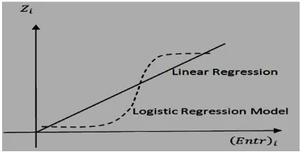
Illustratively, the above expression are depicted in figure 1 as bellows.
Figure 1. Logistic and Linear Regression Model
6. Results and Discussion
Figures 2, 3, are types of carcinoma and benign mammogram images, respectively.
Mammography images had been taken from radiology installations at Dr. Soetomo Hospital,Surabaya,
RSUP Sanglah and RSU. Primamedika, Denpasar, supported by Sony Brand ICR type 3600M
apparatus. All the images in bmp format are stored which sample sized in 2 cm x 2 cm matrices.
Table 1. Physical Quantity Intervals
No Physical Quantity Benign Carcinoma
1 Anguler Secound Moment 0,00015 - 0.01013 0.00013 - 0.08280 2 Inverse Difference Moment 0.01527 - 0.08977 0.01106 - 0.39200
3 Mean 71.55468 - 195.80523 71.16284 - 220.92240
4 Deviation 18.84672 - 56.86512 11.06751 - 93.33126
5 Entropy of the Difference Second Order
Histogram 1.32977 - 2.05756 1.29424 - 2.14790
6 The Second Anguler Moment of the
Difference Second Order Histogram 0.01051 - 0.05878 0.00806 - 0.11134
7 Mean of the Difference Second Order
Histogram 7.65163 - 43.77752 7.27355 - 55.92737
All the physical quantities above formulated bellows:
Z:= -17056.786 + 13939360.273*MA[9] -14975532.439*MA[10] + 79507.135*MD[5] +
123275.512*MD[6] 52858.798 * MD[7] 29317.721*MD[8] 46033.962*MD[9]
84405.247*MD[10] 2616.686*MN[1] + 5924.284*MN[2] 3119.844*MN[3] 453.778*MN[4]
1114.523*MN[5] + 3720.727*MN[6] 3618.971*MN[7] + 11.610*MN[8] + 2152.569*MN[9]
885.095MN[10] 1327.391*D[1] + 2202.098*D[2] 3.143*D[3] 3364.818*D[4] + 7916.137*D[5]
-10676.240*D[6] + 6323.275*D[7] + 415.555*D[8] -1671.692*D[9] + 197.645*D[10] +
23576.501*EH[1] + 7048.037*EH[2] -98617.823*EH[3] + 59177.808*EH[4] -53465.845*EH[5] +
81134.008*EH[6] + 743.337*EH[7] -81311.924*EH[8] + 74475.699*EH[9] -4742.767*EH[10] +
169258.070*MAH[1] + 31944.202*MAH[2] -1083908.718*MAH[3] + 826751.786*MAH[4] +
888282.531*MAH[5] -918006.126*MAH[6] -487749.444*MAH[7] -1455971.004*MAH[8] +
1382311.075*MAH[9] + 844659.147*MAH[10] 124.008*MHD[1] + 74.265*MHD[2]
-744.240*MHD[3] + 1741.103*MHD[4] + 430.726*MHD[5] -116.272*MHD[6] -1461.422*MHD[7]
-1022.368*MHD[8] + 894.912*MHD[9] + 346.808*MHD[10];
The uniformity, local homogeneity, feature represents the nature, feature represents the density, nonuniformity of hdiff, uniformity of hdiff, feature represents the nature of hdiff parameters are optimium to differ carcinoma and benign types in this research.
7. Conclusion
Determination of carcinoma and benign types on breast cancer using physical quantities on the 120
samples in 86.67% sensitivity are resulted with 16 samples are errors. That is the diagnostics
performance of the carcinoma and benign types are increased. The optimum physical parameters :
Angular Second Moment distance between pixels 9, 10, Inverse Difference Moment on the distance
between pixels 5,6,7,8,9,10, Mean, Deviation, Entropy of the Difference Second Order Histogram,
Angular Second Moment of the Difference Second Order Histogram, Mean of the Difference Second
Order Histogram on the distance between pixels 1,2,3,4,5,6,7,8,9,10 are obtained.
Acknowledgments
We would like to thank the Indonesian government has provided funding of research through the
National Competitive Fundamental Grant, Rector of Udayana University and chairman of LPPM Unud
which has provided research funding. Dean of the Faculty Udayana University and Chairman of the
Department of Physics Faculty Udayana University who has given permission to study, also we would
like to thank the director of hospital are Dr. Soetomo Hospital, Surabaya, RSUP Sanglah Hospital and
Primamedika Hospital, Denpasar, Indonesia for the research supporting.
Refference
[1] Muhammed I. Sezan,Ralph Schaetzing, (1988). Digital image processing method employing histogram peak detection, Patent Number: US 4731863.
[2] RyoheiKumagai,( 1991). Method for processing an image byhistogram alteration, Patent Number: US 5077808.
[3] Mark E. Faulhaber, Mark A. Momcilovich,( 1996). Raster image processing with pixel mapping to allow image border density allocation’, Patent Number: US 5485281.
[5] Isaac N. Bankman, William A. Christens-Barry,(1996). Method and system for automated detection of microcalcification clusters in mammograms, Patent Number: US 5574799.
[6] Maria Kallergi,(2008). Computer aided diagnosis of mammographic microcalcification clusters, Patent Number: US 7430308 B1.
[7] V. Suzanne Klimberg, soheliaKorourian, Steven Harms, Gal Shafirstein,(2010). Minimally invasive diagnosis and treatment for breast cancer , Patent Number: US 7769432 B2.
[8] DaoXian H. Zhang, Patrick B. Heffeman, Yue Shen,(2010). Computer aided detection of microcalcification clusters, Patent Number: US 7848555 B2.
[9] Gal Shafirrstein, XiaoWei Xu, Mutlu Mete, (2010). Image processing apparatus and method for histological analysis, Patent Number: US 7853089 B2.
[10] Sheldon Weinbaum, YuliyaVengrenyuk, Luis Cardoso, Lucas Parra, Stephane Carlier, SawasXanthos,(2014). System and method for in vivo imaging of blood vessel walls to detect microcalcifications, Patent Number: US 8,882,674 B2.
[11] A.A.N. Gunawan, Suhariningsih, K.S.P. Triyono, and B. Widodo, (2012). Determination of physical parameter model for the photo film mammographic X-ray results on the breast cancer histology classification, International Journal Of Contemporary Mathematical Sciences, 45, 2235-2244.
[12] A.A.N. Gunawan, Suhariningsih, K.S.P. Triyono, and Yasin, (2013). Conversion of Images into Numerical Modelsto Determine the Condition of Breast Healthon Contralateral, Applied Mathematical Sciences,104, 5185-5191.
[13] A.A.N. Gunawan, (2014). A Novel Model Determination of Breast Cancer Stage Using Physical Parameter, Far East Journal of Matematical Sciences, 87, 23-35.
[14] A. A. N.Gunawan, W.Supardi , I. B.Gede Dharmawan, (2014). Readability Increase Of Mammography X-Ray Photos Results In Determining The Breast Cancer Histopathology Types Using Special Pattern Cropping With Physical Parameter, Advances in Applied Physics, 2, 43-52.